

CD66 receptor specificity exhibited by neisserial Opa variants is controlled by protein determinants in CD66 N-domains
- PMID: 9689124
- PMCID: PMC21382
- DOI: 10.1073/pnas.95.16.9584
CD66 receptor specificity exhibited by neisserial Opa variants is controlled by protein determinants in CD66 N-domains
Abstract
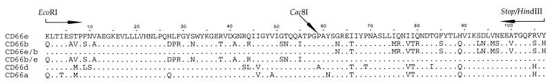
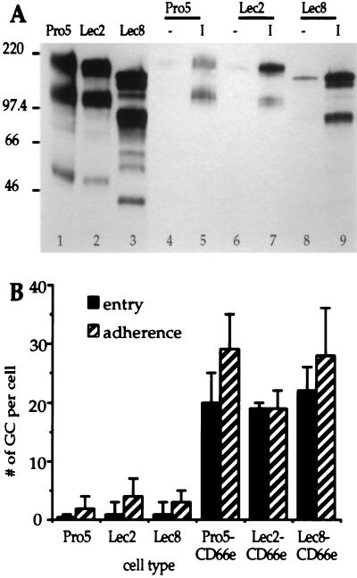
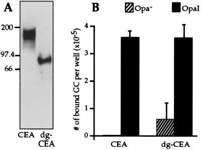
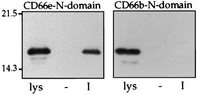
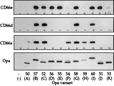
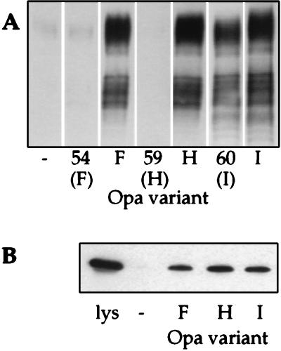
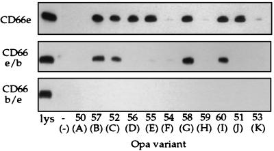
Neisseria gonorrhoeae strain MS11 is able to express 11 different opacity (Opa) proteins on its outer surface. A number of these Opa proteins have been shown to function as adhesins through binding of CD66 receptors present on human cells. CD66 antigens, or carcinoembryonic antigen family members, constitute a family of glycoproteins belonging to the immunoglobulin superfamily. Opa variants recognize this class of receptors in a differential manner such that certain Opa variants recognize up to four different CD66 receptors (CD66a, -c, -d, and -e), whereas others recognize only two (CD66a and -e) or none. We explored the basis for this receptor tropism in the present study. Our data show that glycoforms of CD66e and deglycosylated CD66e are recognized by gonococci in an Opa-specific manner. Binding by Opa variants of recombinant N-terminal domains of CD66 receptors expressed in Escherichia coli reflected the adherence specificities of Opa variants to HeLa cells expressing native CD66 molecules. These data indicate that recognition of CD66 receptors by Opa variants is mediated by the protein backbone of the CD66 N-domains. Furthermore, by using chimeric constructs between different CD66 N-domains we identified distinct binding regions on the CD66e N-domain for specific groups of Opa variants, suggesting that the differential recognition of CD66 receptors by Opa variants is dictated by the presence of specific binding regions on the N-domain of the receptor.
Figures
Similar articles
-
Critical determinants of host receptor targeting by Neisseria meningitidis and Neisseria gonorrhoeae: identification of Opa adhesiotopes on the N-domain of CD66 molecules.Mol Microbiol. 1999 Nov;34(3):538-51. doi: 10.1046/j.1365-2958.1999.01620.x. Mol Microbiol. 1999. PMID: 10564495
-
Homologue scanning mutagenesis reveals CD66 receptor residues required for neisserial Opa protein binding.J Exp Med. 1999 Aug 2;190(3):331-40. doi: 10.1084/jem.190.3.331. J Exp Med. 1999. PMID: 10430622 Free PMC article.
-
Differential Opa specificities for CD66 receptors influence tissue interactions and cellular response to Neisseria gonorrhoeae.Mol Microbiol. 1997 Dec;26(5):971-80. doi: 10.1046/j.1365-2958.1997.6342006.x. Mol Microbiol. 1997. PMID: 9426134
-
The structural basis of CEACAM-receptor targeting by neisserial Opa proteins.Trends Microbiol. 2000 Jun;8(6):258-60; discussion 260-1. doi: 10.1016/s0966-842x(00)01771-6. Trends Microbiol. 2000. PMID: 10838580 Review. No abstract available.
-
The role of neisserial Opa proteins in interactions with host cells.Trends Microbiol. 1998 Dec;6(12):489-95. doi: 10.1016/s0966-842x(98)01365-1. Trends Microbiol. 1998. PMID: 10036728 Review.
Cited by
-
Bacterial protein domains with a novel Ig-like fold target human CEACAM receptors.EMBO J. 2021 Apr 1;40(7):e106103. doi: 10.15252/embj.2020106103. Epub 2021 Feb 1. EMBO J. 2021. PMID: 33522633 Free PMC article.
-
Cell-cell adhesion molecule CEACAM1 is expressed in normal breast and milk and associates with beta1 integrin in a 3D model of morphogenesis.J Mol Histol. 2004 Mar;35(3):287-99. doi: 10.1023/b:hijo.0000032360.01976.81. J Mol Histol. 2004. PMID: 15339048 Free PMC article. Review.
-
Carcinoembryonic antigen family receptor recognition by gonococcal Opa proteins requires distinct combinations of hypervariable Opa protein domains.Infect Immun. 2002 Apr;70(4):1715-23. doi: 10.1128/IAI.70.4.1715-1723.2002. Infect Immun. 2002. PMID: 11895933 Free PMC article.
-
Defining the roles of human carcinoembryonic antigen-related cellular adhesion molecules during neutrophil responses to Neisseria gonorrhoeae.Infect Immun. 2012 Jan;80(1):345-58. doi: 10.1128/IAI.05702-11. Epub 2011 Nov 7. Infect Immun. 2012. PMID: 22064717 Free PMC article.
-
A bacterial siren song: intimate interactions between Neisseria and neutrophils.Nat Rev Microbiol. 2012 Jan 31;10(3):178-90. doi: 10.1038/nrmicro2713. Nat Rev Microbiol. 2012. PMID: 22290508 Free PMC article. Review.
References
-
- Kallstrom H, Liszewski M K, Atkinson J P, Jonsson A B. Mol Microbiol. 1997;25:639–647. - PubMed
-
- Meyer T F, Pohlner J, van Putten J P M. Curr Top Microbiol Immunol. 1994;192:283–317. - PubMed
-
- van Putten J P M, Duensing T D. Rev Med Microbiol. 1997;8:51–59.
-
- Bhat K S, Gibbs C P, Barrera O, Morrison S G, Jähnig F, Stern A, Kupsch E-M, Meyer T F, Swanson J. Mol Microbiol. 1991;5:1889–1901. - PubMed
-
- Stern A, Brown M, Nickel P, Meyer T F. Cell. 1986;47:61–71. - PubMed
Publication types
MeSH terms
Substances
LinkOut - more resources
Full Text Sources
