. 1998 Jun;42(6):836-44.
doi: 10.1136/gut.42.6.836.

Enteric neuronal plasticity and a reduced number of interstitial cells of Cajal in hypertrophic rat ileum

Affiliations

Enteric neuronal plasticity and a reduced number of interstitial cells of Cajal in hypertrophic rat ileum

E Ekblad et al. Gut. 1998 Jun.

Abstract

Background: Partial obstruction of the ileum causes a notable hypertrophy of smooth muscle cells and enteric neurones in the proximally located intestine.

Aims: To study the expression of neuromessengers in the hypertrophic ileum of rat as little is known about neuromessenger plasticity under these conditions. To investigate the presence of interstitial cells of Cajal (ICC) in hypertrophic ileum.

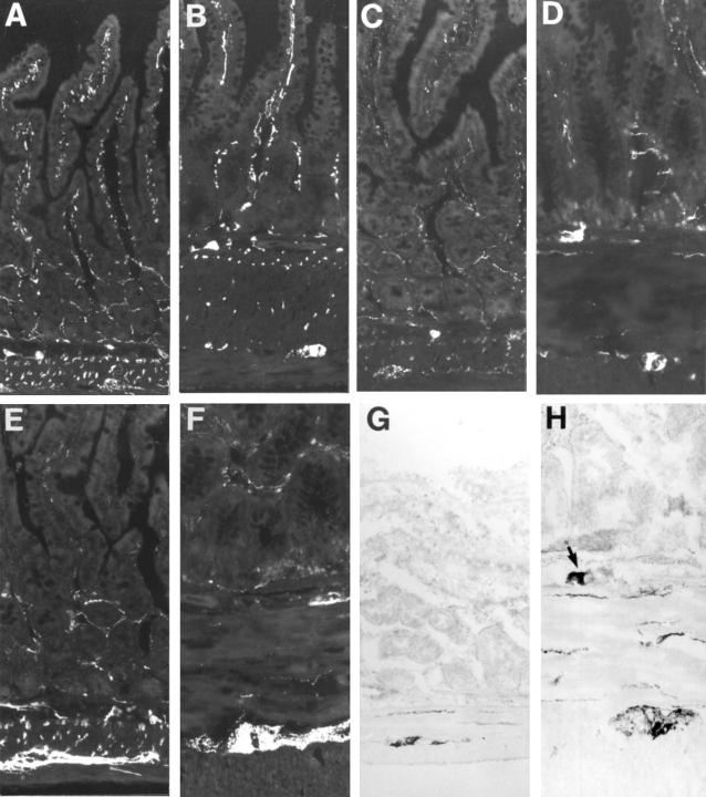
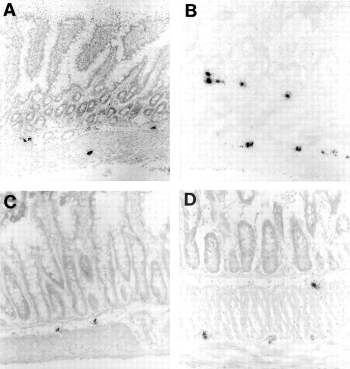
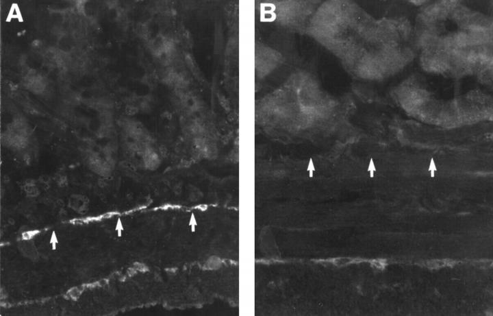
Methods: Ileal hypertrophy was induced by circumferential application of a strip of plastic film for 18-24 days. Immunocytochemistry, in situ hybridisation, nicotinamide adenine dinucleotide phosphate (NADPH) diaphorase histochemistry, and ethidium bromide staining were used to investigate the number of enteric neurones expressing neuropeptides and nitric oxide synthase, and the frequency of ICC.

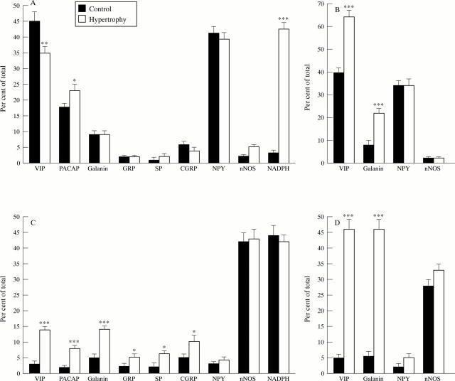
Results: In the hypertrophic ileum several neuronal populations showed changes in their expression of neuromessengers. Myenteric neurones expressing vasoactive intestinal peptide (VIP), pituitary adenylate cyclase activating peptide, and galanin were notably increased in number. In submucous ganglia the number of VIP immunoreactive neurones decreased while those expressing VIP mRNA increased. NADPH diaphorase positive submucous neurones increased dramatically while the number of neuronal type nitric oxide synthase expressing ones was unchanged. The number of ICC decreased notably in hypertrophic ileum.

Conclusion: Enteric neurones change their levels of expression of neuromessengers in hypertrophic ileum. ICC are also affected. The changes are presumably part of an adaptive response to the increased work load.


Figures

Figure 1
Figure 1
Cryostat sections from rat: control (A,C,E,G) and hypertrophic (B,D,F,H) ileum, immunostained for VIP (A,B), PACAP (C,D), and galanin (E,F) or NADPH diaphorase stained (G,H). The notable thickening of the smooth muscle layers in hypertrophic ileum and the decreased frequency of nerve fibres in these layers are obvious. The size of the neuronal cell bodies is increased in hypertrophic ileum. Note in (H) a large NADPH diaphorase positive nerve cell body within the submucosal ganglion (arrow); such cell bodies are commonly seen in hypertrophic ileum but are extremely rare in control tissue. Original magnification ×130.
Figure 2
Figure 2
Cryostat sections from rat: control (A,C) and hypertrophic (B,D) ileum autoradiographically labelled for VIP mRNA (A,B) and galanin mRNA (C,D). VIP mRNA containing nerve cell bodies are larger, more numerous, and more intensely labelled in hypertrophic ileum. The number of galanin mRNA containing nerve cell bodies is particularly increased in the myenteric ganglia where they are larger and more intensely labelled. Original magnification ×130.
Figure 3
Figure 3
Numbers of VIP, PACAP, galanin, GRP, SP, CGRP, NPY, and nNOS immunoreactive, NADPH diaphorase positive (A,C), and VIP mRNA, galanin mRNA, NPY mRNA, and nNOS mRNA expressing (B,D) neuronal cell bodies in submucosal (A,B) and myenteric ganglia (C,D) from control and hypertrophic rat ileum. The numbers of positive cell bodies are expressed as the percentage of the total number of cell bodies, established by ethidium bromide staining. Mean (SEM), *p<0.05, **p<0.01, ***p<0.001.
Figure 4
Figure 4
Cryostat sections from rat: control (A) and hypertrophic (B) ileum, showing the presence of ICC using antiserum against c-kit receptor. Numerous ICC can be found within the deep muscular plexus and at the border of longitudinal and circular muscle in the control (A). In the hypertrophic ileum (B) ICC are notably reduced in number, particularly in the deep muscular plexus. The positions of the deep muscular plexus are indicated by arrows. Original magnification ×160.

References

    1. Gut. 1971 May;12(5):393-8 - PubMed
    1. Gastroenterology. 1995 Nov;109(5):1475-83 - PubMed
    1. J Histochem Cytochem. 1982 Feb;30(2):123-8 - PubMed
    1. J Neurocytol. 1984 Feb;13(1):73-84 - PubMed
    1. FEBS Lett. 1985 Apr 8;183(1):55-9 - PubMed

Publication types

Substances