. 1998 Jul 28;37(30):10746-56.
doi: 10.1021/bi980607g.

Cysteine and disulfide scanning reveals two amphiphilic helices in the linker region of the aspartate chemoreceptor

Affiliations

Cysteine and disulfide scanning reveals two amphiphilic helices in the linker region of the aspartate chemoreceptor

S L Butler et al. Biochemistry. .

Abstract

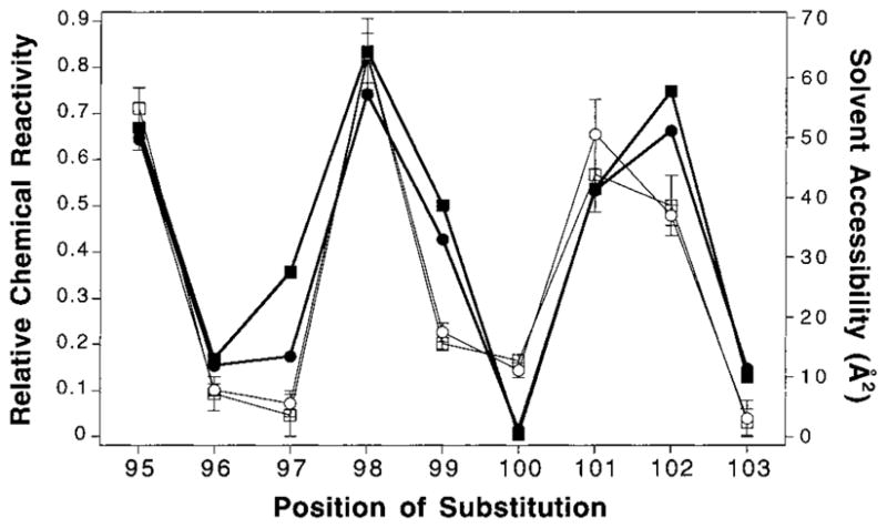
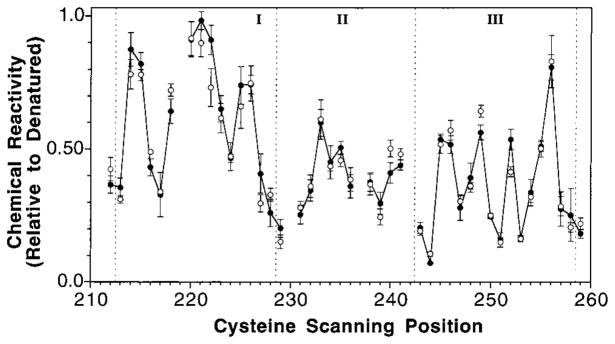
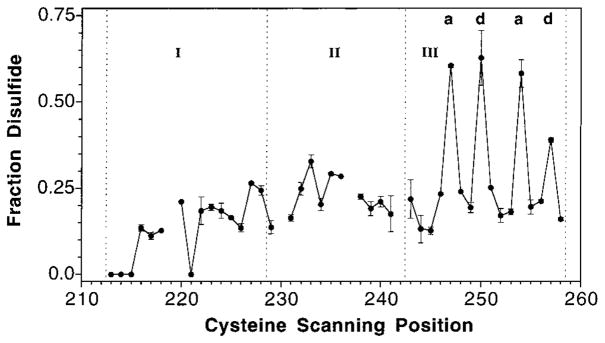
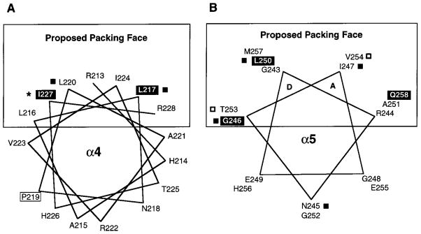
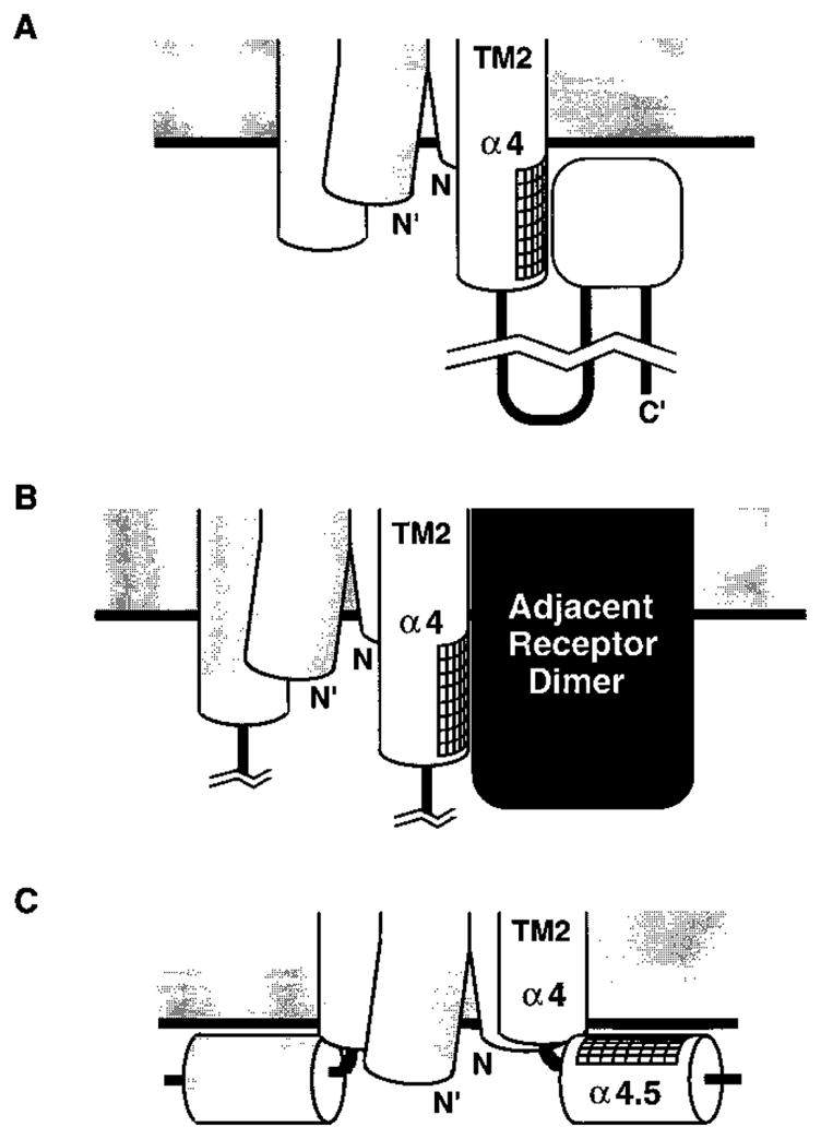
The transmembrane aspartate receptor of E. coli and S. typhimurium mediates cellular chemotaxis toward aspartate by regulating the activity of the cytoplasmic histidine kinase, CheA. Ligand binding results in transduction of a conformational signal through the membrane to the cytoplasmic domain where both kinase regulation and adaptation occur. Of particular interest is the linker region, E213 to Q258, which connects and transduces the conformational signal between the cytoplasmic end of the transmembrane signaling helix (alpha 4/TM2) and the major methylation helix of the cytoplasmic domain (alpha 6). This linker is crucial for stable folding and function of the homodimeric receptor. The present study uses cysteine and disulfide scanning mutagenesis to investigate the secondary structure and packing surfaces within the linker region. Chemical reactivity assays reveal that the linker consists of three distinct subdomains: two alpha-helices termed alpha 4 and alpha 5 and, between them, an ordered region of undetermined secondary structure. When cysteine is scanned through the helices, characteristic repeating patterns of solvent exposure and burial are observed. Activity assays, both in vivo and in vitro, indicate that each helix possesses a buried packing face that is crucial for proper receptor function. The interhelical subdomain is at least partially buried and is also crucial for proper receptor function. Disulfide scanning places helix alpha 4 distal to the central axis of the homodimer, while helix alpha 5 is found to lie at the subunit interface. Finally, sequence alignments suggest that all three linker subdomains are highly conserved among the large subfamily of histidine kinase-coupled sensory receptors that possess methylation sites for use in covalent adaptation.


Figures

Figure 1
Figure 1
Schematic diagram of the full membrane-bound aspartate receptor. The structures of the periplasmic and transmembrane domains have been characterized by crystallographic and disulfide studies (, –29, 46). The cylinders represent helices that were experimentally defined by those studies. Black circles represent sites of adaptive methylation, three of which lie on the major methylation helix α6 and one closer to the C-terminus (believed to be close in space to the first three). The linker region is defined as a segment that begins at R213 where the transmembrane signaling helix (α4/TM2) emerges into the cytoplasm, and extends to the major proteolytic hotspot R259 [arrow, (38)] just N-terminal to the methylation helix α6. The present cysteine and disulfide scanning study encompasses the boxed region including the entire linker (positions R213 to R259).
Figure 2
Figure 2
Comparison of chemical reactivity and calculated solvent accessibility in the known helix α2 of the periplasmic domain. Chemical reactivities at 25 °C were measured for engineered cysteines at positions 95 through 103 in the periplasmic domain of the membrane-bound receptor using 5-iodoacetamidofluorescein as a probe, and were normalized to the corresponding reactivities of the unfolded receptor (see text). In addition, the solvent accessibility of each position in the available crystal structure of the periplasmic domain (24) was calculated using the method of Richards (64). The correlation between these data is shown by plotting the relative chemical reactivities measured with and without saturating aspartate (open squares and open circles, respectively, fine line) along with the calculated solvent accessibility of each position in the aspartate-occupied and apostructures of the periplasmic domain (closed squares and closed circles, respectively, bold line).
Figure 3
Figure 3
Chemical reactivity of cysteine scanning positions in the linker region of the cytoplasmic domain. Chemical reactivities at 25 °C were measured for engineered cysteines in the membrane-bound receptor using 5-iodoacetamidofluorescein as a probe, and were normalized to the corresponding reactivities of the unfolded receptor (see text). Shown are the chemical reactivities obtained in the presence (solid circles, connected by line) or absence (open circles, no line) of saturating aspartate. The gaps at positions 219, 230, 237, and 242 represent positions where cysteine substitution blocked expression or accumulation of the receptor. The three major segments (I–III) defined by vertical dashed lines represent the three distinct structural subdomains of the linker: (i) residues 213 through 228; (ii) residues 229 through 242; and (iii) residues 243 through 258. Criteria for exposure were defined within each segment as buried (lower 30%), intermediate (mid 30%), and exposed (upper 40%) where the highest and lowest reactivities in each segment define the upper and lower limits of the range, respectively (see Results).
Figure 4
Figure 4
Effects of cysteine substitutions on activity in vivo and in vitro. (A) Rates of aspartate-specific chemotactic swarming relative to wild-type receptor (29). Plotted are the chemotactic swarm rates of an E. coli strain overexpressing each of the cysteine-substituted receptors, measured at 30 °C on minimal medium soft agar plates. The open circles indicate positions where cysteine substitution is known to prevent detectable accumulation of membrane-bound receptor. Receptors that yielded swarm rates below 0.3 (dashed line) are designated as inhibitory. For each mutant receptor, the plotted aspartate-specific swarm rate was determined as the difference in swarm rates measured in the presence and absence of aspartate, normalized to the corresponding rate difference measured for wild-type receptor (typically 0.6 mm/h) (B) Rates of CheY phosphorylation by the reduced receptor–CheA–CheW ternary complex (29). The core of the chemotaxis pathway was reconstituted using wild-type receptor and each of the substituted receptors with separately purified ternary complex components CheA and CheW, and the response regulator CheY. The ability of each receptor to regulate the histidine kinase activity of CheA was measured by monitoring the rate of formation of [32P]-phospho-CheY at 25 °C, normalized to the wild-type rate in the absence of aspartate. The wild-type receptor yields a normalized rate of 1.0 in the absence of aspartate (closed circles, fine line) and is down-regulated effectively to 0.0 by the presence of saturating aspartate (open circles, bold line). Mutations that yield over 150% (upper line) or less than 10% (lower line) of the wild-type rate in the absence of aspartate are designated super-activating or inhibitory, respectively.
Figure 5
Figure 5
Efficiency of disulfide formation in the linker region. Formation of each disulfide bond between a symmetric pair of engineered cysteines in the two subunits of the homodimer was catalyzed by the addition of Cu(II)(1,10-phenanthroline)3 to a mild oxidation system (see text). Reactions at 25 °C were quenched after 10 s, and intersubunit disulfide formation was detected by a gel shift in SDS–PAGE. Plotted is the fraction of disulfide formed (dimer/total) for each cysteine substitution in the linker region. The notations a and d above the data refer to positions in the conserved heptad repeating coiled-coil motif seen in sequence alignments (8, 51). The dashed lines indicate the boundaries of the three major subdomains discussed, while breaks in the plot indicate cysteine substitutions that blocked receptor expression or accumulation.
Figure 6
Figure 6
Helical wheel projections of the two proposed helices displaying the chemical reactivity of each cysteine substitution. Filled boxes indicate highly buried positions, and open boxes indicate highly exposed positions (defined in Figure 3), revealing the strong amphiphilic nature of both modeled helices. (A) Helix α4 displays strong segregation of buried and exposed residues to opposite faces of the canonical 3.6 residue per turn helix. It should be noted that R213 is located at the membrane interface, and that the current data do not distinguish whether protection of this residue from solvent results from the defined packing interaction or from partial burial in the membrane. (B) Helix α5 also displays clear buried and exposed faces, and is modeled as a 3.5 residue per turn helix (see Discussion). The a and d positions of this helix are proposed to form a coiled-coil interaction with the symmetric helix α5′ at the dimer interface. Parentheses indicate substitutions that block receptor expression or accumulation in the membrane.
Figure 7
Figure 7
Helical wheel projections of (A) helix α4 and (B) helix α5 displaying effects of cysteine substitutions on receptor activity. Positions where substitution inhibits chemotactic swarming in vivo are shown in black boxes. The open box at P219 indicates a cysteine substitution that prevents expression or accumulation of the receptor. Black squares indicate substitutions that inhibit kinase activation, and open squares indicate those that result in kinase super-activation (defined in Figure 4). The star at position 227 indicates constitutive activation of the kinase upon substitution.
Figure 8
Figure 8
Model of the secondary structural features of the cytoplasmic linker region, as suggested by the currently available data (see Discussion). Helix α4 continues from the transmembrane signaling helix α4/TM2 into the cytoplasm, where it is distal to the central axis of the dimer. This helix is followed by an ordered region of undefined secondary structure (oval) that is also distal to the central axis and becomes helix α5 at its C-terminus. Finally, the two symmetric α5 and α5′ helices of the dimer pack against each other to form a coiled-coil at the subunit interface.
Figure 9
Figure 9
Possible packing arrangements that are consistent with the experimentally determined buried face of helix α4. The fact that helix α4 displays a packing face oriented away from the dimer interface could be explained by: (A) the packing of an undetermined element within the cytoplasmic domain against α4; (B) the packing of another receptor dimer against α4; or (C) the packing of α4 against the surface of the membrane, which would require a break in the helix. Currently, model (A) seems most plausible (see Discussion).

References

    1. Falke JJ, Bass RB, Butler SL, Chervitz SA, Danielson MA. Annu Rev Cell Dev Biol. 1997;13:457. - PMC - PubMed
    1. Wurgler-Murphy SM, Saito H. Trends Biochem Sci. 1997;22:172. - PubMed
    1. Stock AM, Mowbray SL. Curr Opin Struct Biol. 1995;5:744. - PubMed
    1. Stock JB, Surette MG. In: Escherichia coli and Salmonella Cellular and Molecular Biology. Neidhardt FC, editor. ASM Press; Washington, DC: 1996. pp. 1103–1129.
    1. Blair DF. Annu Rev Microbiol. 1995;49:489. - PubMed

Publication types