. 1998 Aug;9(8):1995-2010.
doi: 10.1091/mbc.9.8.1995.

IkappaB is a substrate for a selective pathway of lysosomal proteolysis

Affiliations
Free PMC article

IkappaB is a substrate for a selective pathway of lysosomal proteolysis

A M Cuervo et al. Mol Biol Cell. 1998 Aug.
Free PMC article

Abstract

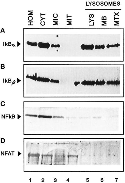
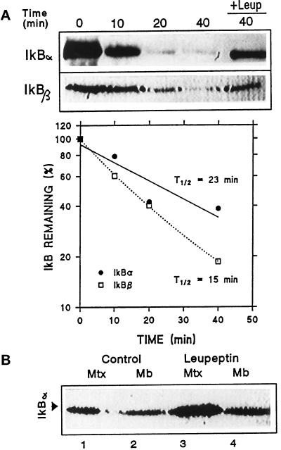
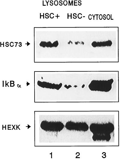
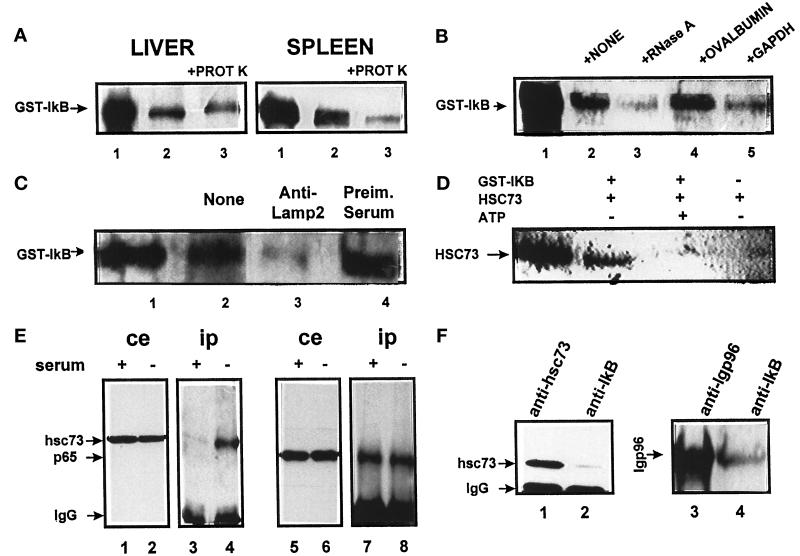
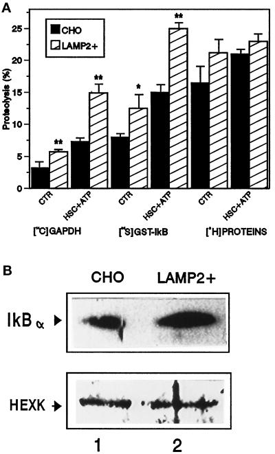
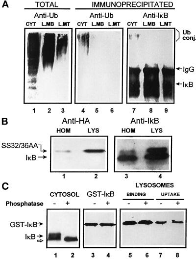
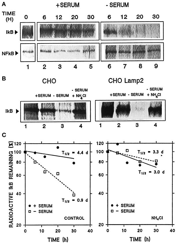
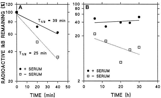
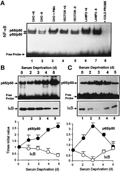
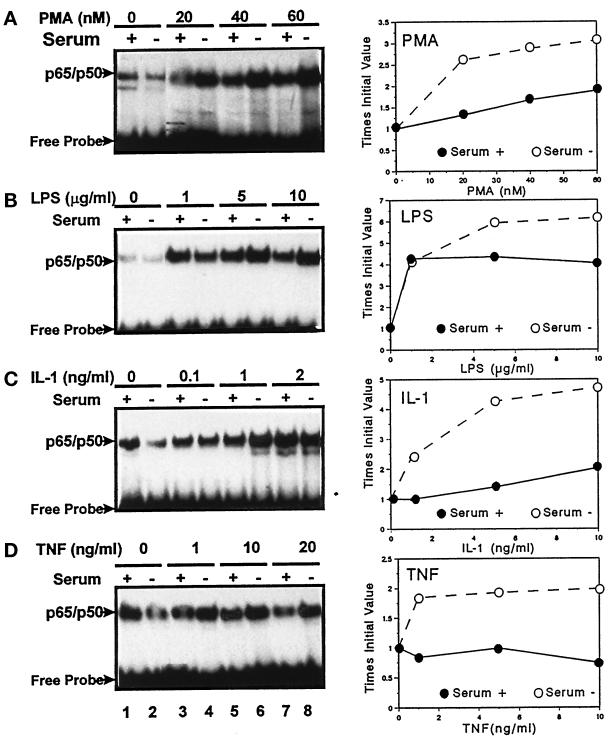
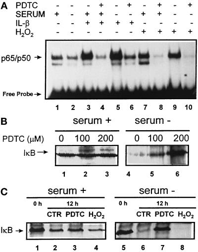
In lysosomes isolated from rat liver and spleen, a percentage of the intracellular inhibitor of the nuclear factor kappa B (IkappaB) can be detected in the lysosomal matrix where it is rapidly degraded. Levels of IkappaB are significantly higher in a lysosomal subpopulation that is active in the direct uptake of specific cytosolic proteins. IkappaB is directly transported into isolated lysosomes in a process that requires binding of IkappaB to the heat shock protein of 73 kDa (hsc73), the cytosolic molecular chaperone involved in this pathway, and to the lysosomal glycoprotein of 96 kDa (lgp96), the receptor protein in the lysosomal membrane. Other substrates for this degradation pathway competitively inhibit IkappaB uptake by lysosomes. Ubiquitination and phosphorylation of IkappaB are not required for its targeting to lysosomes. The lysosomal degradation of IkappaB is activated under conditions of nutrient deprivation. Thus, the half-life of a long-lived pool of IkappaB is 4.4 d in serum-supplemented Chinese hamster ovary cells but only 0.9 d in serum-deprived Chinese hamster ovary cells. This increase in IkappaB degradation can be completely blocked by lysosomal inhibitors. In Chinese hamster ovary cells exhibiting an increased activity of the hsc73-mediated lysosomal degradation pathway due to overexpression of lamp2, the human form of lgp96, the degradation of IkappaB is increased. There are both short- and long-lived pools of IkappaB, and it is the long-lived pool that is subjected to the selective lysosomal degradation pathway. In the presence of antioxidants, the half-life of the long-lived pool of IkappaB is significantly increased. Thus, the production of intracellular reactive oxygen species during serum starvation may be one of the mechanisms mediating IkappaB degradation in lysosomes. This selective pathway of lysosomal degradation of IkappaB is physiologically important since prolonged serum deprivation results in an increase in the nuclear activity of nuclear factor kappa B. In addition, the response of nuclear factor kappa B to several stimuli increases when this lysosomal pathway of proteolysis is activated.


Figures

Figure 1
Figure 1
A portion of intracellular IκB is located in lysosomes. Rat spleens were fractionated after homogenization as described in MATERIALS AND METHODS. After the isolation, part of the lysosomal fraction was subjected to hypotonic shock, and lysosomal membranes and matrix were separated by centrifugation. The same amount of protein (100 μg) of the indicated fractions (and the membrane and matrix derived from 100 μg of lysosomal protein) was then subjected to SDS-PAGE and immunoblotted with specific antibodies against IκB α (A), IκB β (B), p65 (C), or nuclear factor of activated T cells 1 (NFAT, D). HOM, homogenate; CYT, cytosol; MIC, microsomes; MIT, mitochondria; LYS, lysosomes; MB, membrane; MTX, matrix.
Figure 2
Figure 2
Degradation of IκB inside lysosomes. (A) Rat liver lysosomes were incubated in an isotonic medium at 37°C in the absence or presence (+Leup) of leupeptin. At the indicated times, reactions were stopped by addition of Laemmli sample buffer and levels of IκB α and β present in the lysosomal fraction were detected by immunoblot following SDS-PAGE. The graph shows the quantification of four different experiments similar to the one shown and the calculated half-life for both proteins. (B) Lysosomes isolated from liver of nontreated or leupeptin-treated rats (see MATERIALS AND METHODS) were subjected to hypotonic shock and matrix and membranes were separated. Levels of IκB in each lysosomal fraction were detected by immunoblot with a specific antibody following SDS-PAGE.
Figure 3
Figure 3
Distribution of hsc73, IκB, and hexokinase in different lysosomal populations. Two different lysosomal populations were isolated from rat liver as described in MATERIALS AND METHODS. The same amounts of protein (100 μg) of each lysosomal group (HSC+ and HSC−) and the cytosolic fraction were subjected to SDS-PAGE and immunoblotted with specific antibodies for hsc73 (top), IκBα (middle), and hexokinase (HEXK, bottom).
Figure 4
Figure 4
IκB and the hsc73-mediated pathway for protein degradation. (A) Direct uptake of IκB by lysosomes. The fusion protein GST-IκB (5 μg) was incubated with intact rat liver and spleen lysosomes (100 μg of protein) under standard conditions. At the end of the incubation, proteinase K (5 μg) was added to part of the samples, as indicated, and samples were subjected to SDS-PAGE and immunoblotted for IκB. (B) Effect of different proteins on IκB uptake by lysosomes. IκB was incubated under standard conditions with rat liver lysosomes without additions or in the presence of equimolar amounts of the indicated proteins. (C) Effect of blockage of lgp96 on IκB lysosomal uptake. Rat liver lysosomes were preincubated with a specific antibody against lgp96 (lane 3) or with preimmune serum (lane 4). In B and C uptake of IκB was assayed as described in A by adding proteinase K to all of the samples. (D) Binding of hsc73 to IκB. Hsc73 from rat liver was incubated with GST-IκB with the indicated additions for 1 h at 37°C. At the end of the incubation, the sample was subjected to affinity chromatography with glutathione immobilized on agarose beads. Levels of hsc73 associated with GST-IκB or to the beads alone were detected after SDS-PAGE by immunoblot with a specific antibody against hsc73. GST-IκB (1 μg) is in lane 1 in A–C. Hsc73 (0.5 μg) is in lane 1 in D. (E) Coimmunoprecipitation of IκB and hsc73 in CHO cells. CHO cells maintained in the presence or absence of serum for 12 h (as labeled) were subjected to immunoprecipitation with a specific antibody against IκB. Total cellular extracts (ce) and immunoprecipitates (ip) were analyzed by SDS-PAGE and immunoblot for the presence of hsc73 (lanes 1–4) and p65 (lanes 5–8). (F) Association of IκB proteins at the lysosomal membrane. Lysosomes isolated from rat liver were subjected to immunoprecipitation with specific antibodies against hsc73 (lane 1), IκB (lanes 2 and 4), or lgp96 (lane 3). Levels of hsc73 (lanes 1 and 2) or lgp96 (lanes 3 and 4) in the immunoprecipitates were analyzed as in E.
Figure 5
Figure 5
Effect of overexpression of lamp2 in CHO cells on the uptake and degradation of IκB. (A) Proteolytic activity of lysosomes from transfected cells. Intact lysosomes isolated from nontransfected CHO cells or cells transfected with the cDNA for human lamp2 (LAMP2+) were incubated with radiolabeled GAPDH, GST-IκB, or a pool of cytosolic proteins without additions or in the presence of hsc73 (10 μg/ml) and ATP (5 mM) (HSC + ATP) as labeled. Proteolysis rates were measured as described in MATERIALS AND METHODS. Values are the means ± SD of three different experiments. (B) Lysosomal levels of IκB and hexokinase. Intact lysosomes (100 μg of protein) from the cells described in A were subjected to SDS-PAGE and immunoblot with a specific antibody against IκB (top panel) or hexokinase (HEXK, bottom panel).
Figure 6
Figure 6
Lysosome-associated IκB is not ubiquitinated or phosphorylated. (A) Cytosol (100 μg of protein), lysosomal membranes, and lysosomal matrix (derived from 100 μg of lysosomal protein) were isolated as described in MATERIALS AND METHODS and then directly subjected to SDS-PAGE (lanes 1–3) or immunoprecipitated with a specific antibody against IκB (lanes 4–9). Filters were immunoblotted with a specific antibody against ubiquitin (lanes 1–6) or against IκB (lanes 7–9). (B) Homogenate and lysosomes isolated from WEHI231 cells stably transformed with a HA epitope-tagged SS32/36AA IκB. Samples (50 μg of protein) were subjected to SDS-PAGE and immunoblot with a specific antibody against HA (lanes 1 and 2) or against IκB (lanes 3 and 4). (C) Rat liver cytosol (isolated in the presence of phosphatase inhibitors, 100 μg of protein, lanes 1 and 2) or purified GST-IκB (lanes 3–8) were treated with phosphatase as described in MATERIALS AND METHODS and then subjected to SDS-PAGE and immunoblot with a specific antibody against IκB. Binding and uptake of phosphatase-treated and untreated GST-IκB by isolated rat liver lysosomes (lanes 5–8) was analyzed as described in Figure 4A.
Figure 7
Figure 7
Intracellular degradation of IκB and p65 under different conditions. Nontransfected CHO cells (A) or cells transfected with lamp2 as labeled (B) were radiolabeled with [35S]methionine/cysteine for 48 h as described in MATERIALS AND METHODS. After extensive washing, cells were kept in medium with serum (+serum) or without serum (−serum). At indicated times (A) or after 18 h (B), cells were subjected to immunoprecipitation with a specific antibody against IκB (A, top panel, and B) or p65 (A, bottom panel). Immunoprecipitates were resolved by SDS-PAGE and gels were exposed in a PhosphorImager screen. In B (lanes 4), 15 mM NH4Cl was added to the medium during the chase period. (C) Average value of IκB degradation rates of three different experiments similar to the one shown in A (left panel) and similar experiments performed in the presence of 15 mM NH4Cl (right panel). t1/2 were calculated from the formula t1/2 = ln2/degradation rate.
Figure 8
Figure 8
Two different pools of IκB can be detected in CHO cells. Cells were radiolabeled and subjected to immunoprecipitation with anti-IκB antibody as described in Figure 7, with the exception that radiolabeling was shortened to 2 h. (A) Quantification of levels of IκB immunoprecipitated during 40 min of chase in three different experiments. (B) Quantification of levels of IκB immunoprecipitated during 6–30 h of chase in three different experiments.
Figure 9
Figure 9
Effect of overexpression of lamp2 and serum deprivation on NF-κB nuclear translocation. (A) Nuclear extracts were prepared from nontransfected CHO cells or cells transfected with lamp2 (LAMP2) or with an empty vector (vector) maintained in the presence (+S) or absence (−S) of serum for 16 h prior to harvesting. Analysis of NF-κB-binding activity was performed as described in MATERIALS AND METHODS. In lane 3, cells were stimulated with PMA for 6 h. Lane 8 shows a binding assay performed as in lane 7 but in the presence of an excess of unlabeled probe. Results similar to those in lane 8 were obtained when unlabeled probe was added in each of the conditions analyzed (our unpublished results). (B and C) CHO cells (B) or human fibroblasts (C) were maintained in the absence of serum for the indicated times, and after harvesting nuclear extracts and cytosolic fractions were prepared as described in MATERIALS AND METHODS. NF-κB activity in the nuclear extracts (top panel) was analyzed as above. Content of IκB in the cytosolic fractions (middle panel) was detected by immunoblot with a specific antibody for IκB. Bottom panel shows the densitometric quantification of three experiments similar to the ones shown the in top and middle panels.
Figure 10
Figure 10
Effect of serum deprivation on the NF-κB response to different stimuli. CHO cells were maintained in the presence (serum+) or absence (serum−) of serum for 12 h. After that incubation, PMA (A), LPS (B), IL-1 (C), or TNF-α (D) was added to the culture medium at the indicated concentrations for 4 h. At the end of the incubation, cells were harvested and NF-κB activity was assayed in the nuclear extracts as described in MATERIALS AND METHODS. The right side of the figure corresponds to the densitometric quantification of nuclear levels of NF-κB in three or four different experiments similar to the ones shown here.
Figure 11
Figure 11
Reactive oxygen species and lysosomal targeting of IκB. (A) CHO cells were maintained in the presence or absence of serum and 200 μM PDTC (as indicated) during 12 h. Four hours before harvesting, IL-1 (1 ng/ml) or H2O2 (250 μM) were added to some of the samples. NF-κB activity was measured in the nuclear extracts as described in MATERIALS AND METHODS. (B and C) CHO cells were radiolabeled and subjected to immunoprecipitation 10 h after labeling with an anti-IκB antibody as described in Figure 7. Where indicated, PDTC (100 or 200 μM) or H2O2 (125 μM) was added to the incubation medium after the washing postlabeling. The percentage of living cells after the treatment, monitored by trypan blue staining, was comparable to untreated cells. Lanes 1 and 5 in C show initial levels of IκB after labeling.

Similar articles

Cited by

References

    1. Adra CN, Zhu S, Ko J-L, et al. LAPTM5: a novel lysosomal-associated multispanning membrane protein preferentially expressed in hematopoietic cells. Genomics. 1996;35:328–337. - PubMed
    1. Agarraberes F, Terlecky SR, Dice JF. Requirement for an intralysosomal hsc73 for a selective pathway of lysosomal proteolysis. J Cell Biol. 1997;137:825–834. - PMC - PubMed
    1. Alkalay I, Yaron A, Hatzubai A, Orian A, Ciechanover A, Ben-Neriah Y. Stimulation-dependent IκBα phosphorylation marks the NF-κB inhibitor for degradation via the ubiquitin-proteasome pathway. Proc Natl Acad Sci USA. 1995;92:10599–10603. - PMC - PubMed
    1. Aniento F, Roche E, Cuervo AM, Knecht E. Uptake and degradation of glyceraldehyde-3-phosphate dehydrogenase by rat liver lysosomes. J Biol Chem. 1993;268:10463–1070. - PubMed
    1. Aniento F, Papavassiliou AG, Knecht E, Roche E. Selective uptake and degradation of c-fos and v-fos by rat liver lysosomes. FEBS Lett. 1996;390:47–52. - PubMed

Publication types

MeSH terms

LinkOut - more resources