. 1998 Aug;9(8):2037-49.
doi: 10.1091/mbc.9.8.2037.

Cytokinesis and midzone microtubule organization in Caenorhabditis elegans require the kinesin-like protein ZEN-4

Affiliations
Free PMC article

Cytokinesis and midzone microtubule organization in Caenorhabditis elegans require the kinesin-like protein ZEN-4

W B Raich et al. Mol Biol Cell. 1998 Aug.
Free PMC article

Abstract

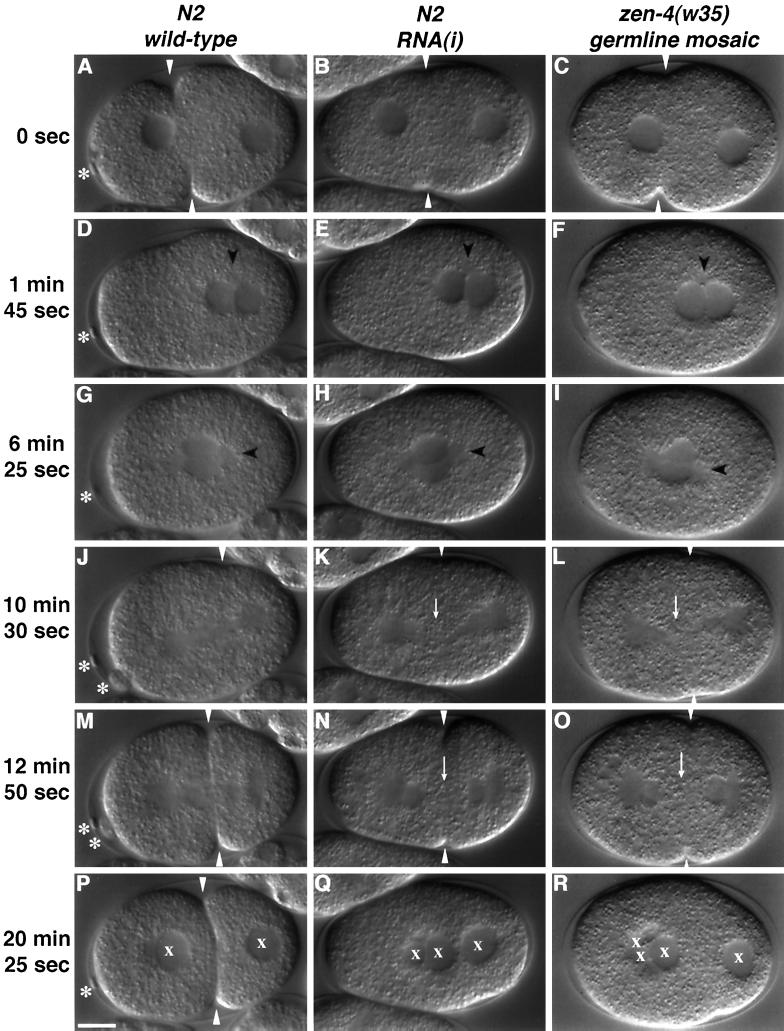
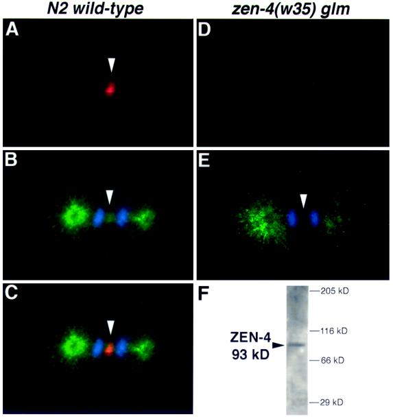
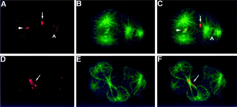
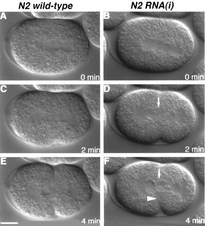
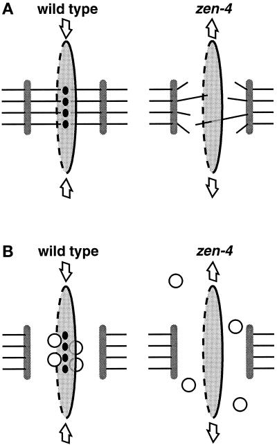
Members of the MKLP1 subfamily of kinesin motor proteins localize to the equatorial region of the spindle midzone and are capable of bundling antiparallel microtubules in vitro. Despite these intriguing characteristics, it is unclear what role these kinesins play in dividing cells, particularly within the context of a developing embryo. Here, we report the identification of a null allele of zen-4, an MKLP1 homologue in the nematode Caenorhabditis elegans, and demonstrate that ZEN-4 is essential for cytokinesis. Embryos deprived of ZEN-4 form multinucleate single-celled embryos as they continue to cycle through mitosis but fail to complete cell division. Initiation of the cytokinetic furrow occurs at the normal time and place, but furrow propagation halts prematurely. Time-lapse recordings and microtubule staining reveal that the cytokinesis defect is preceded by the dissociation of the midzone microtubules. We show that ZEN-4 protein localizes to the spindle midzone during anaphase and persists at the midbody region throughout cytokinesis. We propose that ZEN-4 directly cross-links the midzone microtubules and suggest that these microtubules are required for the completion of cytokinesis.


Figures

Figure 1
Figure 1
Cloning zen-4. (A) Recombination data from three-factor mapping placed zen-4 11/28 of the distance between unc-44(e1260) and bli-6(sc16) on LGIV. zen-4 complements lag-1(q385) and pat-8(st554) but fails to complement the deficiency nDf41. (B) All zen-4 phenotypes are rescued by germline transformation with the cosmid M03D4 and the 7.0-kb XhoI–KpnI subclone pBR938. (C) zen-4 is oriented 5′ to 3′; exons are boxed; exon 7 contains an internal splice site resulting in the formation of two products. The w35 allele contains the insertion GATTT, which is predicted to result in six frameshifted amino acids and a premature stop codon following Arg 91 (asterisk).
Figure 2
Figure 2
zen-4 encodes a member of the MKLP1 subfamily of kinesin-like proteins. Because of alternative splicing, zen-4 is predicted to encode a 775-amino acid protein (ZEN-4a) and a 772-amino acid (ZEN-4b) protein. (A) The predicted sequence of ZEN-4a. The putative nucleotide-binding motifs, N1, N2, and N3, are boxed; the putative microtubule binding sites, DLL and L12, are underlined. The location of the frameshift in the w35 allele is marked with an asterisk. (B) ZEN-4 belongs to the MKLP1 subfamily of kinesin-like proteins. The tree shown was constructed by Moore and Endow (1996) and is based on sequence alignment of the conserved kinesin motor domain. An updated version of the kinesin tree can be found at: http://www.blocks.fhcrc.org/∼kinesin/. (C) ZEN-4 sequence conservation with MKLP1 extends beyond the motor domain. A domain unique to the MKLP1 subfamily is 33% identical in ZEN-4 and MKLP1. While the coiled-coil domain is not well conserved, it contains the heptad repeats characteristic of a coiled coil. The complete sequence data for ZEN-4a and ZEN-4b are available from GenBank/EMBL/DDBJ under accession numbers AF057567 and AF057568.
Figure 3
Figure 3
Nomarski images selected from time-lapse recordings of embryos at successive stages between fertilization and first cleavage. Embryos deprived of ZEN-4 display a late defect in cytokinesis. The left column shows a wt offspring of an N2 animal, the center column shows the offspring of an N2 animal injected with antisense zen-4 RNA, and the right column shows the offspring of a zen-4(w35) homozygote that lost expression of the rescuing transgene pBR938 in the germline. In all panels, anterior is to the left. (A–C) Pseudocleavage and pronuclear migration. The white arrowhead points to the pseudocleavage furrow. The asterisk in panel A highlights the visible polar body; both polar bodies are visible in panel M. Embryos deprived of ZEN-4 fail to extrude polar bodies. (D–F) Pronuclear contact. The timing and position of pronuclear contact in wt (D) and mutant (E and F) embryos is very similar and was used to normalize the time-lapse recordings. Centrosomes are visible as granule-free regions and are labeled with a black arrowhead. (G–I) Pronuclear migration and rotation are unaffected by loss of ZEN-4. (J) Late anaphase and the beginning of first cleavage. A white arrowhead points to the cleavage furrow. (K and L) Spindle elongation is unaffected in embryos deprived of ZEN-4. A white arrowhead points to a region in the central spindle apparently devoid of organized microtubules. (M) Telophase. Microtubules in the spindle midzone can be seen bisecting the maturing cleavage furrow. (N and O) The cleavage furrow fails to propagate in the region of the central spindle. A white arrow points to a granular region at the center of the mitotic spindle lacking organized microtubules. (P) A two-cell embryo. An “x” is used to label each nucleus. (Q–R) The cleavage furrow has completely relaxed. Nuclei aggregate in the center of the embryo. The failure to extrude polar bodies may explain the excess number of nuclei. Note that loss of a rescuing array and RNA interference result in an indistinguishable phenotype. Scale bar, 10 μm.
Figure 4
Figure 4
ZEN-4 is required for normal organization of the midzone microtubules. Embryos are coimmunostained with ZEN-4N to label ZEN-4 (red), N357 to label tubulin (green), and DAPI (blue) to label DNA. (A) ZEN-4 staining during the first cell division. ZEN-4 localizes in discrete short lines in the spindle midzone. (B) Tubulin and DNA staining in the same embryo shown in panel A. A white arrowhead points to organized microtubules in the spindle midzone. (C) An overlay of panels A and B, showing that ZEN-4 colocalizes with the microtubules between the separating chromosomes. (D) ZEN-4N does not stain the offspring of a germ line mosaic mutant, indicating that the w35 allele encodes a protein null. (E) Tubulin and DNA staining of the same embryo shown in panel D. A white arrowhead points to the midzone, which has greatly reduced numbers of microtubules. (F) ZEN-4N recognizes a major band of 93 kDa in extracts of total worm proteins. This is slightly greater than the predicted size of 87 kDa for ZEN-4A and ZEN-4B.
Figure 5
Figure 5
ZEN-4 localization is cell cycle dependent. Embryos are coimmunostained with ZEN-4N to label ZEN-4 (red) and N357 (green) to label the microtubules. Anterior is to the left in all panels. (A) ZEN-4 staining in a two-cell embryo. (B) Tubulin staining in the same embryo shown in panel A. (C) Combined images of panels A and B, showing the overlapping pattern in yellow. ZEN-4N strongly stains the midbody remnant from the first cell division (white arrow), and expression can also be seen at the periphery of the chromosomes in the more posterior cell and in the central region of the midzone in the anterior anaphase cell (white arrowhead). The chromatin can be seen as the black region of the spindle, which excludes both antibodies (white caret). (D) ZEN-4 staining in the midbody of a dividing cell. (E) Tubulin staining of the same embryo shown in panel D. (F) Overlay of panels D and E, showing that ZEN-4 colocalizes with the midzone microtubules at the cytoplasmic bridge between daughter cells.
Figure 6
Figure 6
Time-lapse Nomarski images of first cleavage. The embryo in the left column is wt; the embryo in the right column is the offspring of an animal injected with antisense zen-4 RNA. Anterior is to the left in all panels. (A) wt Metaphase embryo. (B) Metaphase embryo lacking ZEN-4. At this time, the spindle appears normal. (C) wt Anaphase embryo. Initiation of the cleavage furrow alters the ellipsoid shape of the embryo. (D) The cleavage furrow resembles the wt furrow (C), demonstrating that furrow initiation does not require zen-4. The white arrow points to the cytoplasmic granules in the spindle midzone, suggesting a lack of organized midzone microtubules. (E) wt Telophase embryo. The cleavage furrow is approaching the spindle midzone. (F) The white arrowhead points to the limit of furrow progression in an embryo lacking ZEN-4. Scale bar, 10 μm.
Figure 7
Figure 7
Models of ZEN-4 function during cytokinesis. Schematic diagram of the central spindle during cytokinesis in wt embryos and embryos lacking ZEN-4. (A) The microtubule organization model. ZEN-4 (black ovals) localizes to the spindle midzone, where it serves to directly or indirectly cross-link the midzone microtubules (shown as solid lines between the separating chromosomes). In the absence of organized midzone microtubules, the contractile actomyosin ring, shown as a black line ringing the midzone, halts its migration in the region of the mitotic spindle. In this model, the cytokinesis defect seen in embryos deprived of ZEN-4 is a secondary effect of the failure to organize the midzone microtubules. (B) The cargo model. ZEN-4 localizes to the equatorial region of the spindle, where it associates directly or indirectly with proteins localizing to the contracting actomyosin ring. In the diagram, ZEN-4 is shown to interact with the ring via a cargo protein shown as a white circle. In this model, the late cytokinesis defect results because the contractile ring is unable to associate with molecules in the midzone and is thus independent of the presence of absence of organized midzone microtubules. We stress that these models are not mutually exclusive (see DISCUSSION for details).

Comment in

References

    1. Asada T, Shibaoka H. Isolation of polypeptides with microtubule-translocating activity from phragmoplasts of tobacco BY-2 cells. J Cell Sci. 1994;107:2249–2257. - PubMed
    1. Brenner S. The genetics of Caenorhabditis elegans. Genetics, 1974;77:71–94. - PMC - PubMed
    1. Cao LG, Wang YL. Signals from the spindle midzone are required for the stimulation of cytokinesis in cultured epithelial cells. Mol Biol Cell, 1996;7:225–232. - PMC - PubMed
    1. Conrad AH, Paulsen AQ, Conrad GW. The role of microtubules in contractile ring function. J Exp Zool, 1992;262:154–165. - PubMed
    1. Costa M, Raich W, Agbunag C, Leung B, Hardin J, Priess JR. A putative catenin–cadherin system mediates morphogenesis of the Caenorhabditis elegans embryo. J Cell Biol. 1998;141:297–308. - PMC - PubMed

Publication types

MeSH terms

LinkOut - more resources