. 1998 Aug;9(8):2157-71.
doi: 10.1091/mbc.9.8.2157.

Mutations in Drosophila enabled and rescue by human vasodilator-stimulated phosphoprotein (VASP) indicate important functional roles for Ena/VASP homology domain 1 (EVH1) and EVH2 domains

Affiliations
Free PMC article

Mutations in Drosophila enabled and rescue by human vasodilator-stimulated phosphoprotein (VASP) indicate important functional roles for Ena/VASP homology domain 1 (EVH1) and EVH2 domains

S M Ahern-Djamali et al. Mol Biol Cell. 1998 Aug.
Free PMC article

Abstract

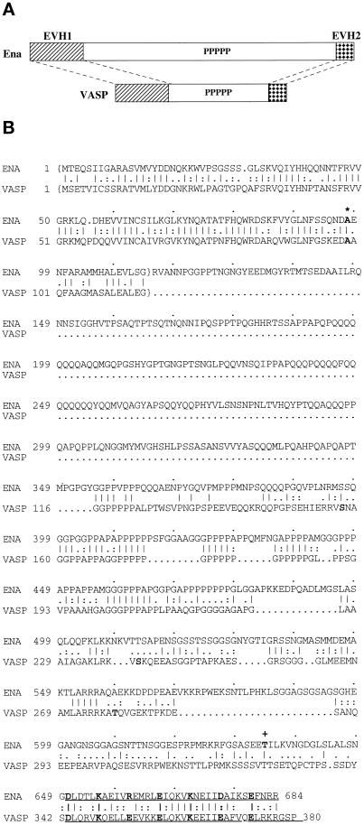
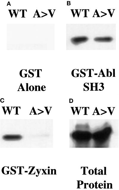
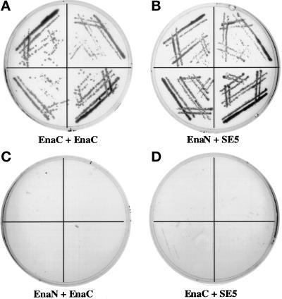
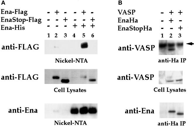
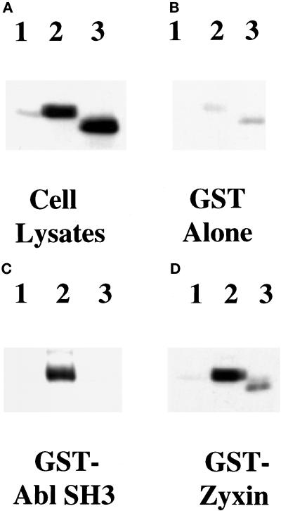
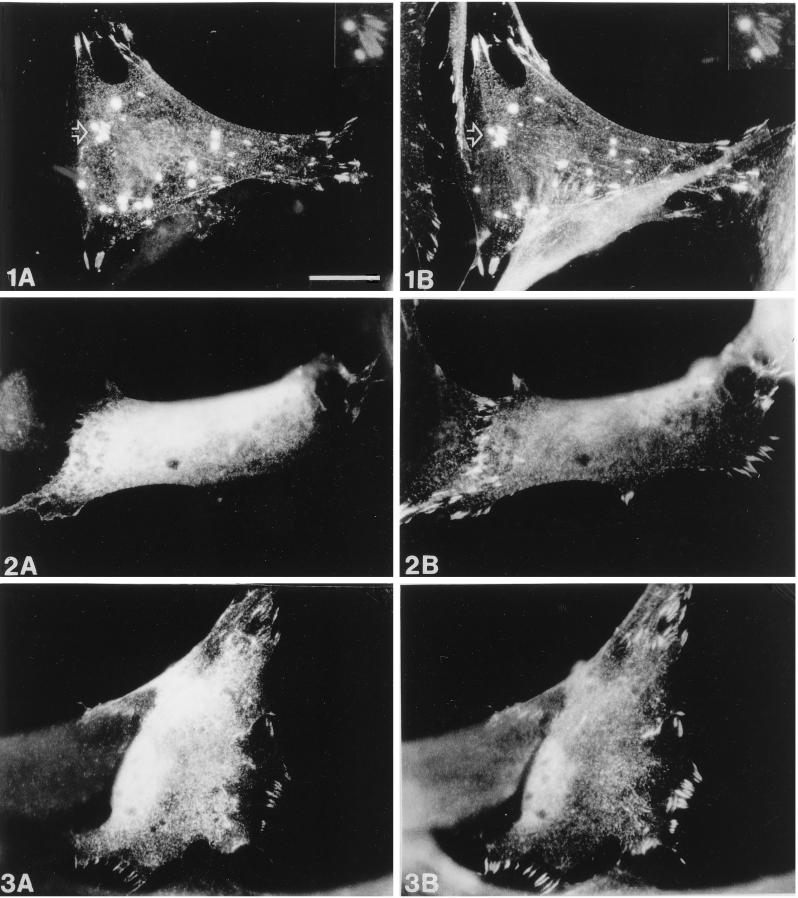
Drosophila Enabled (Ena) was initially identified as a dominant genetic suppressor of mutations in the Abelson tyrosine kinase and, more recently, as a member of the Ena/human vasodilator-stimulated phosphoprotein (VASP) family of proteins. We have used genetic, biochemical, and cell biological approaches to demonstrate the functional relationship between Ena and human VASP. In addition, we have defined the roles of Ena domains identified as essential for its activity in vivo. We have demonstrated that VASP rescues the embryonic lethality associated with loss of Ena function in Drosophila and have shown that Ena, like VASP, is associated with actin filaments and focal adhesions when expressed in cultured cells. To define sequences that are central to Ena function, we have characterized the molecular lesions present in two lethal ena mutant alleles that affected the Ena/VASP homology domain 1 (EVH1) and EVH2. A missense mutation that resulted in an amino acid substitution in the EVH1 domain eliminated in vitro binding of Ena to the cytoskeletal protein zyxin, a previously reported binding partner of VASP. A nonsense mutation that resulted in a C-terminally truncated Ena protein lacking the EVH2 domain failed to form multimeric complexes and exhibited reduced binding to zyxin and the Abelson Src homology 3 domain. Our analysis demonstrates that Ena and VASP are functionally homologous and defines the conserved EVH1 and EVH2 domains as central to the physiological activity of Ena.


Figures

Figure 1
Figure 1
Ena and VASP have structural domain and sequence similarity, and the ena 210 and ena 23 mutations map to these regions of similarity. (A) Comparison of the overall structure and structural domain organization of Ena and VASP. The conserved EVH1 domain (hatched box) and EVH2 domain (checked box) are separated by a central domain of variable length, which contains a region rich in prolines. (B) Comparison of the deduced amino acid sequences and sequence motifs of Drosophila Ena and human VASP. The central regions of both Ena and VASP have multiple polyproline motifs. The C-terminal 35 amino acids contain a mixed charge cluster containing a charged residue at every fifth position (marked in bold). The conserved alanine that is altered in ena 210 mutants is in bold and marked with an asterisk. The EVH1 domains are enclosed in brackets, and the EVH2 domain is underlined. The location of the stop codon in ena23 is marked by a plus sign. Sequence alignment was generated using the Genetics Computer Group (Madison, WI) BestFit program. Amino acid identity is indicated by vertical lines, and similarity is indicated by dots.
Figure 2
Figure 2
Colocalization of Ena and VASP proteins to stress fibers and focal contacts in transfected PtK2 cells. PtK2 cells were transiently transfected with pCMV/ena (A–C) or pCMV/ena and pVSV-VASP (D–F) and processed for double-label immunofluorescence microscopy. Ena was detected with rabbit polyclonal antibodies (A and D), VASP with a monoclonal mouse anti-VSV antibody (E), and F-actin by fluorescein-labeled phalloidin (B). A merge of A and B is shown in C, and a merge of D and E is shown in F. Fluorescent staining was by TRITC for Ena and DTAF for VASP. Ena expressed alone is localized either in focal contacts and stress fibers or is also variably found in spot-like structures, which also stain for F-actin (A–C). Coexpression of Ena and human VASP results in an identical localization of the two proteins in focal contacts and stress fibers (D–F). Controls show no cross-reactivity of the anti-Ena antibodies with VASP. Nuclear fluorescence is nonspecific background because of the secondary antibody.
Figure 3
Figure 3
Analysis of ena mutant alleles. Single-embryo Western blot analysis of heterozygous ena mutant stocks. Embryos were collected for 2 h and then aged 5 h. Single embryos were picked and lysed in sample buffer and Western blotted. For the enaGC8 chromosomal inversion, 25% of the embryos were null for Ena protein (top panel, lanes 1, 5, and 9). This blot was stripped and reprobed with an anti-fax antibody to demonstrate the presence of embryonic proteins in all 12 lanes. The ena 23 allele appears to encode an Ena protein with a small truncation, because 25% of the embryos encode only a smaller protein (small arrow) of ∼80 kDa (middle panel, lanes 3, 9, and 12), and 50% of the embryos encode both full-length Ena (large arrow) and the truncated protein. The ena 210 allele appears to encode a full-length protein, because all embryos picked express full-length Ena.
Figure 4
Figure 4
Binding of Ena and VASP proteins to zyxin and mapping of the binding domain in zyxin. (A) Ena and VASP overlays were carried out essentially as previously described (Reinhard et al., 1995a). Fifty micrograms of total protein from human platelets (lane 1) and 100 ng of purified porcine platelet zyxin (lane 2) were separated by SDS-PAGE and transferred to nitrocellulose. The blot was overlaid with Ena purified from baculovirus-infected cells, and bound Ena was detected by the anti-Ena antibody followed by 125I-protein A (left panel). The control (right panel) was treated identically, except that the overlay solution contained no Ena protein. Ena protein bound to an 83-kDa protein present in platelet lysates (lane 1), which comigrates with full-length zyxin (lane 2), whereas the control panel gave no detectable signal (right panel). (B) S2 cells were transfected with Ena or VASP, and proteins were bound to GST fusion proteins as follows: full-length chicken zyxin (lane 1), zyxin proline domain (lane 2), zyxin LIM domains (lane 3) (Schmeichel and Beckerle, 1994), and GST alone (lane 4). Retained proteins were detected with anti-Ena N-terminal (left panel) or anti-VASP (right panel) polyclonal antibodies. Both proteins bound to full-length zyxin and to the zyxin proline domain (lanes 1 and 2). Neither protein bound to the zyxin LIM domain (lane 3). The faint signal seen in lanes 3 and 4 in the anti-VASP blot is due to background binding between VASP and the GST fusion proteins. Aliquots of transfected cell lysates were analyzed for expression of Ena or VASP protein (lanes 5). These results are representative of three independent experiments.
Figure 5
Figure 5
ena 210 mutation disrupts zyxin binding. Full-length wild-type Ena (WT) or ena210 (A97V) proteins were prepared using an in vitro transcription–translation reaction and bound to GST alone (A), GST-AblSH3 (B), or GST-zyxin (C). Retained proteins were detected with anti-Ena N-terminal polyclonal antibody. Both proteins were expressed equally well (D). As expected, the GST negative control did not bind detectably to either protein (A). Both proteins bound equally well to the GST-AblSH3 domain (B). The wild-type Ena protein bound to the GST-zyxin protein, whereas binding to the A97V mutant protein was reduced. (C) These results are representative of three independent experiments.
Figure 6
Figure 6
Interaction of the Ena EVH2 domain with the C-terminal 243 amino acids of Ena in the yeast two-hybrid system. The first 235 amino acids of Ena (GAL4DB-Ena1–235) and last 243 amino acids of Ena (GAL4DB-Ena441–684) were fused to the sequence encoding the GAL4 DNA-binding domain in the pAS1-CYH2 vector. These two constructs were cotransformed with GAL4AD-Ena628–684, a clone in which amino acids 628–684 of Ena are fused to the sequence encoding the GAL4 activation domain and that was isolated in a yeast two-hybrid screen using the C terminal 243 amino acids of Ena as bait. Four independent colonies from each of the cotransformations were spread on medium lacking histidine and analyzed for growth. GAL4DB-Ena441–684 containing the C-terminal region of the Ena protein can interact with GAL4AD-Ena628–684 (A, EnaC + EnaC), whereas GAL4DB-Ena1–235 cannot (C, EnaN + EnaC). As a control, GAL4DB-Ena1–235 and GAL4DB-Ena401–684 were also tested for their ability to interact with GAL4AD-SE5, a clone identified from the same larval library as GAL4AD-Ena628–684 by its ability to interact with GAL4DB-Ena1–235. As expected, GAL4DB-Ena1–235 can interact with GAL4AD-SE5 (B, EnaN + SE5), whereas GAL4DB-Ena401–684 cannot (D, EnaC + SE5).
Figure 7
Figure 7
The EVH2 domain mediates multimerization. (A) S2 cells were transfected with the following expression constructs: no DNA (lane 1), Ena-FLAG (lane 2), Ena K636Stop-FLAG (EnaStopFlag; lane 3), Ena-His (lane 4), Ena-FLAG + Ena-His (lane 5), and Ena K636Stop-FLAG (EnaStopFlag) + Ena-His (lane 6). Cells were lysed, and extracted proteins were purified on Ni-NTA agarose and blotted with anti-FLAG (top panel) or anti-Ena (bottom panel) antibodies. Aliquots of cell lysates representing <2% of the total protein used in the purifications were analyzed with the anti-FLAG antibody for expression of the FLAG-tagged proteins, and both the Ena-FLAG and EnaK636Stop-FLAG proteins were expressed in the presence of Ena-His (middle panel). However, only full-length Ena-FLAG copurified with the Ena-His on the nickel NTA resin (top panel, compare lanes 5 and 6). A small amount of contaminating Ena-FLAG was purified with the Nickel-NTA resin (top panel, lane 2). (B) S2 cells were transfected with the following expression constructs: no DNA (lane 1), VASP + Ena-Ha (lane 2), and VASP + Ena K636Stop-Ha (EnaStopHa; lane 3). Transfected cell lysates were immunoprecipitated with 5 μg of anti-Ha antibody. Complexes were subsequently analyzed by Western Blot with an anti-VASP antibody (top panel) or anti-Ena antibody (bottom panel). Aliquots of cell lysates representing <2% of the total protein used for the immunoprecipitations were analyzed for expression of the VASP protein with an anti-VASP antibody (middle panel). VASP was expressed in the presence of Ena-Ha and Ena K636Stop-Ha (middle panel, lanes 2 and 3), but VASP only copurified with full-length Ena-Ha in the anti-Ha IPs (compare lanes 2 and 3, top panel). The dark band present in all lanes in the top panel is the heavy-chain antibody from the anti-Ha IPs cross-reacting with the secondary antibody of the Western blot (arrow).
Figure 8
Figure 8
Ena K636Stop is impaired in its ability to bind zyxin and the Abl-SH3 domain. S2 cells were transfected with no DNA (lanes 1), full-length Ena (lanes 2), or Ena K636Stop (lanes 3) expression vectors. Transfected cell lysates were incubated with GST-Zyxin (D) or Abl SH3 (C) domain fusion proteins or with GST alone as a negative control (B). Cells were lysed, and aliquots of the cell lysates were compared to confirm that equal amounts of the full-length and truncated Ena proteins were expressed (A). Lysates were incubated with the GST fusion proteins, and retained proteins were detected with anti-Ena N-terminal polyclonal antibody. Little protein bound to the GST negative control (B). A decrease in binding of the truncated Ena K636Stop protein was seen with both zyxin and Abl SH3 when compared with the full-length Ena protein (C and D, compare lanes 2 and 3). These results are representative of three independent experiments.
Figure 9
Figure 9
Comparison of the subcellular distribution of zyxin and wild-type or mutant Ena. Human fibroblasts were transiently transfected with pCMV/Ena (1), pCMV/EnaA97V (2), or pCMV/EnaK636Stop (3) and processed for double-label immunofluorescence microscopy. Ena staining is shown in A, staining of endogenous zyxin in B. After transfection of wild-type Ena (1A and 1B), colocalization of both Ena and zyxin is found in focal contacts, on microfilaments and in spot-like structures. A magnification of the area indicated by the arrow showing two “spots” and several focal contacts is seen at the top right (insets in 1A and 1B). In contrast, EnaA97V (2A and 2B) is diffusely distributed throughout the cytoplasm and totally absent from focal contacts, which are labeled by the zyxin antibody. EnaK636Stop (3A and 3B) is also diffusely distributed in the cytoplasm but shows some residual focal contact staining, although less pronounced than that of the wild-type Ena protein. Bar in 1A, 20 μm (valid for all panels).

References

    1. Arber S, Caroni P. Specificity of single LIM motifs in targeting and LIM/LIM interactions in situ. Genes Dev. 1996;10:289–300. - PubMed
    1. Bennett RL, Hoffmann FM. Increased levels of the Drosophila Abelson tyrosine kinase in nerves and muscles: subcellular localization and mutant phenotypes imply a role in cell-cell interactions. Development. 1992;116:953–966. - PubMed
    1. Brand AH, Perrimon N. Targeted gene expression as a means of altering cell fates and generating dominant phenotypes. Development. 1993;118:401–415. - PubMed
    1. Chakraborty T, et al. A focal adhesion factor directly linking intracellularly motile Listeria monocytogenes and Listeria ivanovii to the actin-based cytoskeleton of mammalian cells. EMBO J. 1995;14:1314–1321. - PMC - PubMed
    1. Chien C-T, Bartel PL, Sternglanz R, Fields S. The two-hybrid system: a method to identify and clone genes for proteins that interact with a protein of interest. Proc Natl Acad Sci USA. 1991;88:9578–9582. - PMC - PubMed

Publication types

MeSH terms

LinkOut - more resources