Comparative Study
. 1998 Aug;9(8):2231-47.
doi: 10.1091/mbc.9.8.2231.

The Saccharomyces cerevisiae prenylcysteine carboxyl methyltransferase Ste14p is in the endoplasmic reticulum membrane

Affiliations
Free PMC article
Comparative Study

The Saccharomyces cerevisiae prenylcysteine carboxyl methyltransferase Ste14p is in the endoplasmic reticulum membrane

J D Romano et al. Mol Biol Cell. 1998 Aug.
Free PMC article

Abstract

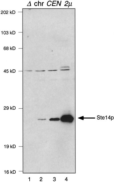
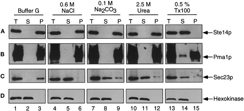
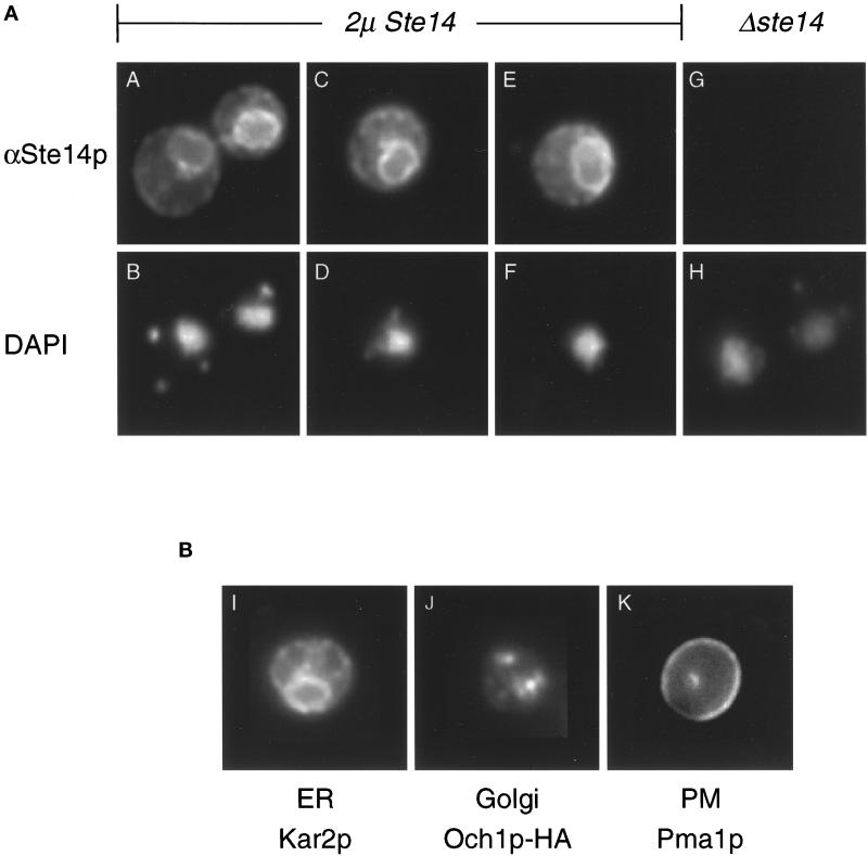
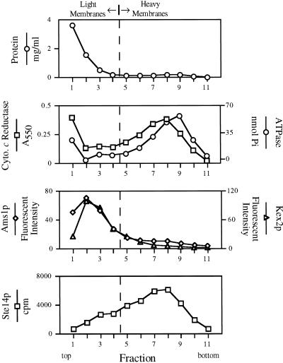
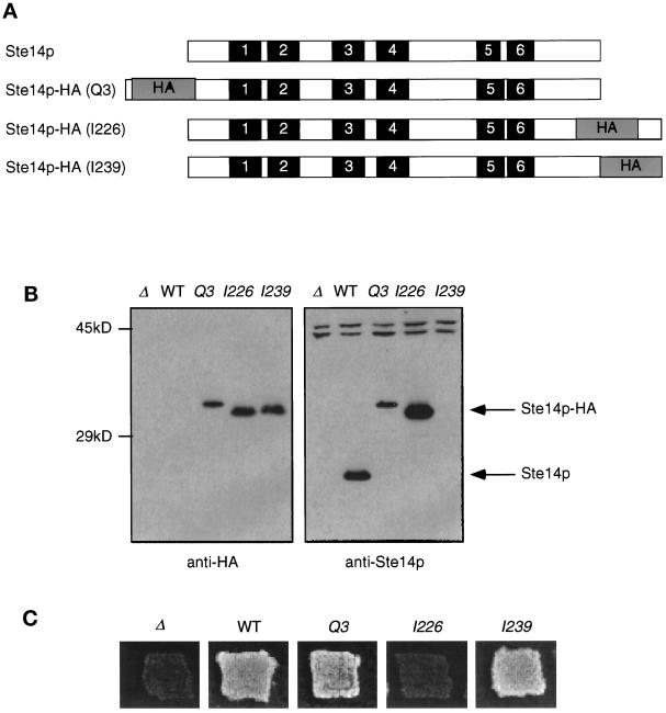
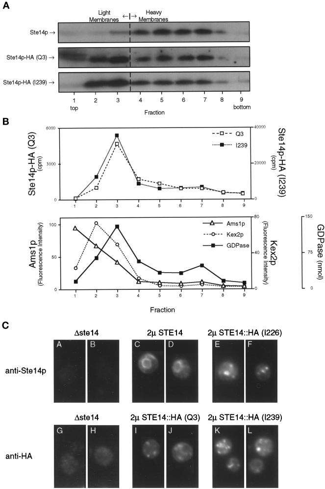
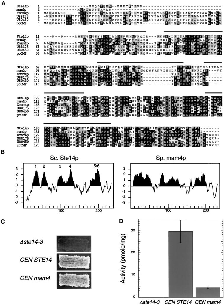
Eukaryotic proteins containing a C-terminal CAAX motif undergo a series of posttranslational CAAX-processing events that include isoprenylation, C-terminal proteolytic cleavage, and carboxyl methylation. We demonstrated previously that the STE14 gene product of Saccharomyces cerevisiae mediates the carboxyl methylation step of CAAX processing in yeast. In this study, we have investigated the subcellular localization of Ste14p, a predicted membrane-spanning protein, using a polyclonal antibody generated against the C terminus of Ste14p and an in vitro methyltransferase assay. We demonstrate by immunofluorescence and subcellular fractionation that Ste14p and its associated activity are localized to the endoplasmic reticulum (ER) membrane of yeast. In addition, other studies from our laboratory have shown that the CAAX proteases are also ER membrane proteins. Together these results indicate that the intracellular site of CAAX protein processing is the ER membrane, presumably on its cytosolic face. Interestingly, the insertion of a hemagglutinin epitope tag at the N terminus, at the C terminus, or at an internal site disrupts the ER localization of Ste14p and results in its mislocalization, apparently to the Golgi. We have also expressed the Ste14p homologue from Schizosaccharomyces pombe, mam4p, in S. cerevisiae and have shown that mam4p complements a Deltaste14 mutant. This finding, plus additional recent examples of cross-species complementation, indicates that the CAAX methyltransferase family consists of functional homologues.


Figures

Figure 1
Figure 1
Detection of Ste14p in whole cell lysates. Crude yeast cell extracts (0.4 OD600 cell equivalents) were resolved by 12.5% SDS-PAGE and transferred to nitrocellulose. Ste14p was detected using rabbit polyclonal anti-Ste14p antibodies that were depleted previously of nonspecific antibodies (described in MATERIALS AND METHODS). Lane 1, SM2926 (Δste14-3); lane 2, SM3041 (chromosomal STE14); lane 3, SM3185 (CEN STE14); and lane 4, SM3495 (2μ STE14).
Figure 2
Figure 2
Ste14p is an integral membrane protein. A crude yeast cell extract from SM3495 (2μ STE14) was prepared and treated with either buffer G, 0.6 M NaCl, 0.1 M Na2CO3 (pH 11), 2.5 M urea, or 0.5% Triton X-100. After incubation on ice for 20 min, one-half of each sample was reserved as total lysate (T). The remaining volume was separated into supernatant (S) and pellet (P) fractions by centrifugation at 200,000 × g. After being resuspended to equivalent volumes with Laemmli sample buffer, equivalent amounts of T, S, and P fractions were subjected to SDS-PAGE and transferred to nitrocellulose. The immunoblots were probed with (A) anti-Ste14p antiserum that had been depleted previously of nonspecific antibodies, (B) anti-Pma1p antiserum, (C) anti-Sec23p antiserum, and (D) anti-hexokinase antiserum.
Figure 3
Figure 3
Ste14p is localized to the ER membrane by immunofluorescence. (A) Immunofluorescence pattern of Ste14p. Indirect immunofluorescence of Ste14p was performed in Δste14-3 strains bearing either pSM1317 (2μ URA3 STE14) (panels A-F) or pRS316 (CEN URA3) (panels G and H). Cells were fixed with formaldehyde, spheroplasted, and probed with a 1:2000 dilution of anti-Ste14p antiserum (αSte14p) that had been depleted previously of nonspecific antibodies, followed by secondary decoration with Cy3-conjugated goat anti-rabbit antiserum (panels A, C, E, and G) and staining with DAPI (panels B, D, F, and H). The images in panels A, C, E, and G were exposed for equivalent amounts of time. (B) Immunofluorescence patterns for a group of marker proteins. The typical immunofluorescence staining pattern of an ER, Golgi, and plasma membrane marker is shown. Fixed and permeabilized cells from an SM1058 background expressing CEN URA3 plasmids or CEN URA3 OCH1::HA (SM3495) were stained with a 1:1000 dilution of anti-Kar2p antiserum (panel I), a 1:1000 dilution of anti-HA antiserum (panel J), or a 1:200 dilution of anti-Pma1p antiserum (panel K). The localization of Kar2p (ER), Och1p-HA (Golgi), and Pma1p (PM) shown here is consistent with previously observed localization data (Rose et al., 1989; Harris et al., 1994; Harris and Waters, 1996).
Figure 4
Figure 4
Ste14p methyltransferase activity cofractionates with ER membranes by subcellular fractionation. A total yeast lysate derived from SM2915 that expresses a chromosomal copy of STE14 was subjected to fractionation on a sucrose step gradient. Equivalent volumes from each fraction were assayed for methyltransferase activity, for various organellar enzyme activities, and for protein concentration. Two populations of membranes can be identified. Light membranes marked by vacuolar α-d-mannosidase (Ams1p) and trans-Golgi network Kex2p activities are distributed at the top of the gradient (with peaks at fractions 2–3); heavier membranes marked by NADPH cytochrome c reductase (ER) and vanadate sensitive ATPase activities (plasma membrane) are distributed near the bottom of the gradient (fractions 6–10) with the peak distribution of these two compartments offset by one fraction. Methyltransferase activity cofractionates with the heavy ER membrane population. The dashed vertical line represents an arbitrary division between light and heavy membranes.
Figure 5
Figure 5
Introduction of epitope tags into Ste14p. (A) Diagram showing the placement of the triple HA epitope tag in Ste14p. Putative membrane spans (solid bars) and the triple HA epitope (gray bars) are indicated and shown in approximate proportions. The epitope tag (HA) was placed at the N terminus (Q3), internally (I226), or at the C terminus of Ste14p (I239). (B) Detection of tagged and untagged Ste14p and Ste14p-HA by anti-HA and anti-Ste14p antisera. Crude cell extracts were subjected to 12.5% SDS-PAGE and transferred to nitrocellulose, and immunoblots were probed with either anti-HA antiserum or anti-Ste14p antiserum depleted previously of nonspecific antibodies as indicated. Lanes contain SM2926 (Δ), SM3185 (WT), SM3187 (Q3), SM3431 (I226), and SM3189 (I239). (C) Patch mating assay. Patches of the indicated MATa strains were replica plated onto a lawn of the MATα mating tester (SM1068) spread on an SD plate and incubated at 30°C for 2 d. Mating is detected by the growth of prototrophic diploids as indicated for WT, Q3, and I239. No mating is observed for Δste14 or I226.
Figure 6
Figure 6
The HA epitope tag mislocalizes Ste14p, as shown by subcellular fractionation and immunofluorescence. (A) Total yeast lysates derived from strains expressing CEN levels of wild-type and epitope-tagged (Q3 and I239) Ste14p (SM3185, SM3187, and SM3189, respectively) were subjected to fractionation on a sucrose step gradient similar to that described for Figure 4. Equivalent volumes from all gradient fractions were subjected to SDS-PAGE, transferred to nitrocellulose, and probed for Ste14p and epitope-tagged Ste14p. Fractions from wild-type and Q3 were probed with anti-Ste14p antiserum, whereas I239 fractions were probed with the anti-HA antibody. As expected, wild-type Ste14p fractionates with the heavy membranes (fractions 5–7). For fractions containing either N- or C-terminal epitope-tagged Ste14p, the majority of the methyltransferase cofractionates with light membranes (fractions 2–3). (B) Equivalent volumes from the Q3- and I239-derived fractions were assayed for methyltransferase activity and three light membrane activities. The Q3 and I239 methyltransferase activities cofractionate with Golgi GDPase activity and not with vacuolar α-d-mannosidase (Ams1p) or trans-Golgi network Kex2p activities. The three marker activities are averaged from the Q3 and I239 gradients. (C) Indirect immunofluorescence of Ste14p and epitope-tagged Ste14p is shown. Fixed and permeabilized cells were stained with either a 1:2000 dilution of rabbit anti-Ste14p antiserum that had been depleted previously of nonspecific antibodies (panels A-F) or a 1:2000 dilution of mouse anti-HA antiserum (12CA5) (panels G-L), followed by secondary decoration with either Cy3-conjugated goat anti-rabbit antiserum (panels A-F) or Cy3-conjugated goat anti-mouse antiserum (panels G-L).
Figure 7
Figure 7
A S. pombe homologue complements a Δste14 defect. (A) Alignments of S. cerevisiae Ste14p, S. pombe mam4p, X. laevis Xmam4p, two C. elegans open reading frames (accession numbers U88175 and U80450), and human pcCMT are shown. Protein sequences were aligned using ClustalW software (Thompson et al., 1994). Black boxes denote amino acid identity, and gray boxes denote amino acid similarity as determined with the Boxshade server. The bars above the sequence denote the putative membrane spans of Ste14p. (B) Comparison of the hydropathy plots of Ste14p and mam4p is shown. Hydropathy plots were generated according to the algorithm of Kyte and Doolittle (1982) with a window of 11 amino acids. Hydrophobic regions are shown in black, and the potential membrane spans of Ste14p are indicated. The hydropathy profile of S. cerevisiae Ste14p and of S. pombe mam4p was calculated using the data of Sapperstein et al. (1994) and of Imai et al. (1997), respectively. (C) The coding sequence of S. pombe mam4 was expressed from the S. cerevisiae STE14 promoter in SM1188 (Δste14-3) and assayed for complementing activity by the plate mating assay (as in Figure 5C). Strains tested were SM2926 (Δste14-3), SM3185 (Δste14-3,CEN STE14), and SM3583 (Δste14-3,CEN mam4). (D) Methyltransferase activity of Δste14, Ste14p, and mam4p is shown. Each reaction performed in triplicate contained 5 μg of membrane proteins, 100 μM AFC, and 0.7 μM [3H]AdoMet and was incubated at 30°C for 30 min. The mean activity ± SD are shown.

References

    1. Abeijon C, Orlean P, Robbins PW, Hirschberg CB. Topography of glycosylation in yeast: characterization of GDP mannose transport and lumenal guanosine diphosphatase activities in Golgi-like vesicles. Proc Natl Acad Sci USA. 1989;86:6935–6939. - PMC - PubMed
    1. Antebi A, Fink GR. The yeast Ca2+-ATPase homologue, PMR1, is required for normal Golgi function and localizes in a novel Golgi-like distribution. Mol Biol Cell. 1992;3:633–654. - PMC - PubMed
    1. Backlund PS. Post-translational processing of RhoA. J Biol Chem. 1997;272:33175–33180. - PubMed
    1. Baker TA, Grossman AD, Gross CA. A gene regulating the heat shock response in Escherichia coli also affects proteolysis. Proc Natl Acad Sci USA. 1984;81:6779–6783. - PMC - PubMed
    1. Berkower C, Loayza D, Michaelis S. Metabolic instability and constitutive endocytosis of STE6, the a-factor transporter of Saccharomyces cerevisiae. Mol Biol Cell. 1994;5:1185–1198. - PMC - PubMed

Publication types

MeSH terms

Substances