

Detection of rabbit haemorrhagic disease virus (RHDV) by in situ hybridisation with a digoxigenin labelled RNA probe
- PMID: 9694329
- PMCID: PMC7120613
- DOI: 10.1016/s0166-0934(98)00030-5
Detection of rabbit haemorrhagic disease virus (RHDV) by in situ hybridisation with a digoxigenin labelled RNA probe
Abstract
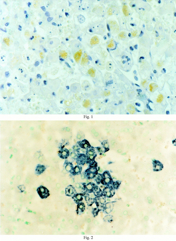
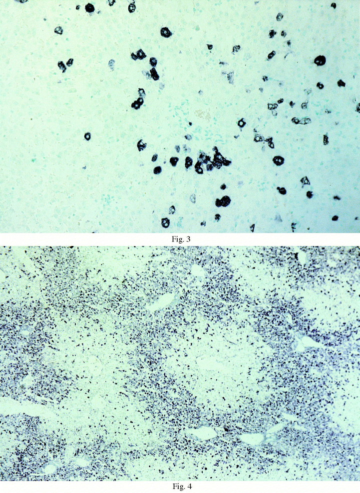
An in-situ hybridisation (ISH) technique for the detection of rabbit haemorrhagic disease virus (RHDV) was developed. Thirteen seronegative adult rabbits were infected oro-nasally using the BS89 RHDV strain. Liver and spleen samples were collected from 4 h post infection (p.i.) and repeated every 4 h till 44 h p.i. Each sample was tested immunohistochemically, by sandwich ELISA and by ISH. A 2.482-kb RNA probe, matching the genomic fragment coding for the VP60 structural protein of RHDV, was arranged. Two RNA probes (sense and antisense) were transcribed in vitro and UTP-digoxigenin-labelled. The antisense probe clearly detected positivity in the cytoplasm of the hepatocytes at 8 h p.i. Labelled hepatocytes were scattered throughout the sections until 24 h p.i. followed by a more diffuse perilobular positive reaction. A much weaker signal of similar distribution was detected up to 24 h p.i. using the sense RNA probe. All spleen samples tested negative for both probes. Liver samples were positive at 32 h p.i. using both ELISA and the immunoperoxidase test. Spleen samples were positive using only the ELISA at 32 h p.i. This study showed that RHDV replication occurred almost immediately after inoculation and that the liver appears to be the main site of replication.
Figures
References
-
- Barbieri, I., Lavazza, A., Brocchi, E., König, M., Capucci, L., 1997. Morphological, structural and antigenic modifications of rabbit haemorrhagic disease virus in the course of the disease. In: Chasey, D., Gaskell, R.M., Clarke, I.N. (Eds.), Proceedings of the First International Symposium on Caliciviruses. ESVV, Reading, UK, 15–17 September 1996, pp. 182–193.
-
- Brown C.C., Meyer R.F., Grubman M.J. Presence of African horse sickness virus in equine tissues, as determined by in situ hybridization. Vet. Pathol. 1994;31:689–694. - PubMed
-
- Capucci L., Scicluna M.T., Lavazza A., Brocchi E. Purification and characterization of the causative agent of viral haemorrhagic disease of rabbit. Sel. Vet. 1990;31:301–312.
Publication types
MeSH terms
Substances
LinkOut - more resources
Full Text Sources
Other Literature Sources
