

doi: 10.1101/gad.12.15.2263.
A new NEDD8-ligating system for cullin-4A
Affiliations
- PMID: 9694792
- PMCID: PMC317039
- DOI: 10.1101/gad.12.15.2263
Item in Clipboard
A new NEDD8-ligating system for cullin-4A
Genes Dev.
.
Abstract
NEDD8 is a ubiquitin (Ub)-like protein. Here we report a novel ubiquitinylation-related pathway for modification by NEDD8. NEDD8 was activated by an E1 (Ub-activating enzyme)-like complex, consisting of APP-BP1 and hUba3 with high respective homologies to the amino- and carboxy-terminal regions of E1 and then linked to hUbc12 (a human homolog of yeast Ub-conjugating enzyme Ubc12p). The major target protein modified by NEDD8 was found to be Hs-cullin-4A (Cul-4A), a member of the family of human cullin/Cdc53 proteins functioning as an essential component of a multifunctional Ub-protein ligase E3 complex that has a critical role in Ub-mediated proteolysis.
Figures
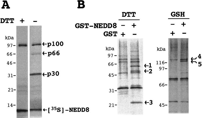
Analysis of proteins interacting with NEDD8 in
rabbit reticulocyte lysates. (A) Analysis of proteins linked
to 35S-labeled NEDD8. After 35S-labeled NEDD8 had
been synthesized for 60 min at 30°C in 5 μl of a reticulocyte
lysate transcription/ translation system, samples (2.5
μl) of the resultant translational products were subjected directly
to SDS-PAGE in the presence (+) or absence (−) of DTT and
autoradiographed. (B) SDS-PAGE analysis of proteins purified
by GST–NEDD8–GSH–Sepharose 4B chromatography. After reticulocyte
lysates had been applied to GSH–Sepharose 4B resin bearing GST or
GST–NEDD8, the bound materials were eluted with 40 mm DTT
(left) and subsequently with 30 mm GSH
(right). Samples were subjected to SDS-PAGE and stained with
silver. Proteins bound specifically to GST–NEDD8 are numbered at
right (1–5).
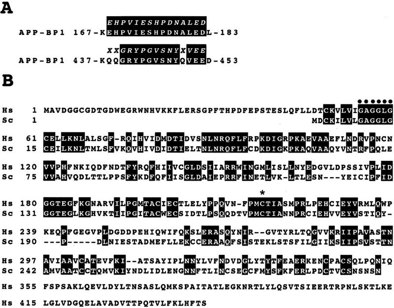
Identification of an E1-like
APP–BP1/hUba3 heterodimer for activation of NEDD8.
(A) Sequence alignment of two peptides of the rabbit 62-kD
protein associated with GST–NEDD8 (band 1 in Fig. 1B) with human
APP–BP1 (Chow et al. 1996). Partial amino acid sequences of the
fragments of the 62-kD band digested with lysylendopeptidase were
determined with a protein sequencer and are shown above the APP–BP1
sequences in italics. (X) An unidentified residue. The
identical amino acids are boxed in black. (B) Primary
structure of human Uba3 (Hs) deduced from the nucleotide sequence of a
human cDNA and its sequence alignment with yeast Uba3p (Sc) (PIR
accession no. S54087). Amino acid residues are numbered from the amino
terminus. Identical amino acids are boxed in black. The motif shown
(•) is the consensus sequence for a nucleotide binding site. The
asterisk indicates the essential Cys residue conserved in E1 enzymes
that becomes linked to Ub in an E1–Ub thioester linkage. (C)
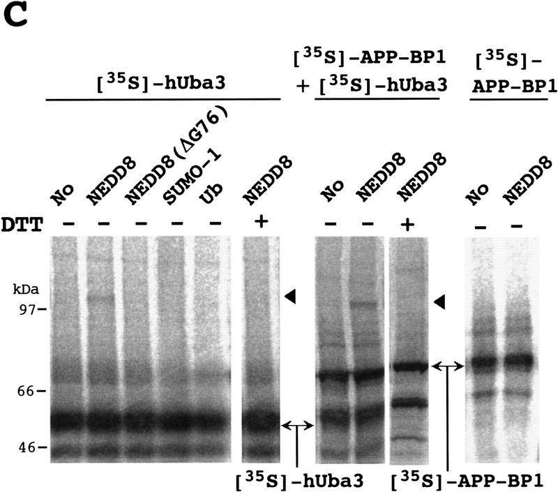
Thioester linkage between 35S-labeled hUba3 and GST–NEDD8.
35S-Labeled hUba3 and/or 35S-labeled
APP–BP1 were cosynthesized for 60 min at 30°C in vitro in a 5
μl reticulocyte lysate transcription/translation
system in the presence of 0.35 μg of unlabeled GST–NEDD8,
GST–NEDD8(Δ76G), GST–SUMO-1, or GST-Ub, and a part of each (2.5
μl) was analyzed as in Fig. 1A. Arrowheads indicate the
35S-labeled hUba3–GST–NEDD8 complex. (D) Complex
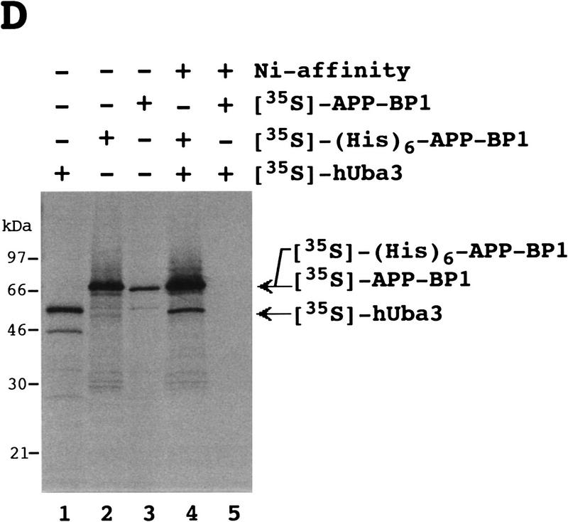
formation between 35S-labeled (His)6–APP–BP1 and
35S-labeled hUba3. 35S-labeled APP–BP1,
35S-labeled (His)6–APP–BP1, and
35S-labeled hUba3 were synthesized individually for 60 min at
30°C in vitro, and 1 μl of each was analyzed (lanes
1–3), as described in Fig. 1A. After samples (5 μl) of
35S-labeled (His)6–APP–BP1 or
35S-labeled APP–BP1 had been incubated with
35S-labeled hUba3 (5 μl) for 30 min at 30°C, half of
each sample was applied onto a Ni-chelate column. After the column had
been washed with 50 mm Na-phosphate buffer (pH 8.0)
containing 0.5 m NaCl, the materials eluted with 100
mm EDTA (lanes 4,5) were analyzed as in Fig.
1A.
Identification of an E1-like
APP–BP1/hUba3 heterodimer for activation of NEDD8.
(A) Sequence alignment of two peptides of the rabbit 62-kD
protein associated with GST–NEDD8 (band 1 in Fig. 1B) with human
APP–BP1 (Chow et al. 1996). Partial amino acid sequences of the
fragments of the 62-kD band digested with lysylendopeptidase were
determined with a protein sequencer and are shown above the APP–BP1
sequences in italics. (X) An unidentified residue. The
identical amino acids are boxed in black. (B) Primary
structure of human Uba3 (Hs) deduced from the nucleotide sequence of a
human cDNA and its sequence alignment with yeast Uba3p (Sc) (PIR
accession no. S54087). Amino acid residues are numbered from the amino
terminus. Identical amino acids are boxed in black. The motif shown
(•) is the consensus sequence for a nucleotide binding site. The
asterisk indicates the essential Cys residue conserved in E1 enzymes
that becomes linked to Ub in an E1–Ub thioester linkage. (C)
Thioester linkage between 35S-labeled hUba3 and GST–NEDD8.
35S-Labeled hUba3 and/or 35S-labeled
APP–BP1 were cosynthesized for 60 min at 30°C in vitro in a 5
μl reticulocyte lysate transcription/translation
system in the presence of 0.35 μg of unlabeled GST–NEDD8,
GST–NEDD8(Δ76G), GST–SUMO-1, or GST-Ub, and a part of each (2.5
μl) was analyzed as in Fig. 1A. Arrowheads indicate the
35S-labeled hUba3–GST–NEDD8 complex. (D) Complex
formation between 35S-labeled (His)6–APP–BP1 and
35S-labeled hUba3. 35S-labeled APP–BP1,
35S-labeled (His)6–APP–BP1, and
35S-labeled hUba3 were synthesized individually for 60 min at
30°C in vitro, and 1 μl of each was analyzed (lanes
1–3), as described in Fig. 1A. After samples (5 μl) of
35S-labeled (His)6–APP–BP1 or
35S-labeled APP–BP1 had been incubated with
35S-labeled hUba3 (5 μl) for 30 min at 30°C, half of
each sample was applied onto a Ni-chelate column. After the column had
been washed with 50 mm Na-phosphate buffer (pH 8.0)
containing 0.5 m NaCl, the materials eluted with 100
mm EDTA (lanes 4,5) were analyzed as in Fig.
1A.
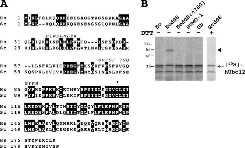
Identification of hUbc12 as a conjugating enzyme
for NEDD8. (A) Primary structure of human Ubc12 (Hs) deduced
from the nucleotide sequence of a human cDNA and sequence alignment
with yeast Ubc12p (Sc) (SWISS-PROT accession no. P52491). The identical
amino acids are boxed in black. The amino acid sequences of three
peptides of the rabbit 22-kD protein associated with GST–NEDD8 (band 3
in Fig. 1B) are shown above the sequence of hUbc12. The asterisk
indicates the essential Cys residue conserved in a family of
Ub-conjugating E2 enzymes that becomes linked to Ub in an E2–Ub
thioester linkage. (B) Thioester linkage between
35S-labeled hUbc12 and GST–NEDD8. 35S-Labeled
hUbc12 synthesized in vitro in the presence of unlabeled GST–NEDD8,
GST–NEDD8(Δ76G), GST–SUMO-1, or GST–Ub was analyzed as in Fig.
2. The arrowhead indicates the 35S-labeled hUbc12–GST–NEDD8
complex.
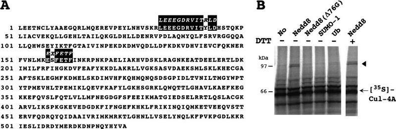
Linkage of NEDD8 to Cul-4A in a DTT-insensitive
fashion. (A) Alignment of amino acid sequences of two peptides
of the rabbit 120-kD protein associated with GST–NEDD8 (protein 5 band
in Fig. 1B) and human Cul-4A that we had cloned. Identical amino acids
are boxed in black. The Cul-4A cDNA was named Cul-4A(524C), as
described in the text. (B) Linkage between
35S-labeled Cul-4A(524C) and GST–NEDD8.
35S-labeled Cul-4A(524C) synthesized in vitro in the presence
of unlabeled GST–NEDD8, GST–NEDD8(Δ76G), GST–SUMO-1, or GST–Ub
was analyzed as in Fig. 2C. The arrowhead indicates the
35S-labeled Cul-4A(524C)–GST–NEDD8 complex. (C)
Linkage between 35S-Labeled Cul-4A(171C) and GST–NEDD8.
35S-labeled Cul-4A(171C) that corresponds to the
carboxy-terminal 171 amino acid residues (see Materials and methods)
was treated or not with unlabeled GST–NEDD8 and analyzed as in
B. The arrowhead indicates the 35S-labeled
Cul-4A(171C)–GST–NEDD8 complex.
References
-
- Chow N, Korenberg JR, Chen X-N, Neve RL. APP-BP1, a novel protein that binds to the carboxyl-terminal region of the amyloid precursor protein. J Biol Chem. 1996;271:11339–11346. - PubMed
-
- Coux O, Tanaka K, Goldberg AL. Structure and functions of the 20S and 26S proteasomes. Annu Rev Biochem. 1996;65:801–847. - PubMed
-
- Dohmen RJ, Sappen R, McGrath JP, Forrova H, Kolarov J, Goffeau A, Varshavsky A. An essential yeast gene encoding a homolog of ubiquitin-activating enzyme. J Biol Chem. 1995;270:18099–18109. - PubMed
-
- Handeli S, Weintraub H. The ts41 mutation in Chinese hamster cells leads to successive S phases in the absence of intervening G2, M, and G1. Cell. 1992;71:599–611. - PubMed
-
- Hass AL, Siepmann TJ. Pathways of ubiquitin conjugation. FASEB J. 1997;11:1257–1268. - PubMed
MeSH terms
Substances
Associated data
- Actions
- Actions
- Actions
- Actions
LinkOut - more resources
Full Text Sources
Other Literature Sources
Molecular Biology Databases
Miscellaneous