. 1998 Aug 1;12(15):2269-77.
doi: 10.1101/gad.12.15.2269.

A histone deacetylase corepressor complex regulates the Notch signal transduction pathway

Affiliations

A histone deacetylase corepressor complex regulates the Notch signal transduction pathway

H Y Kao et al. Genes Dev. .

Abstract

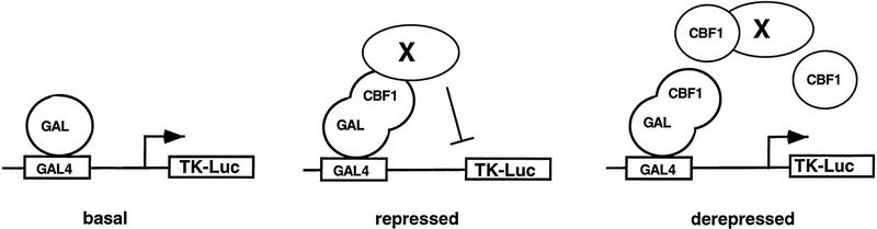
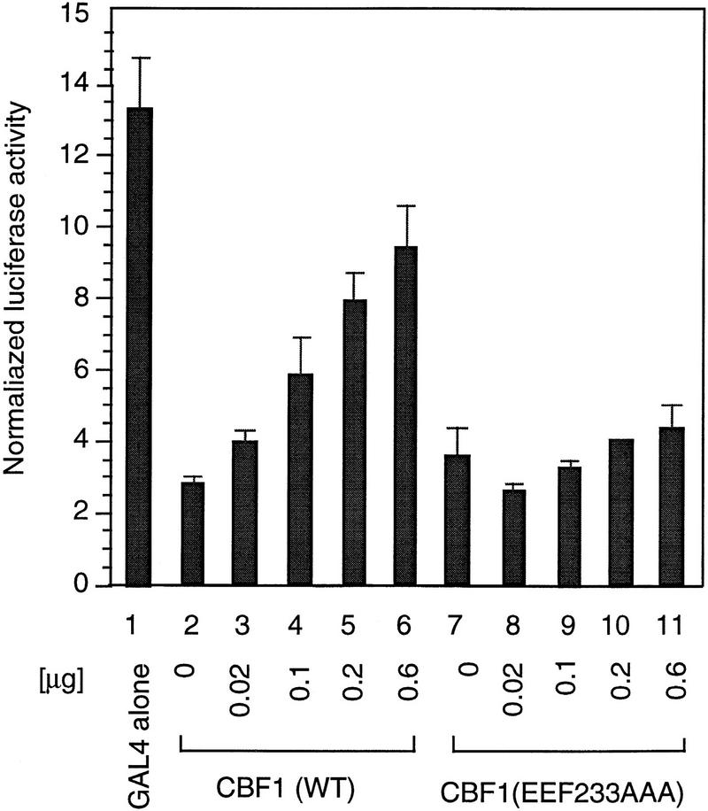
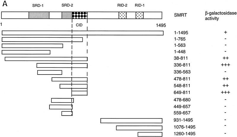
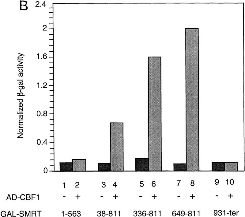
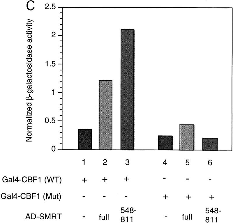
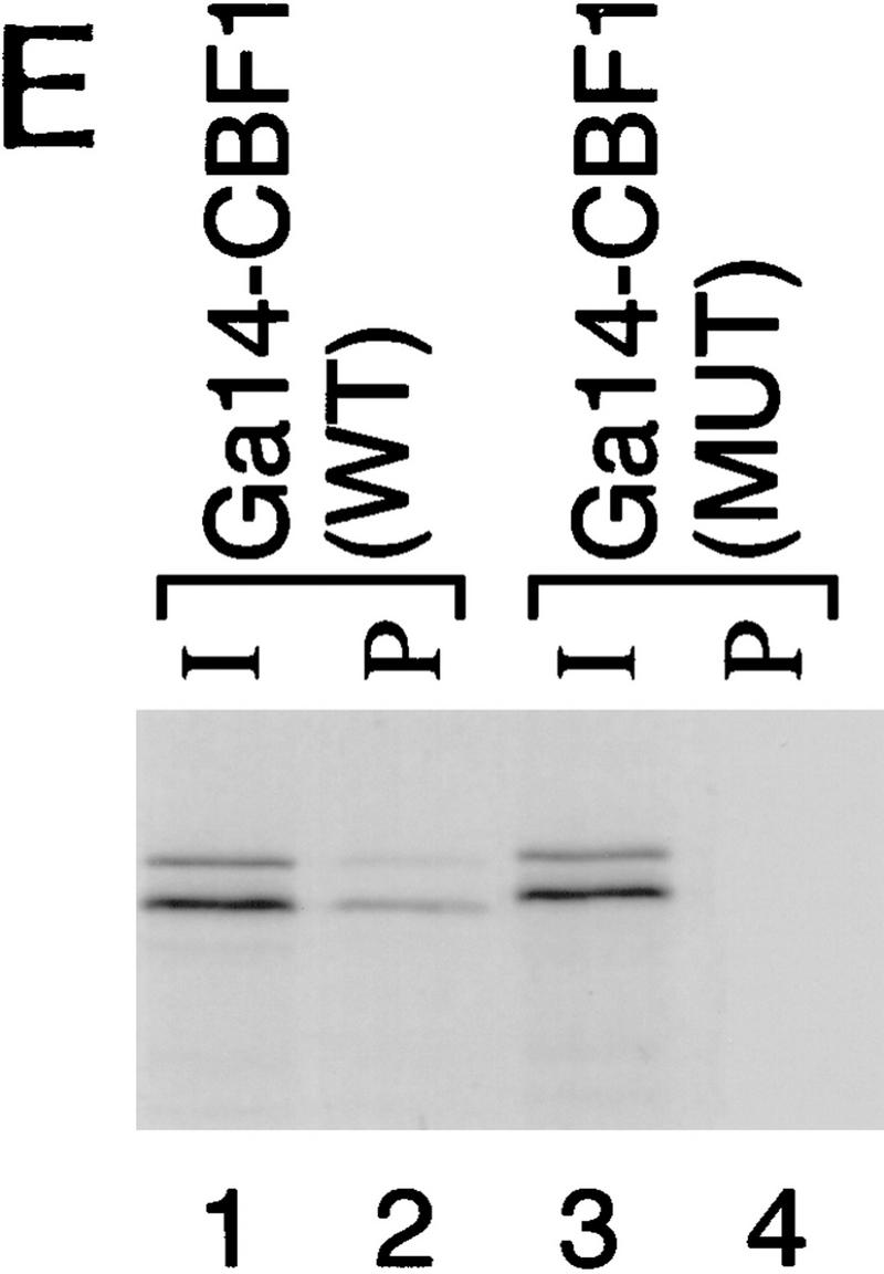
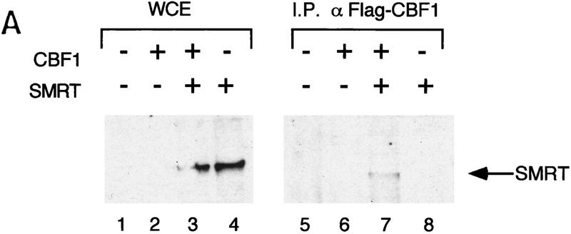
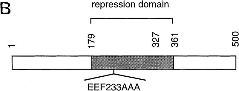
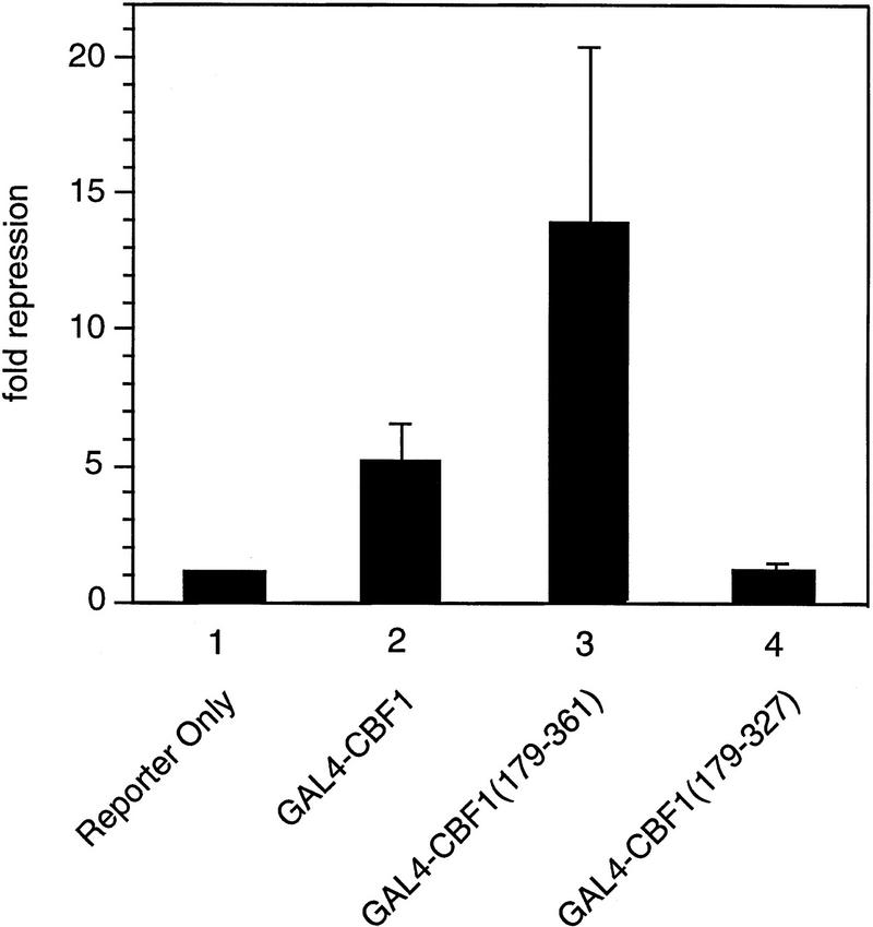
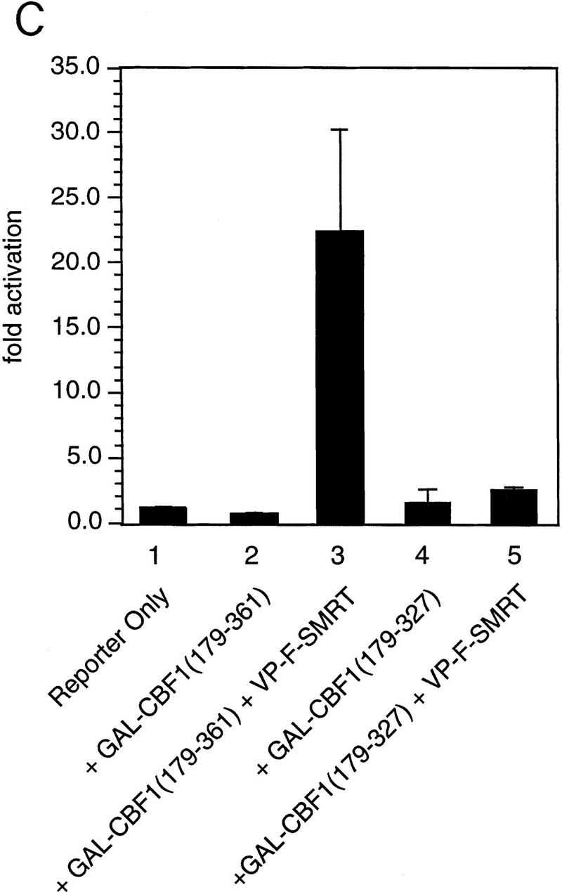
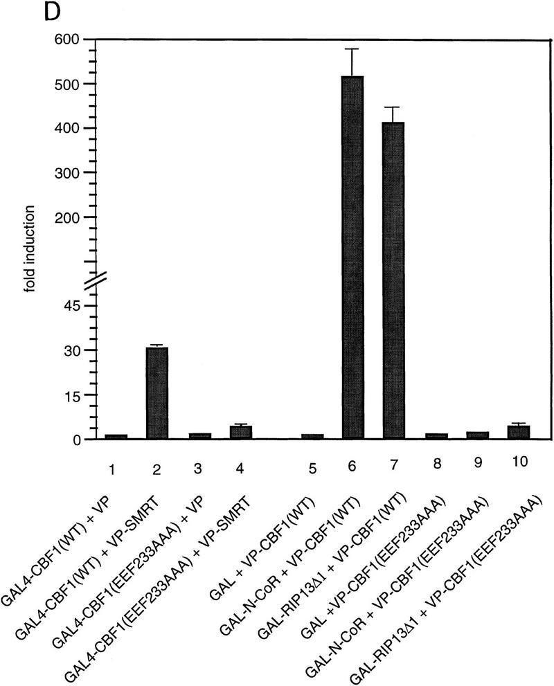
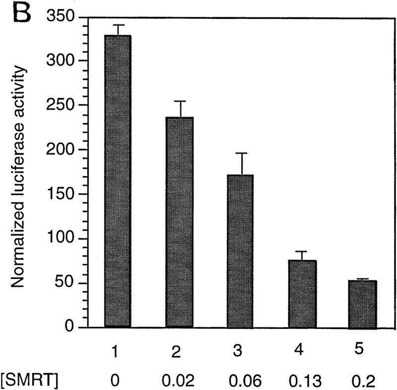
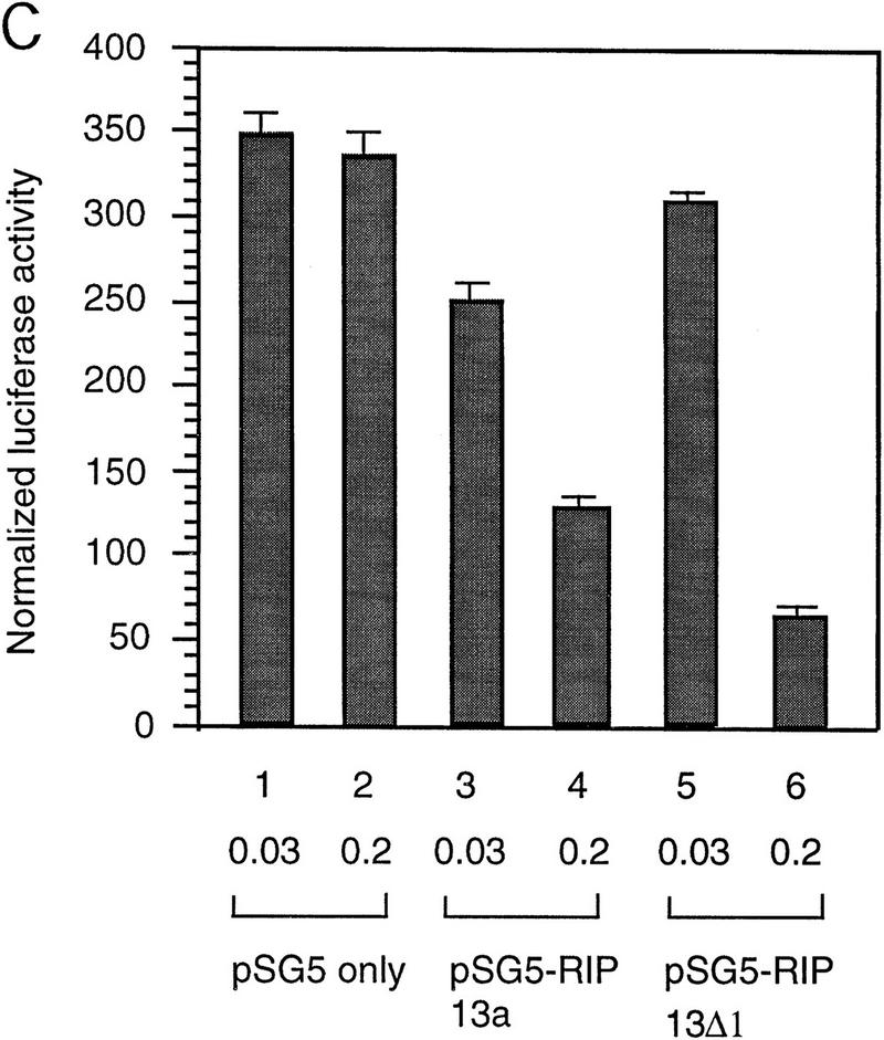
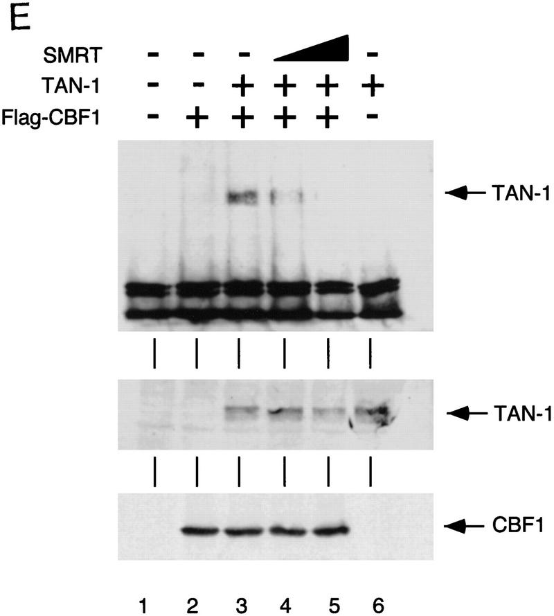
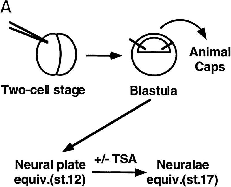
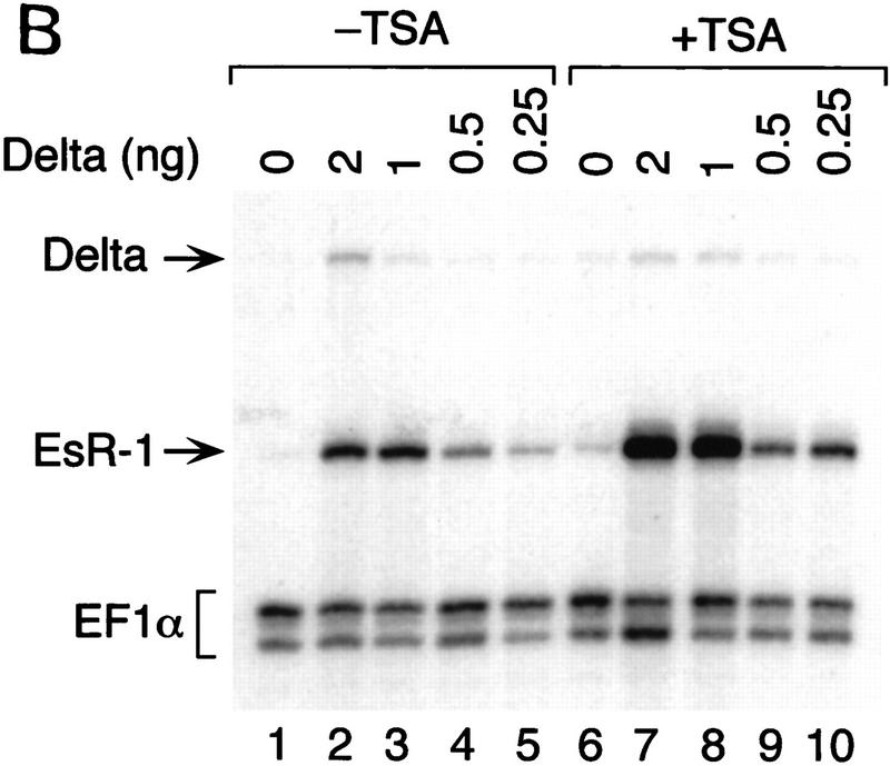
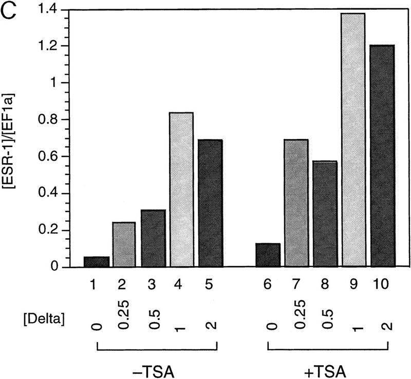
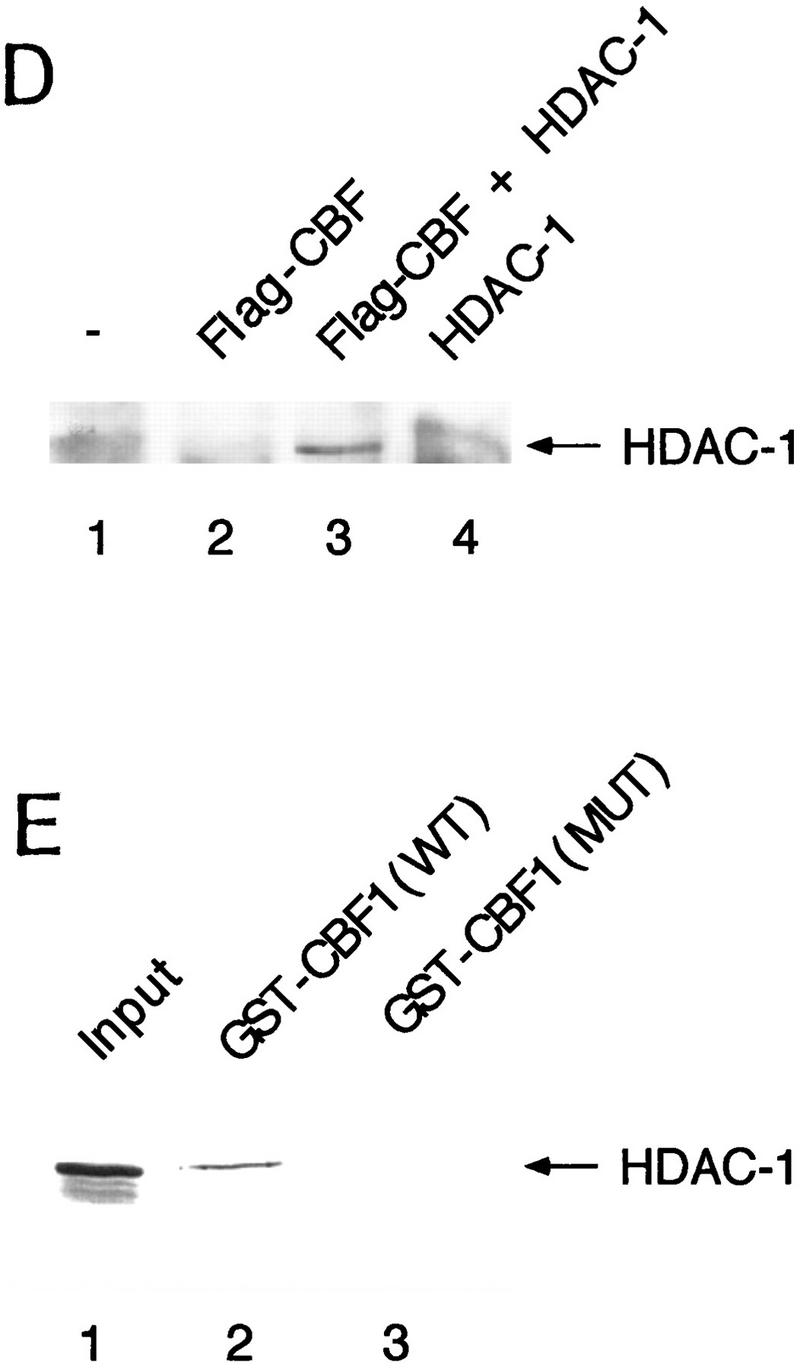
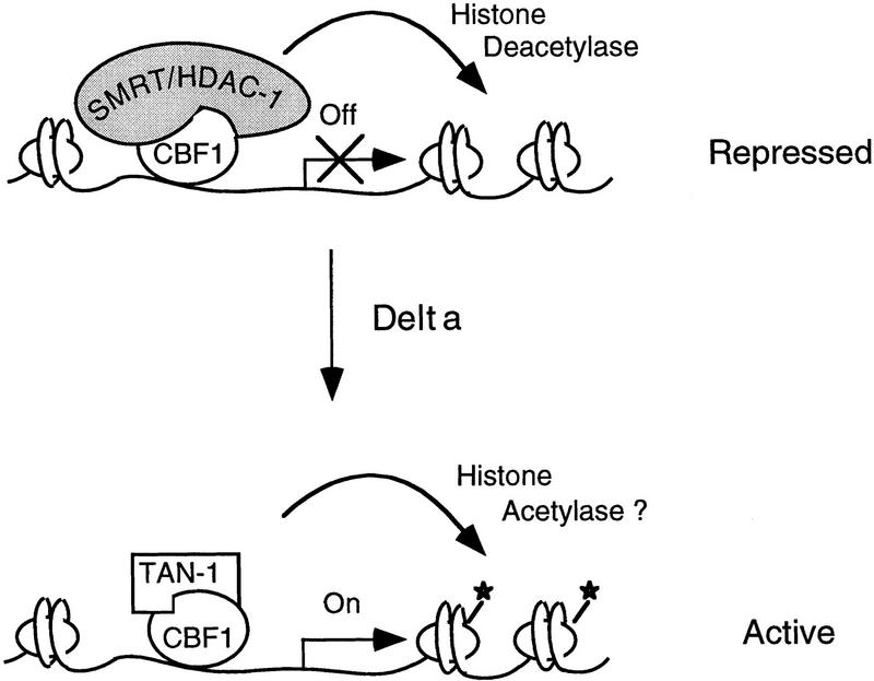
The Delta-Notch signal transduction pathway has widespread roles in animal development in which it appears to control cell fate. CBF1/RBP-Jkappa, the mammalian homolog of Drosophila Suppressor of Hairless [Su(H)], switches from a transcriptional repressor to an activator upon Notch activation. The mechanism whereby Notch regulates this switch is not clear. In this report we show that prior to induction CBF1/RBP-Jkappa interacts with a corepressor complex containing SMRT (silencing mediator of retinoid and thyroid hormone receptors) and the histone deacetylase HDAC-1. This complex binds via the CBF1 repression domain, and mutants defective in repression fail to interact with the complex. Activation by Notch disrupts the formation of the repressor complex, thus establishing a molecular basis for the Notch switch. Finally, ESR-1, a Xenopus gene activated by Notch and X-Su(H), is induced in animal caps treated with TSA, an inhibitor of HDAC-1. The functional role for the SMRT/HDAC-1 complex in CBF1/RBP-Jkappa regulation reveals a novel genetic switch in which extracellular ligands control the status of critical nuclear cofactor complexes.


Figures

Figure 1
Figure 1
Wild-type CBF1, but not mutant CBF1 (EEF233AAA), derepresses GAL4–CBF1. GAL4–CBF1 was transfected along with increasing amounts of either wild-type CBF1 (lanes 2–6) or mutant CBF1 (EEF233AAA) (lanes 7–11) expression plasmids as indicated. Luciferase activity is normalized by β-galactosidase activity (described in Materials and Methods). The diagram illustrates how CBF1 overexpression may result in derepression of GAL4–CBF1.
Figure 1
Figure 1
Wild-type CBF1, but not mutant CBF1 (EEF233AAA), derepresses GAL4–CBF1. GAL4–CBF1 was transfected along with increasing amounts of either wild-type CBF1 (lanes 2–6) or mutant CBF1 (EEF233AAA) (lanes 7–11) expression plasmids as indicated. Luciferase activity is normalized by β-galactosidase activity (described in Materials and Methods). The diagram illustrates how CBF1 overexpression may result in derepression of GAL4–CBF1.
Figure 2
Figure 2
CBF1 interacts with SMRT in yeast and in GST pull-down assays. (A) Mapping of CBF1 interaction domain of SMRT in yeast. SMRT fragments fused to GAL4 DBD (GAL–SMRT) were cotransformed with GAL4 activation domain fused to CBF1(179–500) into yeast strain Y190. (SRD) SMRT repression domain; (RID) receptor interaction domain. (B) Quantitation of CBF1 and SMRT interaction in yeast. (C) Mutant CBF1 (EEF233AAA) fails to interact with SMRT in a yeast two-hybrid assay. Wild-type (lanes 1–3) or mutant GAL4–CBF1 (lanes 4–6) were cotransformed with or without AD–SMRT. (D) CBF1 interacts with GST–SMRT in vitro as shown in lanes 4 and 5. (E) Mutant GAL–CBF1 (EEF233AAA) (cf. lanes 2 and 4) does not interact with GST–SMRT(548–811). (I) Loaded in this experiment. (P) Pellet.
Figure 2
Figure 2
CBF1 interacts with SMRT in yeast and in GST pull-down assays. (A) Mapping of CBF1 interaction domain of SMRT in yeast. SMRT fragments fused to GAL4 DBD (GAL–SMRT) were cotransformed with GAL4 activation domain fused to CBF1(179–500) into yeast strain Y190. (SRD) SMRT repression domain; (RID) receptor interaction domain. (B) Quantitation of CBF1 and SMRT interaction in yeast. (C) Mutant CBF1 (EEF233AAA) fails to interact with SMRT in a yeast two-hybrid assay. Wild-type (lanes 1–3) or mutant GAL4–CBF1 (lanes 4–6) were cotransformed with or without AD–SMRT. (D) CBF1 interacts with GST–SMRT in vitro as shown in lanes 4 and 5. (E) Mutant GAL–CBF1 (EEF233AAA) (cf. lanes 2 and 4) does not interact with GST–SMRT(548–811). (I) Loaded in this experiment. (P) Pellet.
Figure 3
Figure 3
Functional interaction of CBF1 and SMRT. (A) SMRT interacts with CBF1 in vivo. NIH–3T3 cells were cotransfected with pBluescript (lanes 1,5), CBF1–Flag (lanes 2,6), SMRT (lanes 3,7), and CBF1–Flag + SMRT (lanes 4,8). Cells were harvested and lysed in RIPA buffer and extracts were analyzed by Western blotting. For immunoprecipitation, lysates were incubated with anti-Flag antibody and protein A/G–agarose and analyzed by Western blotting. (Left) Whole-cell extract probed with anti-SMRT antibody; (right) immunoprecipitated protein probed with anti-SMRT antibody. (B) NIH–3T3 cells were transfected with GAL5–TK–CAT reporter (1 μg) and the indicated GAL4–CBF1 fusion proteins (1 μg). Full-length CBF1 and CBF1(179–361) repress reporter; CBF1(179–327) does not repress (cf. lanes 2–4). The schematic highlights the minimal repression domain and the mutations that abolish repression activity of CBF1. (C) SMRT interaction with CBF1 requires an intact CBF1 repression domain. NIH–3T3 cells were cotransfected with a GAL4–E1B–luc reporter (0.3 μg), GAL4–CBF1 (1 μg) plasmids as indicated, and VP16–SMRT (1 μg). (D) Interaction with SMRT/N-CoR/RIP13Δ is impaired in a mutant CBF1 (EEF233AAA). CV-1 cells were transfected with a GAL4–E1B–luc reporter (0.1 μg), pCMX–lacZ (0.1 μg), and expression vectors as indicated.
Figure 3
Figure 3
Functional interaction of CBF1 and SMRT. (A) SMRT interacts with CBF1 in vivo. NIH–3T3 cells were cotransfected with pBluescript (lanes 1,5), CBF1–Flag (lanes 2,6), SMRT (lanes 3,7), and CBF1–Flag + SMRT (lanes 4,8). Cells were harvested and lysed in RIPA buffer and extracts were analyzed by Western blotting. For immunoprecipitation, lysates were incubated with anti-Flag antibody and protein A/G–agarose and analyzed by Western blotting. (Left) Whole-cell extract probed with anti-SMRT antibody; (right) immunoprecipitated protein probed with anti-SMRT antibody. (B) NIH–3T3 cells were transfected with GAL5–TK–CAT reporter (1 μg) and the indicated GAL4–CBF1 fusion proteins (1 μg). Full-length CBF1 and CBF1(179–361) repress reporter; CBF1(179–327) does not repress (cf. lanes 2–4). The schematic highlights the minimal repression domain and the mutations that abolish repression activity of CBF1. (C) SMRT interaction with CBF1 requires an intact CBF1 repression domain. NIH–3T3 cells were cotransfected with a GAL4–E1B–luc reporter (0.3 μg), GAL4–CBF1 (1 μg) plasmids as indicated, and VP16–SMRT (1 μg). (D) Interaction with SMRT/N-CoR/RIP13Δ is impaired in a mutant CBF1 (EEF233AAA). CV-1 cells were transfected with a GAL4–E1B–luc reporter (0.1 μg), pCMX–lacZ (0.1 μg), and expression vectors as indicated.
Figure 4
Figure 4
SMRT antagonizes TAN-1 activity. (A) SMRT inhibits TAN-1 activation of GAL4–CBF1. CV-1 cells were transfected with 0.1 μg of TK–MH100x4–Luc, pCMX–lacZ, 0.02 μg of pTAN-1, and pCMX–Gal4 DBD (lanes 1–5) or pCMX–Gal4 DBD–CBF1(179–500) (lanes 6–10). (B) SMRT inhibits Notch-mediated activation of endogenous CBF1. CV-1 cells were transfected with 0.1 μg of a CBF1–luc reporter, pCMX–lacZ, and pTAN-1 (0.02 μg). (C) RIP13a and RIP13Δ1 antagonize TAN-1-mediated activation of endogenous CBF1. Experiments were carried out similar to B and D. SMRT inhibits Notch activation of the HES-1 promoter. Experiments were carried out similar to B using 0.1 μg of a HES-1 promoter construct. (E) SMRT inhibits Notch interaction with CBF1 in vivo. 293T cells were transfected with the indicated plasmids. Flag–CBF1 (4 μg) was immunoprecipitated from whole-cell lysates in RIPA buffer. Immunoprecipitates were washed four times in RIPA buffer and loaded onto an 6% SDS–polyacrylamide gel. (Top) IP reactions probed with TAN-1 antibody; (middle and bottom) Western blots of whole cell extract. Blot was first probed with TAN-1 antibody and subsequently probed with Flag antibody to detect CBF1. Increasing amounts of SMRT were cotransfected in lanes 4 (1 μg) and 5 (2 μg).
Figure 4
Figure 4
SMRT antagonizes TAN-1 activity. (A) SMRT inhibits TAN-1 activation of GAL4–CBF1. CV-1 cells were transfected with 0.1 μg of TK–MH100x4–Luc, pCMX–lacZ, 0.02 μg of pTAN-1, and pCMX–Gal4 DBD (lanes 1–5) or pCMX–Gal4 DBD–CBF1(179–500) (lanes 6–10). (B) SMRT inhibits Notch-mediated activation of endogenous CBF1. CV-1 cells were transfected with 0.1 μg of a CBF1–luc reporter, pCMX–lacZ, and pTAN-1 (0.02 μg). (C) RIP13a and RIP13Δ1 antagonize TAN-1-mediated activation of endogenous CBF1. Experiments were carried out similar to B and D. SMRT inhibits Notch activation of the HES-1 promoter. Experiments were carried out similar to B using 0.1 μg of a HES-1 promoter construct. (E) SMRT inhibits Notch interaction with CBF1 in vivo. 293T cells were transfected with the indicated plasmids. Flag–CBF1 (4 μg) was immunoprecipitated from whole-cell lysates in RIPA buffer. Immunoprecipitates were washed four times in RIPA buffer and loaded onto an 6% SDS–polyacrylamide gel. (Top) IP reactions probed with TAN-1 antibody; (middle and bottom) Western blots of whole cell extract. Blot was first probed with TAN-1 antibody and subsequently probed with Flag antibody to detect CBF1. Increasing amounts of SMRT were cotransfected in lanes 4 (1 μg) and 5 (2 μg).
Figure 5
Figure 5
TSA derepresses expression of the Notch target gene ESR-1. (A) Diagram of the animal cap assay. Each blastomere of a two-cell Xenopus embryo was injected with varying amounts of X-Delta-1 RNA along with RNA (1.0 ng) encoding the neural inducer Noggin. For each assay, eight animal caps were explanted at late blastula stages (st. 9). From stage 12 to 17, treated animal caps were incubated with 400 nm TSA, after which total RNA was extracted. (B) RNase protection assay. RNA isolated from animal caps was assayed simultaneously for the levels of ESR-1, X-Delta-1, and EF-1α (an internal measure of total RNA). Arrows or bracket indicate the position of the protected probe for each RNA. The Xenopus Delta-1 probe detects both injected as well as endogenous RNA. (C) Quantitation of the assay. Protected band intensities were quantitated and normalized for total RNA using a PhosphorImager (Molecular Dynamics). (D) Association of CBF1 and HDAC-1 in vivo. CV-1 cells were cotransfected with CBF1–Flag (lane 3). Cells were harvested and lysed in RIPA buffer, and extracts were analyzed by Western blotting. For immunoprecipitation, lysates were incubated with anti-Flag antibody and protein A/G–agarose and analyzed by Western blotting with anti-HDAC-1 antibody. (E) HDAC-1 interacts with wild-type GST–CBF1 but not mutant GST–CBF1 (EEF233AAA) in vitro.
Figure 5
Figure 5
TSA derepresses expression of the Notch target gene ESR-1. (A) Diagram of the animal cap assay. Each blastomere of a two-cell Xenopus embryo was injected with varying amounts of X-Delta-1 RNA along with RNA (1.0 ng) encoding the neural inducer Noggin. For each assay, eight animal caps were explanted at late blastula stages (st. 9). From stage 12 to 17, treated animal caps were incubated with 400 nm TSA, after which total RNA was extracted. (B) RNase protection assay. RNA isolated from animal caps was assayed simultaneously for the levels of ESR-1, X-Delta-1, and EF-1α (an internal measure of total RNA). Arrows or bracket indicate the position of the protected probe for each RNA. The Xenopus Delta-1 probe detects both injected as well as endogenous RNA. (C) Quantitation of the assay. Protected band intensities were quantitated and normalized for total RNA using a PhosphorImager (Molecular Dynamics). (D) Association of CBF1 and HDAC-1 in vivo. CV-1 cells were cotransfected with CBF1–Flag (lane 3). Cells were harvested and lysed in RIPA buffer, and extracts were analyzed by Western blotting. For immunoprecipitation, lysates were incubated with anti-Flag antibody and protein A/G–agarose and analyzed by Western blotting with anti-HDAC-1 antibody. (E) HDAC-1 interacts with wild-type GST–CBF1 but not mutant GST–CBF1 (EEF233AAA) in vitro.
Figure 6
Figure 6
A model for transcriptional regulation of Notch-activated genes. (Top) In the absence of activated Notch, gene transcription is repressed by binding of CBF1/RBP-Jκ and the recruitment of a corepressor complex containing histone deacetylase activity. (Bottom) Upon activation of Notch signaling by binding of the Notch ligand Delta, an intracellular form of Notch is released by proteolytic cleavage and translocates to the nucleus where it interacts with CBF1/RBP-Jκ. This interaction displaces the corepressor complex and results in activation of transcription presumably by removal of the histone deacetylase activity. It remains to be determined whether transcriptional activation by a Notch : CBF1/RBP-Jκ complex involves the recruitment of histone acetylases.

References

    1. Alland L, Muhle R, Hou H, Potes JJ, Chin L, Schreiber-Agus N, DePinho RA. Role for N-CoR and histone deacetylase in Sin3-mediated transcriptional repression. Nature. 1997;387:49–55. - PubMed
    1. Amakawa R, Jing W, Ozawa K. Human Jk recombination signal binding protein gene (IGKJRB): Comparison with its mouse homologue. Genomics. 1993;17:306–315. - PubMed
    1. Artavanis-Tsakonas S, Matsuno K, Fortini ME. Notch signaling. Science. 1995;268:225–232. - PubMed
    1. Ayer DE, Lawrence QA. Mad-Max transcriptional repression is mediated by ternary complex formation with mammalian homologs of yeast repressor Sin3. Cell. 1995;80:767–776. - PubMed
    1. Bailey AM, Posakony JW. Suppressor of hairless directly activates transcription of enhancer of split complex genes in response to Notch receptor activity. Genes & Dev. 1995;9:2609–2622. - PubMed

Publication types

MeSH terms