. 1998 Sep;72(9):7064-74.
doi: 10.1128/JVI.72.9.7064-7074.1998.

Herpes simplex virus glycoprotein D can bind to poliovirus receptor-related protein 1 or herpesvirus entry mediator, two structurally unrelated mediators of virus entry

Affiliations

Herpes simplex virus glycoprotein D can bind to poliovirus receptor-related protein 1 or herpesvirus entry mediator, two structurally unrelated mediators of virus entry

C Krummenacher et al. J Virol. 1998 Sep.

Abstract

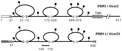
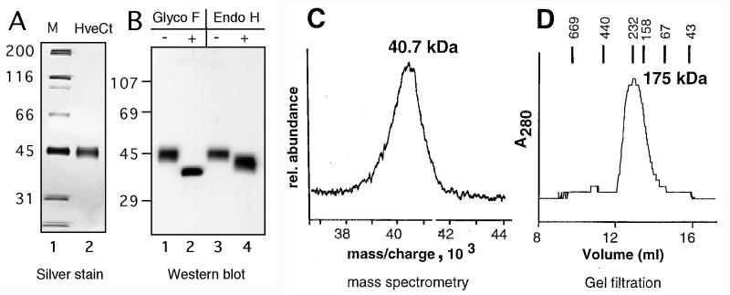
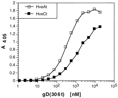
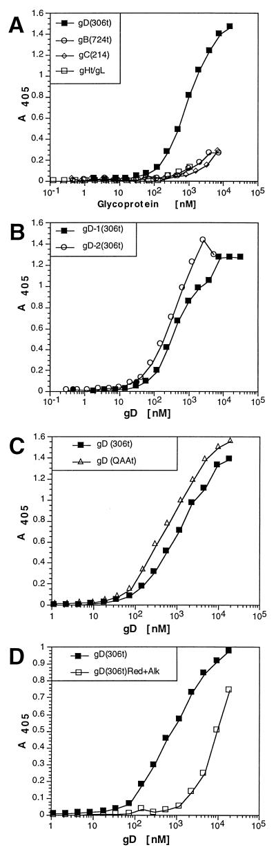
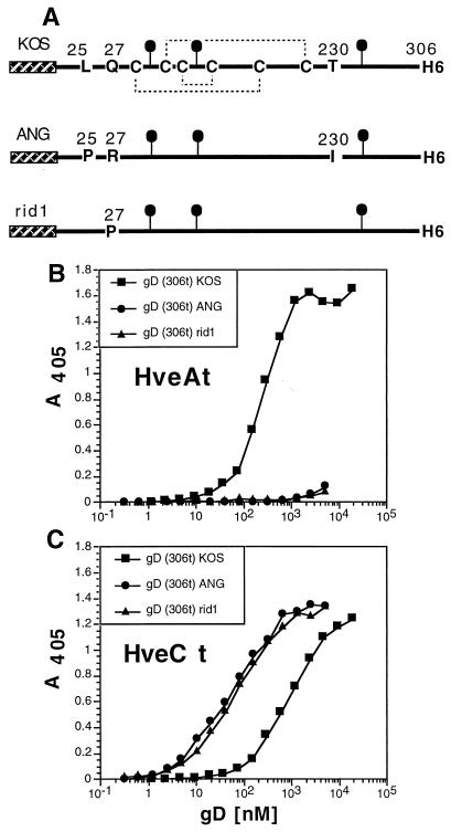
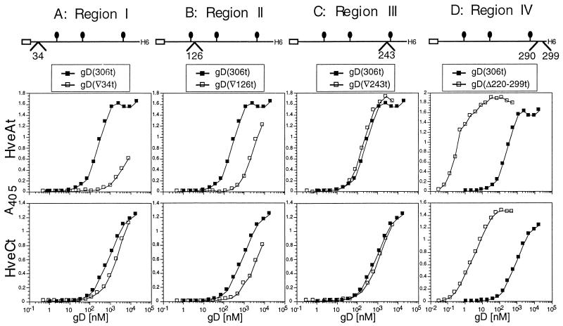
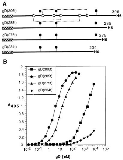
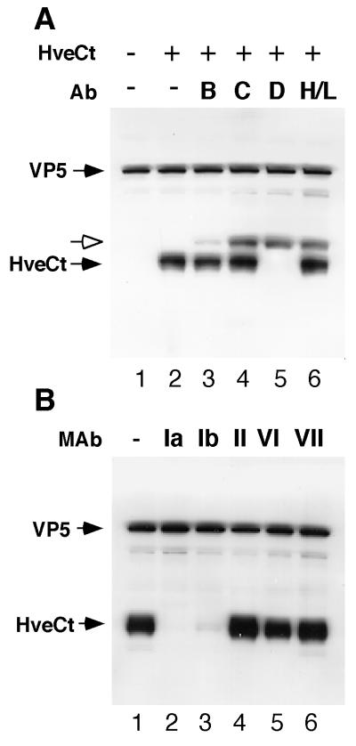
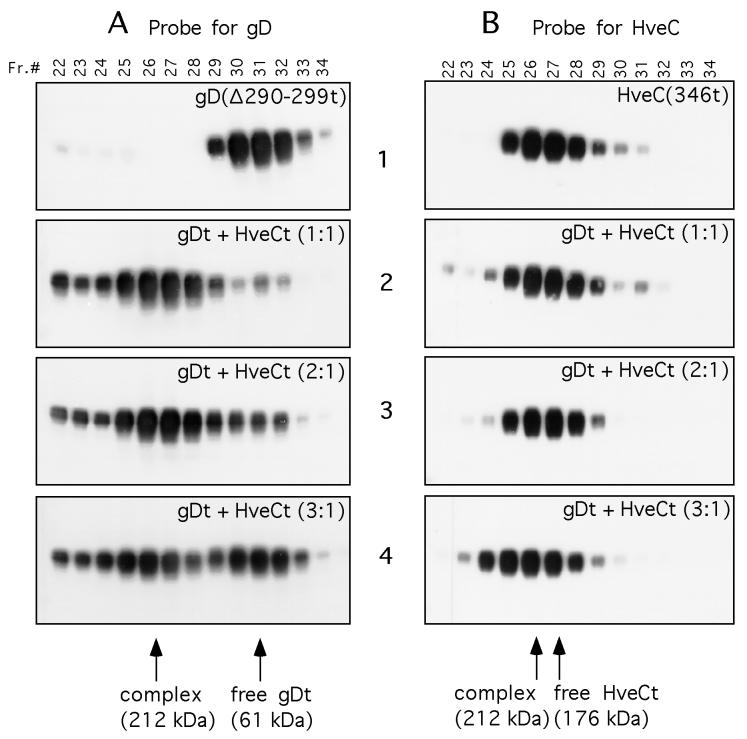
Several cell membrane proteins have been identified as herpes simplex virus (HSV) entry mediators (Hve). HveA (formerly HVEM) is a member of the tumor necrosis factor receptor family, whereas the poliovirus receptor-related proteins 1 and 2 (PRR1 and PRR2, renamed HveC and HveB) belong to the immunoglobulin superfamily. Here we show that a truncated form of HveC directly binds to HSV glycoprotein D (gD) in solution and at the surface of virions. This interaction is dependent on the native conformation of gD but independent of its N-linked glycosylation. Complex formation between soluble gD and HveC appears to involve one or two gD molecules for one HveC protein. Since HveA also mediates HSV entry by interacting with gD, we compared both structurally unrelated receptors for their binding to gD. Analyses of several gD variants indicated that structure and accessibility of the N-terminal domain of gD, essential for HveA binding, was not necessary for HveC interaction. Mutations in functional regions II, III, and IV of gD had similar effects on binding to either HveC or HveA. Competition assays with neutralizing anti-gD monoclonal antibodies (MAbs) showed that MAbs from group Ib prevented HveC and HveA binding to virions. However, group Ia MAbs blocked HveC but not HveA binding, and conversely, group VII MAbs blocked HveA but not HveC binding. Thus, we propose that HSV entry can be mediated by two structurally unrelated gD receptors through related but not identical binding with gD.


Figures

FIG. 1
FIG. 1
Schematic representation of PRR1/HveC proteins. The 517-amino-acid human PRR1/HveC is represented with residues numbered from methionine 1. The open box indicates the HveC signal peptide, and the transmembrane region is abbreviated TMR. The putative N-linked carbohydrates are shown as black lollipops. In the baculovirus construct, the mellitin signal peptide (hatched box) replaced the natural signal peptide (amino acids 1 to 30) from HveC. An additional N-terminal aspartic acid residue was inserted due to the cloning strategy. HveCt was truncated after His346, and five histidine residues were added to generate a six-His tag at the C terminus of HveCt. The synthetic peptide (amino acids 155 to 172) was used to generate rabbit antiserum R145.
FIG. 2
FIG. 2
Biochemical characterization of HveCt. (A) Silver stain of HveCt after nickel chromatography purification and SDS-PAGE under denaturing and reducing conditions. Sizes of the molecular weight markers (M) are indicated in kilodaltons. (B) After SDS-PAGE in denaturing and reducing conditions, proteins were transferred to nitrocellulose and detected with antipeptide serum R145. Lanes 1 and 3, purified HveCt used as mock-digested controls. In lane 2, N-linked carbohydrates of the purified protein were digested by glycopeptidase (Glyco) F; in lane 4, purified HveCt was treated with endoglycosidase (Endo) H. (C) Mass spectrometric analysis of purified HveCt. The calculated mass of singly charged species is indicated in kilodaltons. (D) Purified HveCt (24 μM in PBS) was loaded on a Superdex 200 size exclusion column and eluted with PBS. Elution profiles monitored by A280 is shown. Calculated size is based on positions of molecular size standards, indicated in kilodaltons.
FIG. 3
FIG. 3
Comparison of gD binding to HveCt and HveAt. ELISA plates coated with HveCt or HveAt at 200 nM in PBS were incubated with increasing concentrations of gD(306t). Bound gD was detected with antiserum R7 followed by peroxidase-conjugated secondary antibody and substrate. Absorbance was read at 405 nm.
FIG. 4
FIG. 4
Analysis of binding of gD to HveCt by ELISA. Ninety-six-well plates were saturated with 50 μl of 200 nM HveCt in PBS and incubated with variable concentrations of purified HSV glycoproteins. (A) Glycoproteins bound to immobilized HveC were detected with specific antibodies (R7 for gD, R47 for gC, R69 for gB, and R137 for gH-gL) followed by peroxidase-conjugated secondary antibody and substrate. (B) gD-1(306t)KOS and gD-2(306t)333 at various concentrations were incubated on HveCt-coated plates. (C) Mutant gD (QAAt) lacking N-CHO was compared to the glycosylated control gD(306t) for binding to immobilized HveCt. (D) Purified gD-1(306t) was reduced and alkylated prior to incubation on the HveCt-coated plate. Rabbit polyclonal serum R7 was used to detect any type of gD.
FIG. 5
FIG. 5
Binding of gD(306t) from HSV strains KOS, ANG, and rid1 to HveCt. (A) gDs from both HSV (ANG) and HSV(rid1) were expressed in baculovirus as truncated forms and affinity purified. Cysteine residues on gD(306t) from KOS wild-type strain as well as mutated residues are indicated. The hatched box represents the mellitin signal peptide, and the black lollipops represent N-linked carbohydrates. gDs from these strains were compared to gD KOS for binding to immobilized HveAt (B) or HveCt (C). Antiserum R7 was used to detect bound gD in these ELISAs.
FIG. 6
FIG. 6
Effects of linker insertions in functional regions of gD on binding to HveCt and HveAt. ELISA plates were saturated with HveAt (top panels) or HveCt (bottom panels) and incubated with various concentrations of purified mutant gDs. Binding of each mutant is compared to binding of wild-type gD(306t) (black squares). gD(▿34t) is mutated in functional region I (A), gD(▿126t) is mutated in region II (B), gD(▿243t) is mutated in region III (C), and gD(Δ290-299) is mutated in functional region IV (D). The position of the linker insertion is schematically represented for each mutant. Antiserum R7 was used to detect bound gD.
FIG. 7
FIG. 7
Effect of C-terminal truncation on binding to HveC. (A) Shorter versions of gD-1 KOS were produced in the baculovirus expression system, purified, and tested for binding to HveC. (B) ELISA was performed with HveCt bound to the plate and incubated with variable amounts of gD. Bound gD was detected with antiserum R7.
FIG. 8
FIG. 8
Binding of HveCt to HSV particles is blocked by anti-gD antibodies. (A) Purified HSV-1 KOS virions (107 PFU) were incubated at 4°C for 2 h with (lane 2) or without (lane 1) HveCt (150 μg) and loaded onto a sucrose gradient. The viral band was collected and analyzed by SDS-PAGE and Western blotting. Membranes were probed for presence of VP5 and HveCt (R154 serum). In blocking experiments, virions were preincubated with cocktails of antibodies (Ab) specific for HSV glycoproteins: for gB, SS10 (0.5 μl of ascites), DL16 (5 μg of IgG), DL21 (5 μg of IgG), and R69 (0.5 μl of serum) (lane 3); for gC, MP1 (0.5 μl of ascites), MP5 (5 μg of IgG), 1C8 (5 μg of IgG), and R46 (0.5 μl of serum) (lane 4); for gD, 1D3 (0.5 μl of ascites), DL2 (5 μg of IgG), DL11 (5 μg of IgG), and R7 (0.5 μl of serum) (lane 5); for gH-gL, LP11 (0.5 μl of ascites), 53S (5 μg of IgG), H6 (5 μg of IgG), and R137 (0.5 μl of serum) (lane 6). Rabbit Ig heavy chain is detected by goat anti-rabbit secondary antibody and is indicated with a white arrow. (B) Prior to cosedimentation with HveCt, purified HSV-1 KOS virions (107 PFU) were preincubated with 50 μg of a monoclonal IgG (HD1 [group Ia], DL11 [group Ib], DL6 [group II], DL2 [group VI], or 1D3 [group VII]) during 1 h at 37°C. Untreated control is shown in lane 1.
FIG. 9
FIG. 9
Gel filtration chromatography of the HveC-gD complex. Purified HveCt and gD(Δ290-299t) were loaded independently or mixed at the indicated ratio on a Superdex 200 column. Elution was performed with PBS and monitored by measuring UV absorption at 280 nm. Fractions (Fr.) of 0.5 ml were collected and analyzed by SDS-PAGE in denaturing and reducing conditions. After protein transfer, blots were probed with serum R7 to detect gD (A) or R145 to detect HveC (B). Sizes of complexes were calculated according to elution of standards used to calibrate the column. Purified HveCt (A1) or gD(Δ290-299t) (B1) was diluted to 20 μM in PBS and loaded on the column. Panels A2 and B2 show protein elution from a column loaded with gD(Δ290-299t) (20 μM) and HveCt (20 μM) premixed overnight at 4°C in PBS. The initial molar ratio of gDt monomer to HveCt monomer is 1:1. Panels A3 and B3 show protein elution from a column loaded with gD(Δ290-299t) (20 μM) and HveCt (10 μM) mixed overnight at 4°C in PBS. The initial molar ratio of gD to HveC is 2:1. Panels A4 and B4 show protein elution from a column loaded with gD(Δ290-299t) (30 μM) and HveCt (10 μM) mixed overnight at 4°C in PBS. The initial molar ratio of gD to HveC is 3:1.

Similar articles

Cited by

References

    1. Aoki J, Koike S, Asou H, Ise I, Suwa H, Tanaka T, Miyasaka M, Nomoto A. Mouse homolog of poliovirus receptor-related gene 2 product, mPRR2, mediates homophilic cell aggregation. Exp Cell Res. 1997;235:374–384. - PubMed
    1. Brunetti C R, Burke R L, Hoflack B, Ludwig T, Dingwell K S, Johnson D C. Role of mannose-6-phosphate receptors in herpes simplex virus entry into cells and cell-to-cell transmission. J Virol. 1995;69:3517–3528. - PMC - PubMed
    1. Buckmaster E A, Gompels U, Minson A. Characterisation and physical mapping of an HSV-1 glycoprotein of approximately 115 × 10(3) molecular weight. Virology. 1984;139:408–413. - PubMed
    1. Cai W, Gu B, Person S. Role of glycoprotein B of herpes simplex virus type 1 in viral entry and cell fusion. J Virol. 1988;62:2596–2604. - PMC - PubMed
    1. Campadelli-Fiume G, Arsenakis M, Farabegoli F, Roizman B. Entry of herpes simplex virus 1 in BJ cells that constitutively express viral glycoprotein D is by endocytosis and results in degradation of the virus. J Virol. 1988;62:159–167. - PMC - PubMed

Publication types

MeSH terms

Associated data