. 1998 Sep;72(9):7091-8.
doi: 10.1128/JVI.72.9.7091-7098.1998.

Functional region IV of glycoprotein D from herpes simplex virus modulates glycoprotein binding to the herpesvirus entry mediator

Affiliations

Functional region IV of glycoprotein D from herpes simplex virus modulates glycoprotein binding to the herpesvirus entry mediator

A H Rux et al. J Virol. 1998 Sep.

Abstract

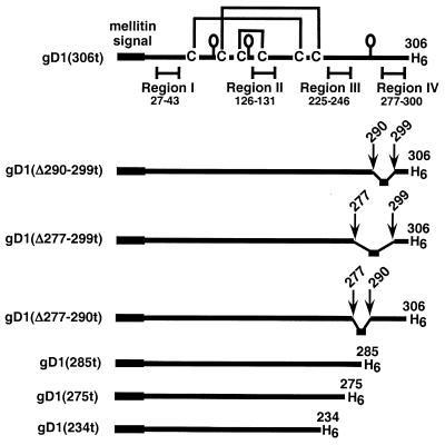
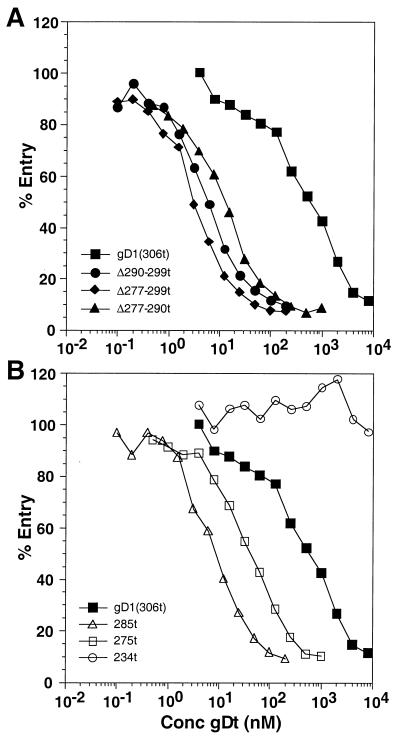
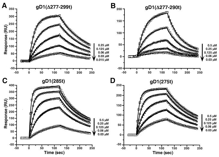
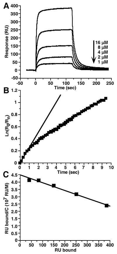
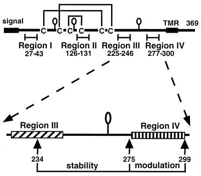
Glycoprotein D (gD) of herpes simplex virus (HSV) is essential for virus entry and has four functional regions (I to IV) important for this process. We previously showed that a truncated form of a functional region IV variant, gD1(Delta290-299t), had an enhanced ability to block virus entry and to bind to the herpesvirus entry mediator (HveAt; formerly HVEMt), a cellular receptor for HSV. To explore this phenotype further, we examined other forms of gD, especially ones with mutations in region IV. Variant proteins with deletions of amino acids between 277 and 300 (region IV), as well as truncated forms lacking C-terminal residues up to amino acid 275 of gD, were able to block HSV entry into Vero cells 1 to 2 logs better than wild-type gD1(306t). In contrast, gD truncated at residue 234 did not block virus entry into Vero cells. Using optical biosensor technology, we recently showed that gD1(Delta290-299t) had a 100-fold-higher affinity for HveAt than gD1(306t) (3.3 x 10(-8) M versus 3.2 x 10(-6) M). Here we found that the affinities of other region IV variants for HveAt were similar to that of gD1(Delta290-299t). Thus, the affinity data follow the same hierarchy as the blocking data. In each case, the higher affinity was due primarily to a faster kon rather than to a slower koff. Therefore, once the gDt-HveAt complex formed, its stability was unaffected by mutations in or near region IV. gD truncated at residue 234 bound to HveAt with a lower affinity (2.0 x 10(-5) M) than did gD1(306t) due to a more rapid koff. These data suggest that residues between 234 and 275 are important for maintaining stability of the gDt-HveAt complex and that functional region IV is important for modulating the binding of gD to HveA. The binding properties of any gD1(234t)-receptor complex could account for the inability of this form of gDt to block HSV infection.


Figures

FIG. 1
FIG. 1
Schematic representations of truncated HSV gD. Each baculovirus-derived form of gD is truncated prior to the C-terminal transmembrane region at the indicated residue. The honeybee mellitin signal peptide replaces the wild-type signal and is cleaved in the fully processed protein. Each mature protein has amino acids DP (not shown) at the N terminus and a six-histidine tag at the C terminus. Each mutant contains all six cysteines and all three N-CHO consensus sites (shown as balloons) except for gD1(234t), which lacks the N-CHO site at residue 262.
FIG. 2
FIG. 2
Effect of gDt on HSV-1 entry into Vero cells. Confluent cells on 96-well plates were incubated with various concentrations of purified gDt at 4°C for 90 min. HSV-1(hrR3) was added at an multiplicity of infection of 0.5 PFU/cell, and the plate was incubated for another 90 min at 4°C. Plates were then shifted to 37°C for 5 h. Cells were lysed, and β-galactosidase activity was measured on aliquots of the cytoplasmic extract, using the substrate chlorophenol red-β-d-galactopyranoside and measuring the increase in absorbance over 50 min at 570 nm; 100% entry corresponds to no added inhibitor. (A) Blocking of entry with gD1(306t) compared to that of the region IV mutants; (B) blocking of entry with gD1(306t) (same curve as in panel A) compared to that of the other truncation mutants.
FIG. 3
FIG. 3
Binding of gDt to HveAt. ELISA plates were coated with 50 μl of 200 nM HveAt in PBS, blocked, and incubated with various concentrations of gDt. Bound gDt was detected with rabbit antiserum R7, followed by peroxidase-conjugated secondary antibody and substrate. The data are the averages of results for duplicate wells, and each experiment was repeated twice with similar results. (A) Binding of gD1(306t) compared with that of the three region IV mutants. (B) Binding of gD1(306t) (same curve as in panel A) compared with that of the other truncation mutants. Abs, absorbance.
FIG. 4
FIG. 4
Overlay of sensorgrams showing the interaction of decreasing concentrations of the indicated gDt variants with immobilized HveAt. Data points were collected every 0.2 or 0.4 s, but for simplicity selected points at every 5 s are shown as open symbols. The solid lines are the best global fits to the simple bimolecular model.
FIG. 5
FIG. 5
Binding of gD1(234t) to immobilized HveAt. (A) Sensorgram overlays. (B) Maximum koff determination using the dissociation phase of the sensorgram at 16 μM gD1(234t). Squares show the data for the first 10 s of dissociation, and the straight line is the fit to the initial second of data. The slope of the line gives a maximum koff of 2 × 10⁻¹ s−1. r2 for the linear fit is 0.989. (C) Scatchard analysis. Equilibrium binding levels (RU bound) were obtained from sensorgrams in panel A. C is the molar concentration of gD1(234t). The negative inverse of the slope of the line gives a KD of 2 × 10⁻⁵ M. r2 for the linear fit is 0.978.
FIG. 6
FIG. 6
Top, schematic drawing of gD from HSV-1 with the four functional regions. The balloons represent the N-CHOs. TMR is the transmembrane region. Bottom, functional regions III and IV of gD. Residues between 234 and 275 help to stabilize the gD-HveA complex, while residues between 275 and 299 down-modulate the binding of gD to HveA.

Similar articles

Cited by

References

    1. Biacore AB. BIAevaluation software handbook, version 3.0. Uppsala, Sweden: Biacore AB; 1997.
    1. Campadelli-Fiume G, Arsenakis M, Farabegoli F, Roizman B. Entry of herpes simplex virus 1 in BJ cells that constitutively express viral glycoprotein D is by endocytosis and results in degradation of the virus. J Virol. 1988;62:159–167. - PMC - PubMed
    1. Chiang H-Y, Cohen G H, Eisenberg R J. Identification of functional regions of herpes simplex virus glycoprotein gD by using linker-insertion mutagenesis. J Virol. 1994;68:2529–2543. - PMC - PubMed
    1. Cohen G H, Dietzschold B, Ponce de Leon M, Long D, Golub E, Varrichio A, Pereira L, Eisenberg R J. Localization and synthesis of an antigenic determinant of herpes simplex virus glycoprotein D that stimulates production of neutralizing antibody. J Virol. 1984;49:102–108. - PMC - PubMed
    1. Cohen G H, Isola V J, Kuhns J, Berman P W, Eisenberg R J. Localization of discontinuous epitopes of herpes simplex virus glycoprotein D: use of a nondenaturing (“native” gel) system of polyacrylamide gel electrophoresis coupled with Western blotting. J Virol. 1986;60:157–166. - PMC - PubMed

Publication types

MeSH terms

Substances