. 1998 Sep;72(9):7115-24.
doi: 10.1128/JVI.72.9.7115-7124.1998.

The polyserine tract of herpes simplex virus ICP4 is required for normal viral gene expression and growth in murine trigeminal ganglia

Affiliations

The polyserine tract of herpes simplex virus ICP4 is required for normal viral gene expression and growth in murine trigeminal ganglia

P A Bates et al. J Virol. 1998 Sep.

Abstract

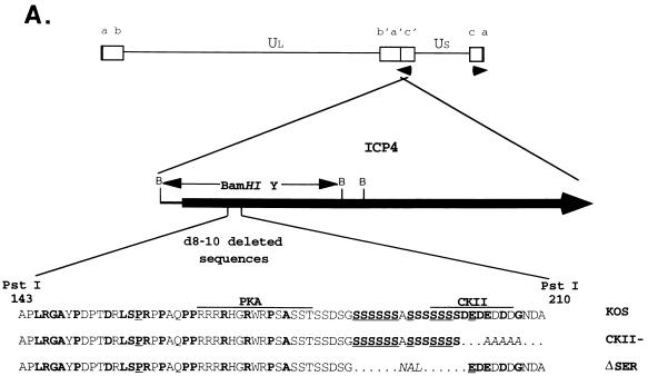
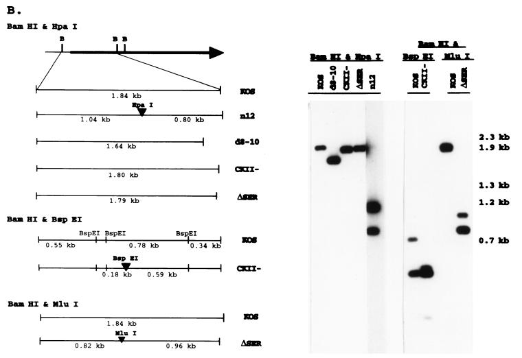
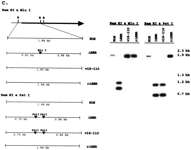
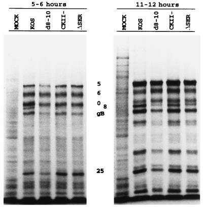
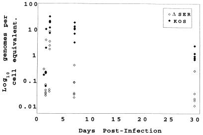
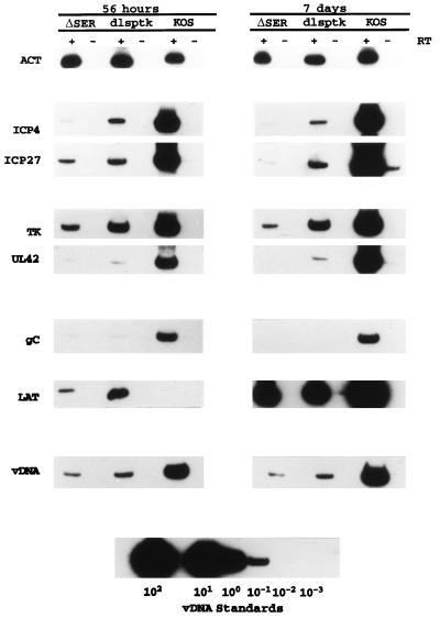
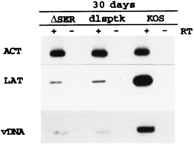
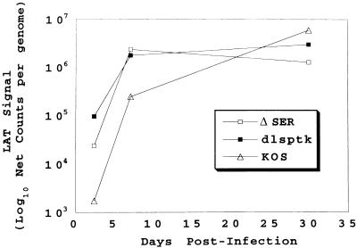
ICP4 of herpes simplex virus (HSV) is essential for productive infection due to its central role in the regulation of HSV transcription. This study identified a region of ICP4 that is not required for viral growth in culture or at the periphery of experimentally inoculated mice but is critical for productive growth in the trigeminal ganglia. This region of ICP4 encompasses amino acids 184 to 198 and contains 13 nearly contiguous serine residues that are highly conserved among the alphaherpesviruses. A mutant in which this region is deleted (DeltaSER) was able to grow on the corneas of mice and be transported back to the trigeminal ganglia. DeltaSER did not grow in the trigeminal ganglia but did express low levels of several immediate-early (ICP4 and ICP27) and early (thymidine kinase [tk] and UL42) genes. It expressed very low levels of the late gC gene and did not appear to replicate DNA. This pattern of gene expression was similar to that observed for a tk mutant, dlsptk. Both DeltaSER and dlsptk expressed higher levels of the latency-associated transcript (LAT) per genome earlier in infected ganglia than did the wild-type virus, KOS. However, infected ganglia from all three viruses accumulated the same level of LAT per genome at 30 days postinfection (during latency). The data suggest that the polyserine tract of ICP4 provides an activity that is required for lytic infection in ganglia to progress to viral DNA synthesis and full lytic gene expression. In the absence of this activity, higher levels of LAT per genome accumulate earlier in infection than with wild-type virus.


Figures

FIG. 1
FIG. 1
ICP4 serine tract mutations. (A) The genomic location and direction of transcription (arrowheads) of ICP4 are shown along with the BamHI restriction sites in reference to the ICP4 coding region. The BamHI Y fragment and the serine tract region (aa 143 to 210) deleted in mutant d8-10 (68, 77) are indicated. The corresponding wt (KOS) and mutant (CKII and ΔSER) sequences are compared, showing the deleted (dots) and substituted (italics) residues in reference to the consensus sites for the cellular kinases PKA and CKII. Amino acids that are conserved in the HSV-1 and pseudorabies virus (8) or varicella-zoster virus (54) ICP4 homologs are designated in boldface; underlined residues are conserved in all three. (B) Fine map of the BamHI Y fragment in the wt (KOS) and mutant viruses is shown with the relevant restriction sites for determining the presence of the CKII and ΔSER mutations. Southern blots of viral DNA digested with the indicated restriction enzymes and hybridized to labeled BamHI-Y probe are shown on the right along with the sizes of the expected fragments. (C) Fine map of the BamHI Y fragment is shown with the relevant restriction sites for determining the structures of the rΔSER and vi8-i10 viruses. Southern blots of viral DNA digested with the indicated restriction enzymes and hybridized to labeled BamHI-Y probe are shown on the right along with the sizes of the expected fragments.
FIG. 1
FIG. 1
ICP4 serine tract mutations. (A) The genomic location and direction of transcription (arrowheads) of ICP4 are shown along with the BamHI restriction sites in reference to the ICP4 coding region. The BamHI Y fragment and the serine tract region (aa 143 to 210) deleted in mutant d8-10 (68, 77) are indicated. The corresponding wt (KOS) and mutant (CKII and ΔSER) sequences are compared, showing the deleted (dots) and substituted (italics) residues in reference to the consensus sites for the cellular kinases PKA and CKII. Amino acids that are conserved in the HSV-1 and pseudorabies virus (8) or varicella-zoster virus (54) ICP4 homologs are designated in boldface; underlined residues are conserved in all three. (B) Fine map of the BamHI Y fragment in the wt (KOS) and mutant viruses is shown with the relevant restriction sites for determining the presence of the CKII and ΔSER mutations. Southern blots of viral DNA digested with the indicated restriction enzymes and hybridized to labeled BamHI-Y probe are shown on the right along with the sizes of the expected fragments. (C) Fine map of the BamHI Y fragment is shown with the relevant restriction sites for determining the structures of the rΔSER and vi8-i10 viruses. Southern blots of viral DNA digested with the indicated restriction enzymes and hybridized to labeled BamHI-Y probe are shown on the right along with the sizes of the expected fragments.
FIG. 2
FIG. 2
Polypeptide profiles of the serine tract mutants. Vero cells were infected (MOI of 10) with the indicated viruses, pulsed with [35S]methionine at the indicated times postinfection, and processed for SDS-PAGE analysis on an SDS–9% polyacrylamide gel. The positions of selected viral proteins are designated.
FIG. 3
FIG. 3
Accumulation of viral DNA in ΔSER- and KOS-infected ganglia. Viral DNA content at the designated time points in KOS- or ΔSER-infected trigeminal ganglia was determined by PCR as described in Materials and Methods (41). Each data point was determined from an individual ganglion.
FIG. 4
FIG. 4
Viral gene expression in the trigeminal ganglia during acute infection. RT-PCR products of viral and cellular transcripts from ganglia infected with ΔSER, KOS, or the tk mutant dlsptk (10) from pooled ganglia harvested at 56 h and 7 days postinfection are shown. PCR products of viral DNA (vDNA) for each sample are also shown along with the PCR signals used to establish a viral DNA standard curve. ACT, β-actin.
FIG. 5
FIG. 5
LAT expression at 30 days postinfection. RT-PCR products of LAT and β-actin (ACT) prepared from ganglia infected with the designated viruses and harvested at 30 days postinfection are shown in the top two panels. The bottom panel shows the PCR products of viral DNA (vDNA) for each virus. These data represent an animal experiment different from that used for Fig. 4 but are typical of previous results.
FIG. 6
FIG. 6
LAT expression per genome during acute and latent infection. The 56-h- and 7-day-postinfection LAT RT-PCR and viral DNA PCR data from Fig. 4 were combined with the corresponding 30-day-postinfection LAT RT-PCR and viral DNA PCR data to illustrate LAT expression per genome as a function of time. As the absolute amount of LAT was not quantitatively determined with standards, the LAT RT-PCR signal per genome is represented simply as net counts per genome.

References

    1. Anderson A S, Francesconi A, Morgan R W. Complete nucleotide sequence of the Marek’s disease virus ICP4 gene. Virology. 1992;189:657–667. - PubMed
    1. Baradaran K, Dabrowski C E, Schaffer P A. Transcriptional analysis of the region of the herpes simplex virus type 1 genome containing the UL8, UL9, and UL10 genes and identification of a novel delayed-early gene product, OBPC. J Virol. 1994;68:4251–4261. - PMC - PubMed
    1. Batchelor A H, O’Hare P. Regulation and cell-type-specific activity of a promoter located upstream of the latency associated transcript of herpes simplex virus type 1. J Virol. 1990;64:3269–3279. - PMC - PubMed
    1. Batchelor A H, O’Hare P. Localization of cis-acting sequence requirements in the promoter of the latency-associated transcript of herpes simplex virus type 1 required for cell-type-specific activity. J Virol. 1992;66:3573–3582. - PMC - PubMed
    1. Bohenzky R A, Papavassiliou A G, Gelman I H, Silverstein S. Identification of a promoter mapping within the reiterated sequences that flank the herpes simplex virus type 1 UL region. J Virol. 1993;67:632–642. - PMC - PubMed

Publication types

LinkOut - more resources