Modulation of human immunodeficiency virus type 1-induced syncytium formation by the conformational state of LFA-1 determined by a new luciferase-based syncytium quantitative assay
- PMID: 9696806
- PMCID: PMC109934
- DOI: 10.1128/JVI.72.9.7125-7136.1998
Modulation of human immunodeficiency virus type 1-induced syncytium formation by the conformational state of LFA-1 determined by a new luciferase-based syncytium quantitative assay
Abstract
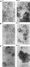
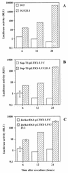
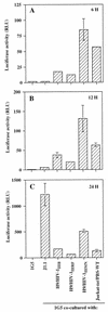
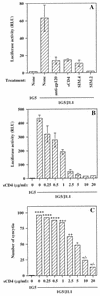
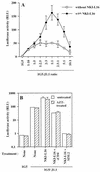
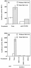
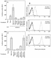
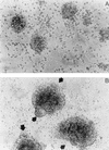
The ICAM-1/LFA-1 interaction has been clearly demonstrated to play an active role in syncytium formation induced by human immunodeficiency virus type 1 (HIV-1). Since it is known that a high-affinity state of LFA-1 for ICAM-1 can be induced through conformational change, such a high-affinity state may also contribute to the process of syncytium formation. In this study, we have investigated the involvement of the conformational status of LFA-1 in HIV-1-dependent syncytium formation by using the anti-LFA-1 antibody NKI-L16, which is known to activate the high-affinity state. Initial visual observations by light microscopy indeed suggested that the addition of the NKI-L16 antibody led to bigger and more numerous syncytia when different cell lines were tested. To further analyze this NKI-L16-dependent increment of syncytium formation in a quantitative assay, a new luciferase-based assay was developed by using a T-cell line containing an HIV-1 long terminal repeat (LTR)-driven luciferase construct (1G5) in coincubation with an HIV-1-positive cell line (J1.1). Upon fusion, the viral Tat protein could diffuse to the 1G5 cells, leading to a transcriptional increase of the HIV-1 LTR-driven luciferase gene. Initial evaluation of this assay showed a good correlation between the level of syncytium formation determined by microscopic observation and the level of measured luciferase activity. In addition, this assay showed a greater induction of enzymatic activity correlating with syncytium formation in comparison to a similar incubation with the HeLa-CD4-LTR-beta-gal indicator cell line. By using this test, NKI-L16 treatment of 1G5/J1.1 cells led to a three- to sevenfold increase in HIV-1 LTR-driven luciferase activity. The syncytium-dependent luciferase activity in NKI-L16-treated cells could be blocked by classical syncytium inhibitors such as soluble CD4, anti-CD4, and anti-gp120 antibodies. Inhibition could also be observed with specific blocking agents for the chemokine receptor CXCR4, as well as with soluble ICAM-1, anti-LFA-1, anti-ICAM-1, and anti-ICAM-2 blocking antibodies, indicating the requirement for the LFA-1/ICAM interaction. Treatment of peripheral blood mononuclear cells with NKI-L16 resulted in a higher level of syncytium formation in the presence of the cell line J1.1. Conversely, when PBMCs were infected with two different syncytium-inducing HIV-1 primary isolates, coincubation with NKI-L16-pretreated 1G5 cells led to higher levels of luciferase activity for both virus isolates. Our results therefore show for the first time a direct role for the LFA-1 high-affinity state in virus-mediated syncytium formation. Based on the demonstration that an increase in ICAM-1 binding is induced by T-cell activation, these data suggest an in vivo involvement of the high-affinity state of LFA-1 in HIV-1-induced syncytium formation. Moreover, syncytia might preferentially occur in lymph nodes, since this microenvironment harbors a high proportion of activated T cells.
Figures



Similar articles
-
Role of the leukocyte function antigen-1 conformational state in the process of human immunodeficiency virus type 1-mediated syncytium formation and virus infection.Virology. 1999 Apr 25;257(1):228-38. doi: 10.1006/viro.1999.9687. Virology. 1999. PMID: 10208936
-
T cells expressing activated LFA-1 are more susceptible to infection with human immunodeficiency virus type 1 particles bearing host-encoded ICAM-1.J Virol. 1998 Mar;72(3):2105-12. doi: 10.1128/JVI.72.3.2105-2112.1998. J Virol. 1998. PMID: 9499066 Free PMC article.
-
Inhibition of HIV-1-mediated syncytium formation and virus replication by the lipophosphoglycan from Leishmania donovani is due to an effect on early events in the virus life cycle.Clin Exp Immunol. 2001 Apr;124(1):32-42. doi: 10.1046/j.1365-2249.2001.01492.x. Clin Exp Immunol. 2001. PMID: 11359440 Free PMC article.
-
Role of cellular adhesion molecules in HIV type 1 infection and their impact on virus neutralization.AIDS Res Hum Retroviruses. 1998 Oct;14 Suppl 3:S247-54. AIDS Res Hum Retroviruses. 1998. PMID: 9814951 Review.
-
Induction of lymphomonocyte activation by HIV-1 glycoprotein gp120. Possible role in AIDS pathogenesis.J Biol Regul Homeost Agents. 1996 Oct-Dec;10(4):83-91. J Biol Regul Homeost Agents. 1996. PMID: 9604776 Review.
Cited by
-
LFA-1 antagonists as agents limiting human immunodeficiency virus type 1 infection and transmission and potentiating the effect of the fusion inhibitor T-20.Antimicrob Agents Chemother. 2009 Nov;53(11):4656-66. doi: 10.1128/AAC.00117-09. Epub 2009 Aug 31. Antimicrob Agents Chemother. 2009. PMID: 19721069 Free PMC article.
-
Monitoring of HIV-1 envelope-mediated membrane fusion using modified split green fluorescent proteins.J Virol Methods. 2009 Nov;161(2):216-22. doi: 10.1016/j.jviromet.2009.06.017. Epub 2009 Jun 25. J Virol Methods. 2009. PMID: 19559731 Free PMC article.
-
Binding of LFA-1 (CD11a) to intercellular adhesion molecule 3 (ICAM-3; CD50) and ICAM-2 (CD102) triggers transmigration of human immunodeficiency virus type 1-infected monocytes through mucosal epithelial cells.J Virol. 2002 Jan;76(1):32-40. doi: 10.1128/jvi.76.1.32-40.2002. J Virol. 2002. PMID: 11739669 Free PMC article.
-
Human immunodeficiency virus type 1 replication in dendritic cell-T-cell cocultures is increased upon incorporation of host LFA-1 due to higher levels of virus production in immature dendritic cells.J Virol. 2007 Jul;81(14):7672-82. doi: 10.1128/JVI.02810-06. Epub 2007 May 9. J Virol. 2007. PMID: 17494076 Free PMC article.
-
Sodium lauryl sulfate abrogates human immunodeficiency virus infectivity by affecting viral attachment.Antimicrob Agents Chemother. 2001 Aug;45(8):2229-37. doi: 10.1128/AAC.45.8.2229-2237.2001. Antimicrob Agents Chemother. 2001. PMID: 11451679 Free PMC article.
References
-
- Aguilar-Cordova E, Chinen J, Donehower L, Lewis D E, Belmont J W. A sensitive reporter cell line for HIV-1 tat activity, HIV-1 inhibitors, and T cell activation effects. AIDS Res Hum Retroviruses. 1994;10:295–301. - PubMed
-
- Alkhatib G, Combadiere C, Broder C C, Feng Y, Kennedey P E, Murphy P M, Berger E A. CC CKR5: a RANTES, MIP-1α, MIP-1β receptor as a fusion cofactor for macrophage-tropic HIV-1. Science. 1996;272:1955–1959. - PubMed
-
- Allen A D, Hart D N, Hechinger M K, Slattery M J, Chesson C V, Vidikan P. Leukocyte adhesion molecules as a cofactor in AIDS: basic science and pilot study. Med Hypotheses. 1995;45:164–168. - PubMed
-
- Barbeau B, Bernier R, Dumais N, Briand G, Olivier M, Faure R, Posner B I, Tremblay M. Activation of HIV-1 LTR transcription and virus replication via NF-κB-dependent and -independent pathways by potent phosphotyrosine phosphatase inhibitors, the peroxovanadium compounds. J Biol Chem. 1997;272:12968–12977. - PubMed
-
- Bazil V, Stefanova I, Hilgert I, Kristofova H, Vanek S, Horejsi V. Monoclonal antibodies against leukocytes antigens IV. Antibodies against the LFA-1 (CD11a/CD18) leukocyte-adhesion glycoprotein. Folia Biol. 1990;36:41–50. - PubMed
Publication types
MeSH terms
Substances
LinkOut - more resources
Full Text Sources
Other Literature Sources
Research Materials
Miscellaneous

