. 1998 Sep;72(9):7160-9.
doi: 10.1128/JVI.72.9.7160-7169.1998.

Interactions between the structural domains of the RNA replication proteins of plant-infecting RNA viruses

Affiliations

Interactions between the structural domains of the RNA replication proteins of plant-infecting RNA viruses

E K O'Reilly et al. J Virol. 1998 Sep.

Abstract

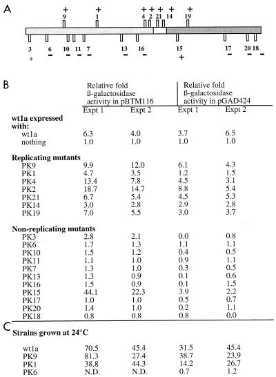
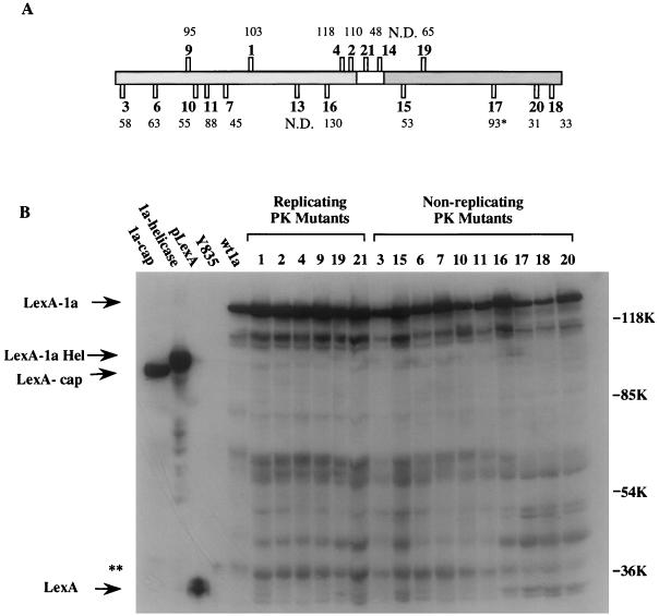
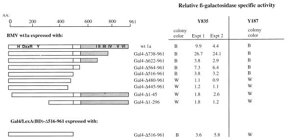
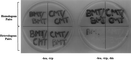
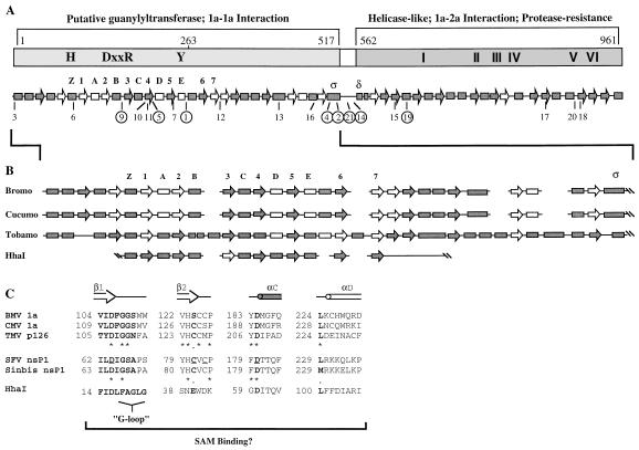
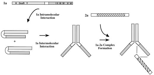
Brome mosaic virus (BMV), a positive-strand RNA virus, encodes two replication proteins: the 2a protein, which contains polymerase-like sequences, and the 1a protein, with N-terminal putative capping and C-terminal helicase-like sequences. These two proteins are part of a multisubunit complex which is necessary for viral RNA replication. We have previously shown that the yeast two-hybrid assay consistently duplicated results obtained from in vivo RNA replication assays and biochemical assays of protein-protein interaction, thus permitting the identification of additional interacting domains. We now map an interaction found to take place between two 1a proteins. Using previously characterized 1a mutants, a perfect correlation was found between the in vivo phenotypes of these mutants and their abilities to interact with wild-type 1a (wt1a) and each other. Western blot analysis revealed that the stabilities of many of the noninteracting mutant proteins were similar to that of wt1a. Deletion analysis of 1a revealed that the N-terminal 515 residues of the 1a protein are required and sufficient for 1a-1a interaction. This intermolecular interaction between the putative capping domain and itself was detected in another tripartite RNA virus, cucumber mosaic virus (CMV), suggesting that the 1a-1a interaction is a feature necessary for the replication of tripartite RNA viruses. The boundaries for various activities are placed in the context of the predicted secondary structures of several 1a-like proteins of members of the alphavirus-like superfamily. Additionally, we found a novel interaction between the putative capping and helicase-like portions of the BMV and CMV 1a proteins. Our cumulative data suggest a working model for the assembly of the BMV RNA replicase.


Figures

FIG. 1
FIG. 1
(A) Locations of insertion mutations and their replication phenotypes in protoplasts. The box represents the 1a open reading frame. The lightly shaded N-terminal portion contains sequences putatively involved in capping, while the darkly shaded C-terminal portion contains helicase-like sequences. The vertical bars represent the positions of the 2- to 3-aa insertions made and characterized by Kroner and colleagues (17). Bars pointing up represent viable mutants, while bars pointing down represent mutants unable to replicate in barley protoplasts. PK1, -4, and -19 were found to be temperature sensitive for replication at 35°C. The ability (+) or inability (−) of each PK mutant to interact with wt1a in the two-hybrid system is indicated. (B) β-Galactosidase activity detected when each of the PK mutants is coexpressed with wt1a in yeast strain Y835. The results from two independent experiments (Expt) are shown. These activities are presented as fold activity over that of wt1a in the DNA binding domain plasmid. β-Galactosidase activity is shown as micromoles of O-nitrophenyl galactoside hydrolyzed per minute per milligram of protein. (C) β-Galactosidase activity detected for strains grown at 24°C. PK1, a replication-competent mutant in protoplasts which did not interact with wt1a as a LexA fusion at 30°C, did interact at 24°C. PK9, a replicating mutant, and PK6, a nonreplicating mutant, serve as positive and negative controls, respectively. N.D., not determined.
FIG. 2
FIG. 2
Relative abundance of wt and mutant 1a proteins as observed in Western blots. (A) The basic diagram is the same as that in Fig. 1A. The numbers above or below each PK mutant indicate the percent abundance of each protein relative to that of wt1a. The results shown are an average of the quantitations from two independent Western blots. Quantitation was performed by laser densitometry (Molecular Dynamics). The abundance of PK17 (∗) was somewhat inconsistent between trials, being 150 and 35% that of wt1a. N.D., not determined. (B) Scan of a Western blot showing the abundance of the ca. 140-kDa LexA-PK mutant fusions. The presumed full-length protein is indicated along with the presumed helicase and capping fusions. For unknown reasons the migration of the capping domain was reproducibly faster than expected based on its predicted mass. Lower-molecular-mass bands are presumed to be degradation products. A ca. 40-kDa cellular protein (∗∗) cross-reacted with the antibodies and was used to normalize the amount of protein loaded.
FIG. 3
FIG. 3
Mapping the 1a-1a binding domain by deletion analysis. The domains of the 1a protein are depicted along with various deletions in either the N or C terminus. Fold β-galactosidase levels detected when each of the deletions was expressed with wt1a from two independent experiments are shown relative to those of wt1a alone. For strain Y187 (Clonetech Laboratories, Inc.), only the results of the qualitative filter assays are shown. Δ516-966 retains the ability to interact with both itself and wt1a. B, blue colonies; W, normal yeast colony color. AA, amino acid.
FIG. 4
FIG. 4
Assay for induction of the HIS3 gene in homologous or heterologous pairings of BMV and/or CMV 1a methyltransferase fusions. The plate on the left is supplemented with histidine, while the one on the right is not. Each doubly transformed strain was written onto plates to indicate the identity of the 1a fusion proteins. The LexA fusion is indicated above the slash, and the GAL4 fusion is indicated below the slash (i.e., LexA/GAL4).
FIG. 5
FIG. 5
Summary of the predicted secondary structures of BMV and related 1a proteins. Boxes, α-helices; arrows, β-sheets; solid shapes, >80% accuracy; open shapes, >50% accuracy. (A) Diagram of the BMV 1a protein and its predicted secondary structures. The previously identified sequences involved in capping and helicase function are indicated. Consistent with Fig. 1A, the putative capping domain required for 1a-1a interaction is lightly shaded while the protease-resistant helicase-like domain required for 1a-2a interaction is darkly shaded. The locations of the PK mutants in the predicted secondary structures are indicated, with the replication-competent mutants circled. The secondary structures which correspond to those found in DNA methyltransferases are numbered according to the system of Schluckebier et al. (28). We also labeled helices ς and δ to facilitate the flow of the text. (B) Expanded picture of the predicted secondary structure of the putative capping domain of the bromo-, cucumo-, and tobamovirus families compared to that of the DNA methyltransferase HhaI. β3 of HhaI, shown as an unshaded arrow, was not present in the originally solved crystal structure and was not predicted by PHD analysis (6). This strand was later added upon comparison of the structure HhaI to other methyltransferase crystal structures (19, 28). Also, there is a 4-aa β-sheet (β4′) following β4 which we have not included in our diagram for simplicity (6). (C) Functional residues involved in SAM binding in HhaI and their putative functional homologs in the SAM binding capping proteins of RNA viruses. Residues conserved between the DNA and RNA SAM binding proteins, including the G-loop, are shown in bold letters. The underlined residues of SFV nsP1 have been mutated to alanine and all, except D180A, have effects on methyltransferase and guanylyltransferase functions. Asterisks indicate identical amino acids, and periods indicate similar amino acids.
FIG. 6
FIG. 6
Working model for the assembly of the 1a-2a complex. The putative intramolecular interaction in 1a prevents the formation of the 1a-2a complex until the intermolecular 1a-1a interaction has occurred. The 2a protein interacts with the helicase-like domain of 1a through its N terminus.

References

    1. Ahlquist P, Strauss E G, Rice C M, Strauss J H, Haseloff J, Zimmern D. Sindbis virus proteins nsP1 and nsP2 contain homology to nonstructural proteins from several RNA plant viruses. J Virol. 1985;53:536–542. - PMC - PubMed
    1. Ahola T, Laakkonen P, Vihinen H, Kääriäinen L. Critical residues of Semliki Forest virus RNA capping enzyme involved in methyltransferase and guanylyltransferase-like activities. J Virol. 1997;71:392–397. - PMC - PubMed
    1. Argos P. A sequence motif in many polymerases. Nucleic Acids Res. 1988;16:9909–9916. - PMC - PubMed
    1. Bao Y, Carter S A, Nelson R S. The 126- and 183-kilodalton proteins of tobacco mosaic virus, and not their common nucleotide sequence, control mosaic symptom formation in tobacco. J Virol. 1996;70:6378–6383. - PMC - PubMed
    1. Blumenthal T, Carmichael G. RNA replication: function and structure of Qβ-replicase. Annu Rev Biochem. 1979;48:525–548. - PubMed

Publication types

LinkOut - more resources