. 1998 Sep;72(9):7201-12.
doi: 10.1128/JVI.72.9.7201-7212.1998.

Control of murine cytomegalovirus in the lungs: relative but not absolute immunodominance of the immediate-early 1 nonapeptide during the antiviral cytolytic T-lymphocyte response in pulmonary infiltrates

Affiliations

Control of murine cytomegalovirus in the lungs: relative but not absolute immunodominance of the immediate-early 1 nonapeptide during the antiviral cytolytic T-lymphocyte response in pulmonary infiltrates

R Holtappels et al. J Virol. 1998 Sep.

Abstract

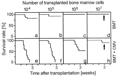
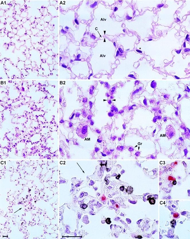
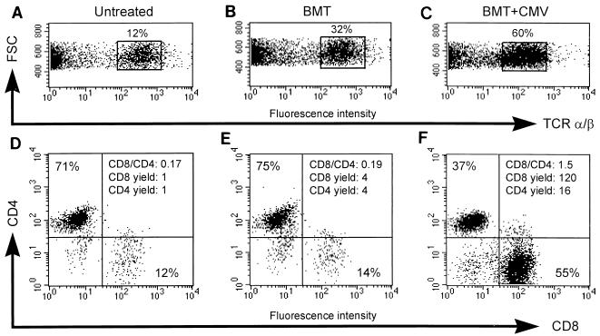
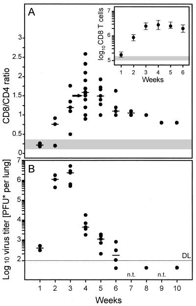
The lungs are a major organ site of cytomegalovirus (CMV) infection, pathogenesis, and latency. Interstitial CMV pneumonia represents a critical manifestation of CMV disease, in particular in recipients of bone marrow transplantation (BMT). We have employed a murine model for studying the immune response to CMV in the lungs in the specific scenario of immune reconstitution after syngeneic BMT. Control of pulmonary infection was associated with a vigorous infiltration of the lungs, which was characterized by a preferential recruitment and massive expansion of the CD8 subset of alpha/beta T cells. The infiltrate provided a microenvironment in which the CD8 T cells differentiated into mature effector cells, that is, into functionally active cytolytic T lymphocytes (CTL). This gave us the opportunity for an ex vivo testing of the antigen specificities of CTL present at a relevant organ site of viral pathogenesis. The contribution of the previously identified immediate-early 1 (IE1) nonapeptide of murine CMV was evaluated by comparison with the CD3epsilon-redirected cytolytic activity used as a measure of the overall CTL response in the lungs. The IE1 peptide was detected by pulmonary CTL, but it accounted for a minor part of the response. Interestingly, no additional viral or virus-induced antigenic peptides were detectable among naturally processed peptides derived from infected lungs, even though infected fibroblasts were recognized in a major histocompatibility complex-restricted manner. We conclude that the antiviral pulmonary immune response is a collaborative function that involves many antigenic peptides, among which the IE1 peptide is immunodominant in a relative sense.


Figures

FIG. 1
FIG. 1
Syngeneic BMT with high doses of BMC prevents lethal CMV disease. Kaplan-Meier survival plots documenting the influence of the number of transplanted BMC on the survival rates (ordinate) as a function of time (abscissa) after BMT (a to d) or BMT and concurrent murine CMV infection (e to h) for groups of 20 recipients are shown. The arrows mark the time point of the histological analysis depicted in Fig. 2.
FIG. 2
FIG. 2
In situ colocalization of CD3ɛ-positive T lymphocytes and foci of infection. (A) Histology of the lung tissue 3 weeks after BMT with no infection (corresponding to data shown in Fig. 1d). (A1) Overview; (A2) detail. HE staining. Alv, alveoli; C, capillary. An asterisk indicates an elongated nucleus of a parenchymal lung cell; opposed arrowheads indicate an alveolar septum. Note the absence of infiltrates and the normal architecture of the lung tissue. (B) Pathohistology of the lung tissue 3 weeks after BMT and murine CMV infection (corresponding to data shown in Fig. 1h). (B1) Overview; (B2) detail. HE staining. AM, alveolar macrophages; Gr, granulocytes. Opposed arrowheads indicate a widened alveolar septum with interstitial mononuclear leukocytes. Note the infiltration and the widening of the alveolar septa. (C) Detection of infiltrating CD3ɛ-positive lymphocytes and of infected cells 3 weeks after BMT and murine CMV infection, corresponding to the HE-stained tissue shown in panel B. Infected cells are visualized by red (APAAP-new fuchsin) IHC staining of the intranuclear viral IE1 protein. Infiltrating lymphocytes are visualized by black (ABC-diaminobenzidine-nickel) IHC staining of CD3ɛ antigen. (C1) Overview; (C2) Detail of panel C1. The arrow indicates a focus of infection, surrounded by CD3ɛ-positive infiltrating cells. (C3 and C4) CD3ɛ-positive cells located in intimate proximity to infected cells. Bars, 20 μm.
FIG. 3
FIG. 3
Preferential enrichment of CD8 T cells in lung infiltrates. Three-color cytofluorometric analysis of interstitial pulmonary T lymphocytes was done. After perfusion and bronchoalveolar lavage were done for groups of three to five mice, mononuclear leukocytes were recovered from the lung parenchyma by collagenase-DNase digestion and were enriched by Ficoll gradient centrifugation. A lymphocyte gate was set in the forward versus side scatter plot (data not shown). (A and D) Pulmonary lymphocytes recovered as a control from lung tissue of untreated, adult BALB/c mice; (B and E) pulmonary lymphocytes recovered from lung tissue 4 weeks after performance of BMT with 107 syngeneic BMC; (C and F) pulmonary lymphocytes recovered from lung tissue 4 weeks after BMT (same conditions as those described for panels B and E) and concurrent murine CMV infection; (A to C) dot plot of forward scatter (FSC, ordinate) versus PE (TCR α/β, abscissa) fluorescence intensity, with TCR α/β cells enclosed in a rectangle and their percentage among the cells in the lymphocyte gate indicated; (D to F) dot plots of RED613 (CD4, ordinate) versus FITC (CD8, abscissa) fluorescence intensities for TCR α/β-expressing cells. The percentages of CD4 and CD8 T cells are given in the respective quadrants, and the CD8/CD4 ratio is indicated in the upper right quadrant. The yield of CD8 and CD4 T cells is expressed as a multiple of the respective yield from normal, uninfected lungs.
FIG. 4
FIG. 4
The peak of virus production in the lungs precedes the peak of infiltration. (A) Kinetics of the infiltration expressed as CD8/CD4 cell ratios documenting the preferential engagement of CD8 T cells. A cumulative plot of results compiled over a period of 2 years from 12 independent but analogous transplantations is shown. Each dot represents one time point of one transplantation. The median values are indicated by dashes. An arrow marks the particular transplantation and time point for which the determination of the ratio is documented as an example in Fig. 3. The shaded region indicates the range of CD8/CD4 cell ratios observed 4 weeks after BMT with no infection. The inset shows the median values of the absolute numbers of CD8 T cells per lung. Ranges are indicated by vertical bars. The shaded region indicates the range of the CD8 T-cell yields obtained 4 weeks after BMT with no infection. (B) Kinetics of virus production in the lungs. Each dot represents the median value of the virus titers of five mice of one transplantation and time point. The median values for independent transplantations are indicated by dashes. DL and dotted line, detection limit of the assay; n.t.: not tested.
FIG. 5
FIG. 5
Characterization of the pulmonary effector cells of CD3ɛ-redirected lysis. Throughout, pulmonary lymphocytes were isolated from lung infiltrates at 4 weeks after BMT and murine CMV infection. E/T, effector/target cell ratio. (A) Redirected lysis was assayed with P815 target cells carrying Fc receptor-bound antibodies directed against murine CD3ɛ, TCR α/β, or TCR γ/δ. (B) The pulmonary infiltrate cell population was depleted of either CD8- or CD4-positive lymphocytes by treatment with the respective MAbs and complement before the assay of CD3ɛ-redirected lysis. (C) The state of activity of pulmonary lymphocytes was tested by providing B7-1 on the target cells for the B7-CD28 costimulatory interaction. The inset shows the cytofluorometric analysis of B7-1 expression by the P815-B7 transfectant and the absence of B7-1 on parental P815. FL, log10 FITC fluorescence intensity. (D) Sensitivity of CD3ɛ-redirected lysis by pulmonary lymphocytes to concanamycin A (CMA), with P815 as the target (E/T, 100). The solvent dimethyl sulfoxide (DMSO) was titrated as a control.
FIG. 6
FIG. 6
Cytolytic activity of pulmonary T lymphocytes. (A) Kinetics of cytolytic activity in lung infiltrates as determined by CD3ɛ-redirected lysis for an effector/target cell ratio (E/T) of 100, with P815 mastocytoma cells as targets. Dots represent results compiled from independent but analogous transplantations. The median values are marked by dashes. The shaded area represents the activity measured with pulmonary lymphocytes recovered from lung tissue 4 weeks after BMT with no infection. This activity never exceeded the upper 95% confidence limit of the spontaneous lysis measured in the absence of effector cells. To serve as a positive reference, CD3ɛ-redirected lysis and murine CMV IE1 peptide-specific lysis are compared for an IE1 peptide-specific long-term CTLL in the insets. (B) Kinetics of the IE1 peptide-specific cytolytic activity in lung infiltrates for an E/T of 100. Target cells were P815 pulsed with a saturating concentration [10−8 M] of the synthetic IE1 peptide, a nonapeptide of the sequence YPHFMPTNL. Symbols are as described for panel A. IE1 peptide dose dependence of IE1-specific cytolytic activity of pulmonary effector cells recovered 4 weeks after BMT and infection is shown in the left inset. P815-B7 transfectants and parental P815 served as target cells at an E/T of 100. Titration of the pulmonary effector cells is shown in the right inset. Target cells were pulsed with a saturating concentration [10−8 M] of synthetic IE1 peptide.
FIG. 7
FIG. 7
Antigen processing and presentation in infected lungs. (A) Kinetics of IE1 peptide processing in infected lungs after BMT. Naturally processed peptides were acid extracted from lung tissue and separated by HPLC. The HPLC fractions were used at the dilutions indicated in the left panel for the exogenous peptide pulsing of P815 target cells. The presentation of the IE1 peptide by the targets, and thus the presence of the IE1 peptide in the respective HPLC fraction, was probed with the IE1-specific CTLL at an effector/target cell ratio (E/T) of 10. The sensitivity of the CTLL tested by titration of the synthetic IE1 nonapeptide YPHFMPTNL is shown in the inset. (B) Spectrum of all antigenic peptides, viral and nonviral, presented in the infected lungs and detected by pulmonary effector cells. HPLC fractions (undiluted 0.1 ml thereof) from the separation performed in week 3 after BMT and infection (see panel A) were used to pulse P815 or P815-B7 target cells for testing the specificity of pulmonary CTL that were isolated at the peak of infiltration. The assay was performed at an E/T of 200. The shaded area indicates the 95% confidence limits (two sided) of the spontaneous lysis. The cytolytic activities of the pulmonary CTL in the CD3ɛ-redirected assay and on P815 target cells pulsed with 10−8 M of the synthetic IE1 peptide are shown in the insets.
FIG. 8
FIG. 8
Recognition of infected target cells by pulmonary CTL. (A) Kinetics of cytolytic activity specific for the phases of the viral replicative cycle. Lung infiltrate cells were isolated at the indicated time points after BMT and infection and tested for cytolytic activity against infected MEF at an effector/target cell ratio (E/T) of 200. Data for week 4 are compiled from three analogous transplantations. Median values are indicated by dashes. MEF were infected with 4 PFU* per cell and used for the cytolytic assay in the three phases of viral gene expression: IE phase, cycloheximide (CH; 50 μg/ml) added for 3 h, replaced by actinomycin D (ActD; 5 μg/ml) for 2 h; E phase, no inhibitor for 12 h; L phase, no inhibitor for 24 h. S indicates exogenous loading of virion proteins from inoculum doses of 4 to 40 PFU* per cell, with ActD (5 μg/ml) added for 5 h. The shaded area represents the upper 95% confidence limit of the highest spontaneous lysis, which was that of the late-phase target. (B) MHC restriction of the E-phase-specific lysis. Pulmonary effector cells were recovered 4 weeks after BMT and infection and tested at an E/T of 200 on syngeneic BALB/c (H-2d) and allogeneic C57BL/6 (H-2b) MEF, which were either left uninfected or infected with murine CMV under E-phase conditions.

References

    1. Altman J D, Moss P A H, Goulder P J R, Barouch D H, McHeyzer-Williams M G, Bell J I, McMichael A J, Davis M M. Phenotype analysis of antigen-specific T lymphocytes. Science (Washington, DC) 1996;274:94–96. - PubMed
    1. Azuma M, Cayabyab M, Buck D, Philipps J H, Lanier L L. CD28 interaction with B7 costimulates primary allogeneic proliferative responses and cytotoxicity mediated by small, resting T lymphocytes. J Exp Med. 1992;175:353–360. - PMC - PubMed
    1. Azuma M, Cayabyab M, Phillips J H, Lanier L L. Requirements for CD28-dependent T cell-mediated cytotoxicity. J Immunol. 1993;150:2091–2101. - PubMed
    1. Bancroft G J, Shellam G R, Chalmer J E. Genetic influence on the augmentation of natural killer (NK) cells during murine cytomegalovirus infection: correlation with patterns of resistance. J Immunol. 1981;126:988–994. - PubMed
    1. Bukowski J F, Woda B A, Welsh R M. Pathogenesis of murine cytomegalovirus infection in natural killer cell-depleted mice. J Virol. 1984;52:119–128. - PMC - PubMed

Publication types

Substances