. 1998 Sep;72(9):7245-54.
doi: 10.1128/JVI.72.9.7245-7254.1998.

The disulfide-bonded structure of feline herpesvirus glycoprotein I

Affiliations

The disulfide-bonded structure of feline herpesvirus glycoprotein I

J D Mijnes et al. J Virol. 1998 Sep.

Abstract

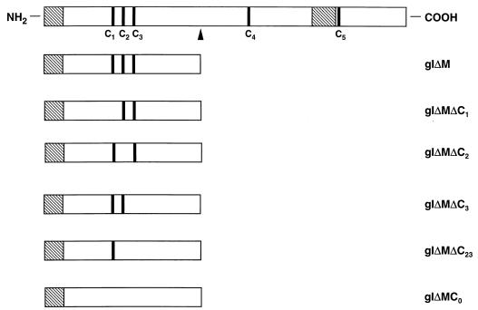
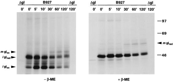
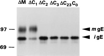
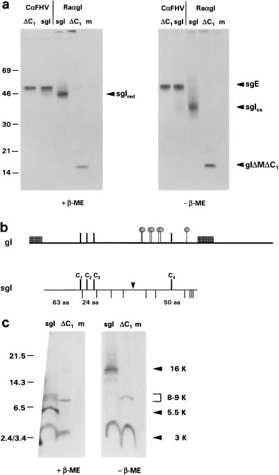
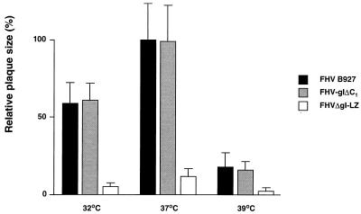
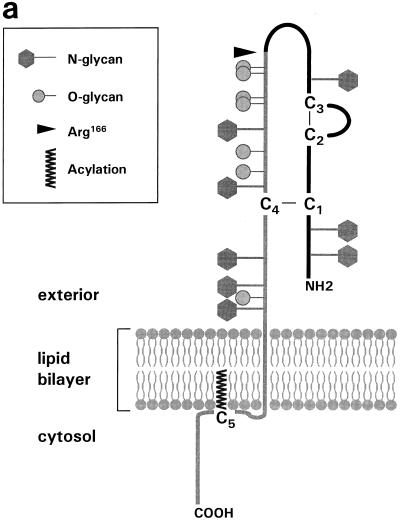
Alphaherpesvirus glycoproteins E and I (gE and gI, respectively) assemble into a hetero-oligomeric complex which promotes cell-to-cell transmission, a determining factor of virulence. Focusing on gI of feline herpesvirus (FHV), we examined the role of disulfide bonds during its biosynthesis, its interaction with gE, and gE-gI-mediated spread of the infection in vitro. The protein's disulfide linkage pattern was determined by single and pairwise substitutions for the four conserved cysteine residues in the ectodomain. The resulting mutants were coexpressed with gE in the vaccinia virus-based vTF7-3 system, and the formation and endoplasmic reticulum (ER)-to-Golgi transport of the hetero-oligomeric complex were monitored. The results were corroborated biochemically by performing an endoproteinase Lys-C digestion of a [35S]Cys-labeled secretory recombinant form of gI followed by tricine-sodium dodecyl sulfate-polyacrylamide gel electrophoresis analysis of the peptides under reducing and nonreducing conditions. We found that (i) gI derivatives lacking Cys79 (C1) and/or Cys223 (C4) still assemble with gE into transport-competent complexes, (ii) mutant proteins lacking Cys91 (C2) and/or Cys102 (C3) bind to gE but are retained in the ER, (iii) radiolabeled endoproteinase Lys-C-generated peptide species containing C1 and C4 are linked through disulfide bonds, and (iv) peptides containing both C2 and C3 are not disulfide linked to any other peptide. From these findings emerges a model in which C1 and C4 as well as C2 and C3 form intramolecular disulfide bridges. Since the cysteines in the ectodomain have been conserved during alphaherpesvirus divergence, we postulate that the model applies for all gI proteins. Analysis of an FHV recombinant with a C1-->S substitution confirmed that the C1-C4 disulfide bond is not essential for the formation of a transport-competent gE-gI complex. The mutation affected the posttranslational modification of gI and caused a slight cold-sensitivity defect in the assembly or the intracellular transport of the gE-gI complex but did not affect plaque size. Thus, C1 and the C1-C4 bond are not essential for gE-gI-mediated cell-to-cell spread, at least not in vitro.


Figures

FIG. 1
FIG. 1
Schematic representation of FHV gI, gIΔM, and the various gIΔM derivatives carrying Cys→Ser substitutions. The proteins are represented by open boxes. The N-terminal signal peptide and the transmembrane region of each protein are indicated by hatched boxes. C1, C2, C3, and C4, representing the cysteine residues in the ectodomain of gI at positions 79, 91, 102, and 223, respectively, are indicated by black bars. A fifth cysteine residue, C5, present in the cytoplasmic domain at position 322, is also shown. The position of Arg166 is marked by an arrowhead.
FIG. 2
FIG. 2
Analysis of gI and gIΔC1 under reducing and nonreducing conditions. Subconfluent monolayers of CRFK cells were infected with either wild-type FHV strain B927 (B927) or FHV-gIΔC1 (gIΔC1). Cells infected with the gI-deficient recombinant FHVΔgI-LZ (ΔgI) (38) served as a negative control. The cells were metabolically labeled from 7 to 8 h p.i. at 37°C. The cell lysates were prepared in the presence of 20 mM NEM to block free sulfhydryl groups and were then subjected to RIPA with the gI-specific antiserum Ra-αgI. The proteins were either treated with PNGase F (+) or mock treated (−) and separated in SDS–7.5% polyacrylamide gels in the absence and presence of β-ME (− β-ME and + β-ME, respectively). The closed arrowheads indicate immature reduced and oxidized gI species (igIred and igIox, respectively) and mature reduced and oxidized gI species (mgIred and mgIox, respectively). In the left-hand panel, mature gIΔC1 is indicated by an open arrowhead. Molecular sizes are in kilodaltons.
FIG. 3
FIG. 3
Pulse-chase analysis of FHV gI under reducing and nonreducing conditions. Subconfluent monolayers of CRFK cells were infected with wild-type FHV strain B927 (B927). At 8 h p.i., the cells were metabolically labeled for 5 min at 37°C and harvested either immediately (0’) or after chase periods of 5, 10, 30, 60, and 120 min (5’, 10’, 30’, 60’, and 120’, respectively). To assure quantitative precipitation of labeled gI also after a 60-min or 120-min chase, the amount of unlabeled gI was reduced by adding 0.5 mM cycloheximide to the culture supernatant 30 min after labeling. Cells infected with FHVΔgI-LZ (ΔgI) and harvested after a 5-min pulse-labeling (0’) or following a subsequent 120-min chase period (120’) served as negative controls. Cell lysates, prepared in the presence of 20 mM NEM, were subjected to RIPA with Ra-αgI. To facilitate the analysis, all samples were treated with PNGase F. The proteins were separated in SDS–7.5% polyacrylamide gels in the absence (−) and presence (+) of β-ME. Immature reduced and immature oxidized gI species (igIred and igIox, respectively) and mature reduced and mature oxidized gI species (mgIred and mgIox, respectively) are indicated. Molecular sizes are in kilodaltons.
FIG. 4
FIG. 4
Coexpression of gE with cysteine mutants of gIΔM. vTF7-3-infected OST7-1 cells were cotransfected with plasmid pBS-gE and a plasmid encoding gIΔM or one of its derivatives (gIΔMΔC1, -ΔC2, -ΔC3, -ΔC23, or -C0). The cells were metabolically labeled from 5 to 6 h p.i., followed by a 2-h chase. The cell lysates were subjected to RIPA with an FHV-specific feline hyperimmune serum, Cat-αFHV. The samples were analyzed in SDS–7.5% polyacrylamide gels. The immature, EndoH-sensitive 83-kDa gE species (igE) and the mature, EndoH-resistant, 95-kDa gE species (mgE) are indicated with arrowheads. Molecular sizes are in kilodaltons.
FIG. 5
FIG. 5
Protein-chemical analysis of the disulfide-bonded structure of FHV gI. (a) Secretory forms of gE and gI (sgI) were coexpressed in vTF7-3-infected OST7-1 cells. Mock-transfected cells (m) and cells coexpressing gIΔMΔC1 and gE (ΔC1) were used as controls. The cells were metabolically labeled with [35S]Cys from 5 to 8 h p.i. The culture supernatants were harvested, treated with SDS to dissociate the sgE-sgI hetero-oligomers, and subjected to RIPA with either Cat-αFHV (CαFHV) or Ra-αgI (RaαgI). N-linked oligosaccharides were removed by treating the immunoprecipitates with PNGase F. The samples were analyzed in SDS–15% polyacrylamide gels in the presence (+) or absence (−) of β-ME. Of each Ra-αgI precipitate, only one-quarter was analyzed; the remainder was digested with EndoLys-C (see below). Mature reduced and oxidized sgI (sgIred and sgIox, respectively), mature sgE (sgE), and gIΔMΔC1 are all indicated. (b) The EndoLys-C restriction map of sgI. The upper panel shows a schematic representation of full-length gI. The signal peptide and the transmembrane anchor are indicated by hatched boxes, the cysteine residues in the ectodomain (C1 to C4) are indicated by black bars, and potential O-glycosylation sites are indicated by “lollipops.” The lower panel shows a schematic representation of sgI. The protein is depicted as a horizontal bar, and the relative positions of the four cysteine residues and Arg166 (arrowhead) are indicated. EndoLys-C cuts C terminal to lysine residues. The positions of these residues in sgI are marked by vertical lines below the horizontal bar. The predicted sizes of the fragments containing C1 to C4 are given in amino acids (aa). (c) Schägger-von Jagow SDS-PAGE analysis of EndoLys-C-generated radiolabeled peptides. Immunoprecipitated, PNGase F-treated sgI and gIΔMΔC1 (see panel a) were digested with EndoLys-C (sgI and ΔC1, respectively). Material immunoprecipitated from mock-infected cells was used as a control (m). The samples were analyzed under reducing (+ β-ME) and nonreducing (− β-ME) conditions in 16.5% T–6% C polyacrylamide gels, employing the discontinuous tricine-SDS-PAGE system (47). Radiolabeled peptides were visualized by fluorography. The sgI-derived peptides are indicated at the right. Peptide sizes were estimated by using the Rainbow low-molecular-weight marker (Amersham). Molecular sizes are in kilodaltons.
FIG. 6
FIG. 6
Biosynthesis of gE and gI in FHV-gIΔC1-infected cells. (a) Comparative analysis of gIΔC1. Subconfluent monolayers of CRFK cells were infected at 37°C with wild-type FHV strain B927 (WT), with FHV-gIΔC1 (ΔC1), or with FHVΔgI-LZ (ΔgI). The cells were metabolically labeled from 7 to 8 h p.i. at 32, 37, or 39°C. Following a 2-h chase period at the respective temperatures, the cells were harvested and the cell lysates were subjected to RIPA with Ra-αgI. The proteins were treated with PNGase F (+) or mock treated (−) and subsequently analyzed in SDS–7.5% polyacrylamide gels. The immature and mature gI forms are indicated (igI and mgI, respectively), as are the corresponding PNGase F-treated species (igI′ and mgI′, respectively). (b) Maturation of gE in FHV-gIΔC1-infected cells. The experiment was performed as described above, except that RIPA was done with the gE-specific antiserum, Ra-αgE. The immature and mature gE forms are indicated (igE and mgE, respectively), as are the corresponding PNGase F-treated species (igE′ and mgE′, respectively). Molecular sizes are in kilodaltons.
FIG. 7
FIG. 7
Plaque phenotype of FHV-gIΔC1. A plaque assay was performed by infecting CRFK cells at 37°C with FHV B927, FHV-gIΔC1, or FHVΔgI-LZ and then incubating for 72 h at 32, 37, or 39°C. Plaques were visualized immunohistochemically, and plaque sizes were quantitated by measuring 25 randomly selected plaques along the x and y axes at a 20-fold magnification. The average plaque size in square millimeters was then calculated from the mean radius (r) by using the term πr2. The histogram shows the plaque sizes relative to that of FHV B927 incubated at 37°C. Standard deviations are indicated by bars. The experiment was performed three times, yielding essentially identical results.
FIG. 8
FIG. 8
(a) The disulfide-bonded structure of FHV gI. Shown is a schematic model in which the gI polypeptide is represented by a thick line. The cysteine residues at positions 79, 91, 102, and 223 are indicated by C1, C2, C3, and C4, respectively. Disulfide bonds between C1 and C4 as well as between C2 and C3 are indicated by thin lines. The arrowhead indicates the position of Arg166. N-linked oligosaccharides are indicated by hexagons on sticks, and putative O-linked glycans are depicted as “lollipops.” Also shown is cysteine residue C5 at position 322 in the cytoplasmic domain of gI; we speculate that this residue is a target for acylation. (b) Comparison of the disulfide linkage patterns of HSV gD (32) and FHV gI. Proteins are depicted by horizontal bars, and the transmembrane regions are indicated by hatched boxes. The C-X11-C-X8–10-C motifs in gD and gI are aligned. Disulfide bonds are indicated by brackets.
FIG. 8
FIG. 8
(a) The disulfide-bonded structure of FHV gI. Shown is a schematic model in which the gI polypeptide is represented by a thick line. The cysteine residues at positions 79, 91, 102, and 223 are indicated by C1, C2, C3, and C4, respectively. Disulfide bonds between C1 and C4 as well as between C2 and C3 are indicated by thin lines. The arrowhead indicates the position of Arg166. N-linked oligosaccharides are indicated by hexagons on sticks, and putative O-linked glycans are depicted as “lollipops.” Also shown is cysteine residue C5 at position 322 in the cytoplasmic domain of gI; we speculate that this residue is a target for acylation. (b) Comparison of the disulfide linkage patterns of HSV gD (32) and FHV gI. Proteins are depicted by horizontal bars, and the transmembrane regions are indicated by hatched boxes. The C-X11-C-X8–10-C motifs in gD and gI are aligned. Disulfide bonds are indicated by brackets.

Similar articles

Cited by

References

    1. Alconada A, Bauer U, Hoflack B. A tyrosine-based motif and a casein kinase II phosphorylation site regulate the intracellular trafficking of the varicella-zoster virus glycoprotein I, a protein localized in the trans-Golgi network. EMBO J. 1996;15:6096–6110. - PMC - PubMed
    1. Audonnet J C, Winslow J, Allen G, Paoletti E. Equine herpesvirus type 1 unique short fragment encodes glycoproteins with homology to herpes simplex virus type 1 gD, gI and gE. J Gen Virol. 1990;71:2969–2978. - PubMed
    1. Ausubel F M, Brent R, Kingston R E, Moore D D, Seidman J G, Smith J A, Struhl K, editors. Current protocols in molecular biology. New York, N.Y: Greene Publishing Associates and Wiley-Interscience; 1989.
    1. Balan P, Davis Poynter N, Bell S, Atkinson H, Browne H, Minson T. An analysis of the in vitro and in vivo phenotypes of mutants of herpes simplex virus type 1 lacking glycoproteins gG, gE, gI or the putative gJ. J Gen Virol. 1994;75:1245–1258. - PubMed
    1. Baucke R B, Spear P G. Membrane proteins specified by herpes simplex viruses. V. Identification of an Fc-binding glycoprotein. J Virol. 1979;32:779–789. - PMC - PubMed

Publication types

LinkOut - more resources