

CD4(+) and CD8(+) T cells make discrete contributions to demyelination and neurologic disease in a viral model of multiple sclerosis
- PMID: 9696828
- PMCID: PMC109956
- DOI: 10.1128/JVI.72.9.7320-7329.1998
CD4(+) and CD8(+) T cells make discrete contributions to demyelination and neurologic disease in a viral model of multiple sclerosis
Abstract
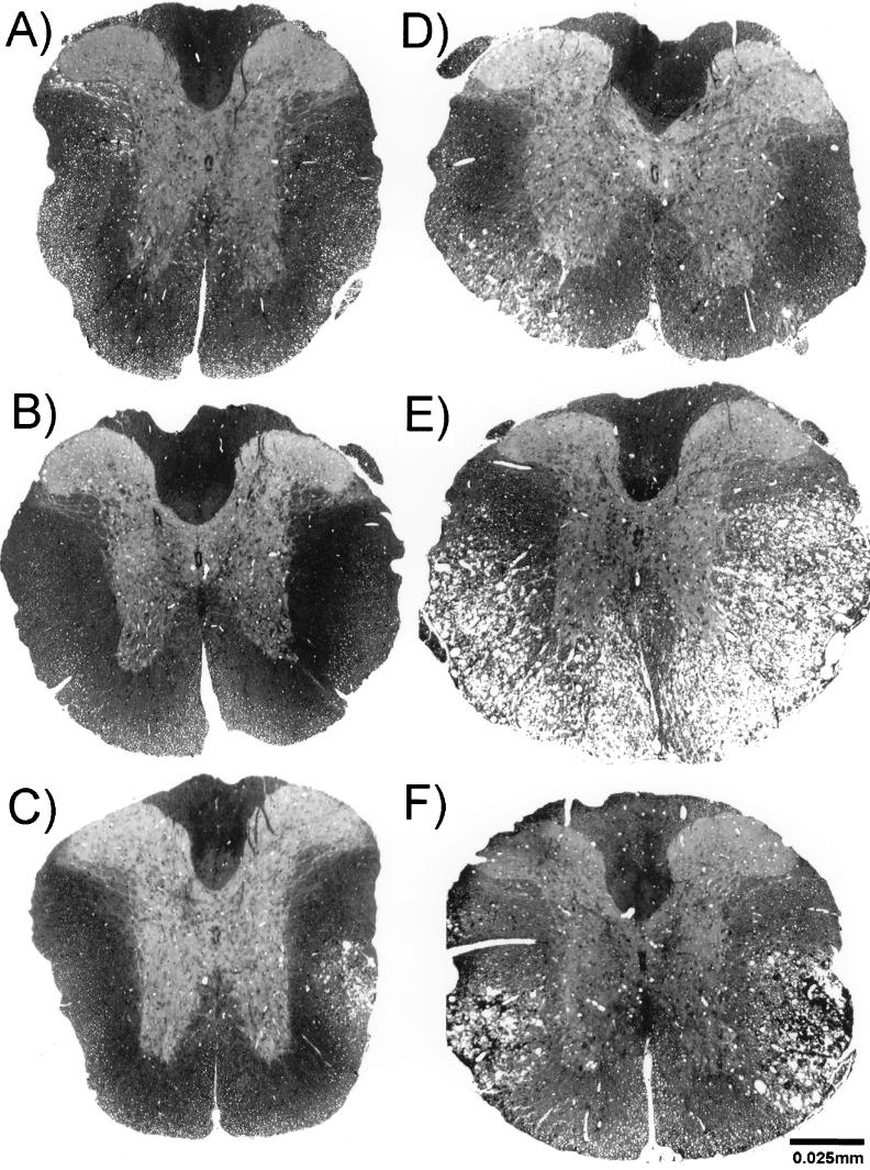
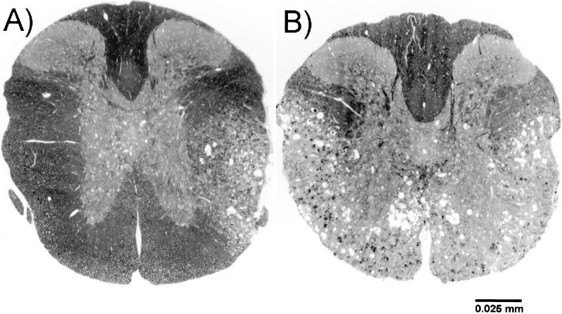
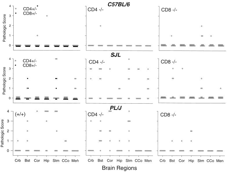
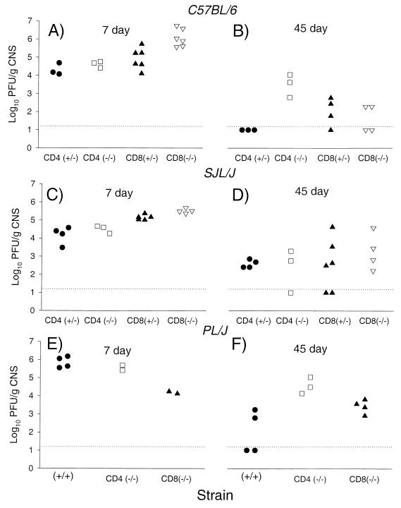
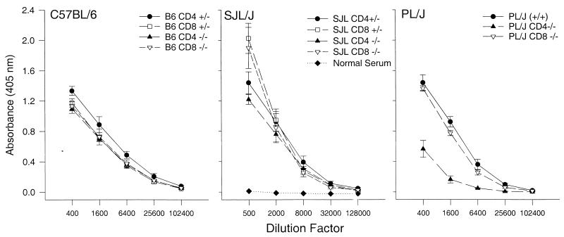
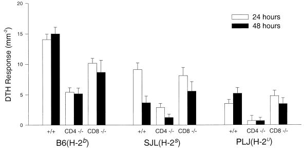
Following intracerebral infection with Theiler's murine encephalomyelitis virus (TMEV), susceptible strains of mice (SJL and PLJ) develop virus persistence and demyelination similar to that found in human multiple sclerosis. Resistant strains of mice (C57BL/6) clear virus and do not develop demyelination. To resolve the controversy about the role of CD4(+) and CD8(+) T cells in the development of demyelination and neurologic deficits in diseases of the central nervous system, we analyzed TMEV infection in CD4- and CD8-deficient B6, PLJ, and SJL mice. Genetic deletion of either CD4 or CD8 from resistant B6 mice resulted in viral persistence and demyelination during the chronic stage of disease. Viral persistence and demyelination were detected in all strains of susceptible background. Although genetic deletion of CD8 had no effect on the extent of demyelination in susceptible strains, deletion of CD4 dramatically increased the degree of demyelination observed. Whereas strains with deletions of CD4 showed severe neurologic deficits, mice with deletions of CD8 showed minimal or no deficits despite demyelination. In all strains, deletion of CD4 but not CD8 resulted in a decreased delayed-type hypersensitivity response to viral antigen. We conclude that each T-cell subset makes a discrete and nonredundant contribution to protection from viral persistence and demyelination in resistant strains. In contrast, in susceptible strains, CD8(+) T cells do not provide protection against chronic demyelinating disease. Furthermore, in persistent TMEV infection of the central nervous system, neurologic deficits appear to result either from the absence of a protective class II-restricted immune response or from the presence of a pathogenic class I-restricted response.
Figures
References
-
- Altintas A, Cai Z, Pease L R, Rodriguez M. Differential expression of H-2K and H-2D in the central nervous system of mice infected with Theiler’s virus. J Immunol. 1993;151:2803–2812. - PubMed
-
- Aubert C, Chamorro M, Brahic M. Identification of Theiler’s virus infected cells in the central nervous system of the mouse during demyelinating disease. Microb Pathog. 1987;3:319–326. - PubMed
-
- Blakemore W F, Welsh C J, Tonks P, Nash A A. Observations on demyelinating lesions induced by Theiler’s virus in CBA mice. Acta Neuropathol. 1988;76:581–589. - PubMed
-
- Clatch R J, Melvold R W, Miller S D, Lipton H L. Theiler’s murine encephalomyelitis virus (TMEV)-induced demyelinating disease in mice is influenced by the H-2D region: correlation with TMEV-specific delayed-type hypersensitivity. J Immunol. 1985;135:1408–1414. - PubMed
Publication types
MeSH terms
Substances
Grants and funding
LinkOut - more resources
Full Text Sources
Medical
Molecular Biology Databases
Research Materials
