. 1998 Sep;72(9):7397-406.
doi: 10.1128/JVI.72.9.7397-7406.1998.

Genomic structure of three phenotypically different isolates of peach latent mosaic viroid: implications of the existence of constraints limiting the heterogeneity of viroid quasispecies

Affiliations

Genomic structure of three phenotypically different isolates of peach latent mosaic viroid: implications of the existence of constraints limiting the heterogeneity of viroid quasispecies

S Ambrós et al. J Virol. 1998 Sep.

Abstract

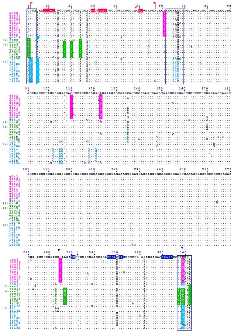
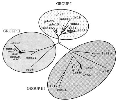
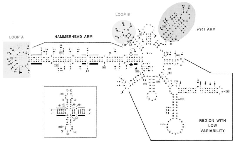
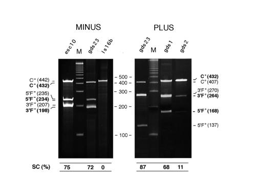
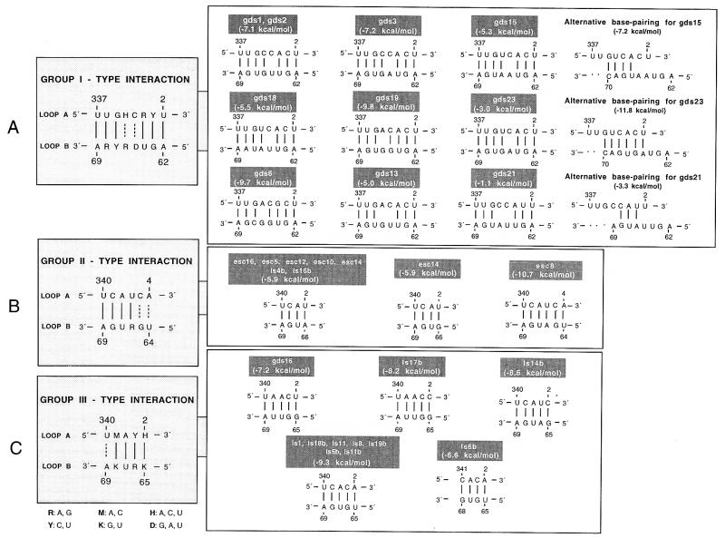
The peach latent mosaic viroid (PLMVd) is used to study the interactions between a viroid containing hammerhead ribozymes and its natural host, peach. To gain insight into the molecular basis of the phenotypic effects observed upon viroid infection, sequence variants from three PLMVd isolates that differ in symptom expression on the peach indicator GF-305 have been characterized. Analysis of the primary structures of a total of 29 different sequence variants derived from a severe and two latent isolates has revealed a large number of polymorphic positions in the viroid molecule. The variability pattern indicates that preservation of the stability of both hammerhead structures and conservation of a branched secondary structure of the viroid molecule may be factors limiting sequence heterogeneity in PLMVd. Moreover, compensatory mutations in two hairpin loops of the proposed secondary structure, suggesting that a pseudoknot-like interaction may exist between them, have also been observed. Phylogenetic analysis has allowed the allocation of PLMVd molecules into three major groups. This clustering does not strictly correlate with the source isolate from which the variants were obtained, providing insights into the complex mixture of molecules which make up each isolate. Bioassays of individual PLMVd sequence variants on GF-305 peach seedlings have shown that the biological properties of the PLMVd isolates may be correlated with both the complexity of their viroid populations and the presence of specific sequence variants.


Figures

FIG. 1
FIG. 1
Sequence alignment of 29 molecular variants of PLMVd derived from three different isolates. The reference sequence of PLMVd is included for comparative purposes and is shown at the top with two corrections, a deletion of one of the three Cs at positions 117 to 119 and a duplication of the G at position 257, with respect to that published previously (23). Dots indicate residues identical to the reference sequence, and dashes denote gaps. Three sequence variants (esc8, esc5, and ls1) are represented by several clones, whose number is given in parentheses. Regions involved in forming plus- and minus-polarity hammerhead structures are flanked by flags, the conserved nucleotides present in most natural hammerhead structures are indicated on a colored background, and the self-cleavage sites are shown by arrows; dark blue and red refer to plus and minus polarities, respectively. Informative changes dividing PLMVd variants into groups I, II, and III are on magenta, green, and light blue backgrounds, respectively. Other nucleotide variations present in most sequences of groups II and III are in green and light blue, respectively. Primers used for RT-PCR amplification cover positions 178 to 225. Residues involved in a potential pseudoknot-like element are boxed.
FIG. 2
FIG. 2
Phylogenetic tree of PLMVd variants. The analysis was based on the genetic distances calculated between the 29 PLMVd sequence variants characterized in this study. Three phylogenetic groups are delineated. Asterisks near nodes indicate their statistical significance as determined by bootstrap analysis after 100 replicates. ∗∗, node detected in 90 to 100% of replicates; ∗, node detected in ≥50% of replicates.
FIG. 3
FIG. 3
Predicted secondary structure of PLMVd showing the distribution of polymorphic positions along the molecule. The most stable folding for the reference positions in the PLMVd molecule (substitutions, insertions, or deletions) are marked by circles, and the number of sequence variants in which one specific position is affected is indicated in each case. Regions involved in forming plus- and minus-polarity hammerhead structures are flanked by flags, the conserved nucleotides present in most natural hammerhead structures are indicated by bars, and the self-cleavage sites are indicated by arrows. Solid and open symbols refer to plus and minus polarities, respectively. The reference sequence is marked every 20 nt with boxed numbers. (Inset) Alternative cruciform conformation adopted by the hammerhead arm in the most stable secondary structures predicted for all gds variants, most ls variants (except ls16b and ls4b), and esc8.
FIG. 4
FIG. 4
Hammerhead structures of the plus and minus strands of PLMVd variants. Sequence heterogeneity has been indicated on the self-cleaving domains of the PLMVd reference sequence (23). The nucleotide variations found between variants belonging to groups I (A), II (B), and III (C), as defined in Fig. 1, are shown. Nucleotide substitutions are indicated within circles, insertions are indicated within squares, and deletions are indicated by triangles pointing out in each case the PLMVd sequence variants affected by the corresponding mutation. Arrows indicate the predicted self-cleavage sites. The 13 conserved nucleotides present in all natural hammerhead structures (with the exception of the plus strand of sBYDV RNA [36] and the minus strand of a viroid-like RNA found recently in cherry [12], in which only 11 nt are conserved), are boxed. The same numbering is used for the plus and minus polarities and corresponds to that of the alignment shown in Fig. 1. Asterisks denote nucleotide changes present in all sequence variants of the corresponding group. (Inset) Scheme of PLMVd hammerhead structures with the proposed numbering system (26).
FIG. 5
FIG. 5
Self-cleavage during in vitro transcription of monomeric plus- and minus-strand RNAs from several PLMVd variants. The extension of self-cleavage was monitored by electrophoresis in denaturing polyacrylamide gels that were either stained with ethidium bromide or dried and scanned with a bioimage analyzer (not shown). The positions, polarities, and sizes of the complete transcripts C and of the self-cleavage fragments 5′F and 3′F are indicated on both sides. Boldface and regular type refer to transcription products derived from PLMVd cDNAs inserted in different orientations or cloning sites (see Materials and Methods). The sizes of some DNA markers (M) are indicated between the panels. The extent of self-cleavage (SC) is shown at the bottom of the figure.
FIG. 6
FIG. 6
Potential pseudoknot-like element in the PLMVd molecule. Shown are the proposed long-range interactions between nucleotides of loops A and B (Fig. 2) of the predicted secondary structures of variants belonging to groups I (A), II (B), and III (C), as defined in Fig. 1. The type interaction of each group is given in the left panel, with continuous and broken lines indicating the existence of a base pair in all and some cases, respectively. Free energy values (at 25°C) for the proposed interactions are shown in parentheses.

References

    1. Ambrós S, Desvignes J C, Llácer G, Flores R. Peach latent mosaic and pear blister canker viroids: detection by molecular hybridization and relationships with specific maladies affecting peach and pear trees. Acta Hortic. 1995;386:515–521.
    1. Branch A D, Robertson H D. A replication cycle for viroids and other small infectious RNAs. Science. 1984;223:450–455. - PubMed
    1. Branch A D, Benenfeld B J, Robertson H D. Evidence for a single rolling circle in the replication of potato spindle tuber viroid. Proc Natl Acad Sci USA. 1988;85:9128–9132. - PMC - PubMed
    1. Bruening G. Compilation of self-cleaving sequences from plant virus satellite RNAs and other sources. Methods Enzymol. 1989;180:546–558. - PubMed
    1. Côte F, Perrault J P. Peach latent mosaic viroid is locked by a 2′,5′-phosphodiester bond produced by in vitro self-ligation. J Mol Biol. 1997;273:533–543. - PubMed

Publication types

Associated data

LinkOut - more resources