. 1998 Sep;72(9):7420-7.
doi: 10.1128/JVI.72.9.7420-7427.1998.

Measles virus spread and pathogenesis in genetically modified mice

Affiliations

Measles virus spread and pathogenesis in genetically modified mice

B Mrkic et al. J Virol. 1998 Sep.

Abstract

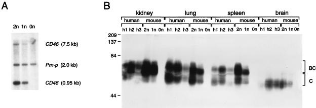
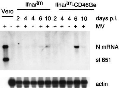
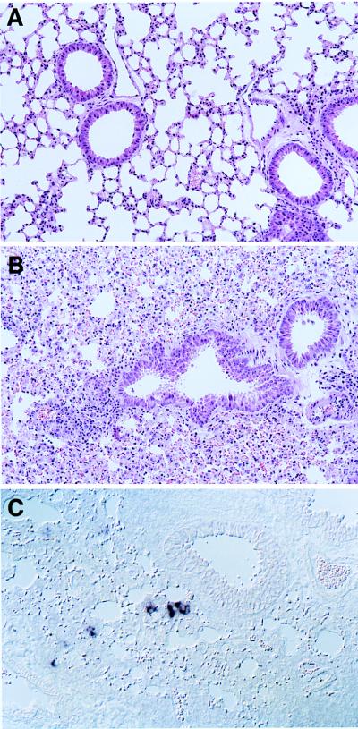
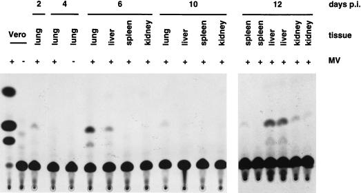
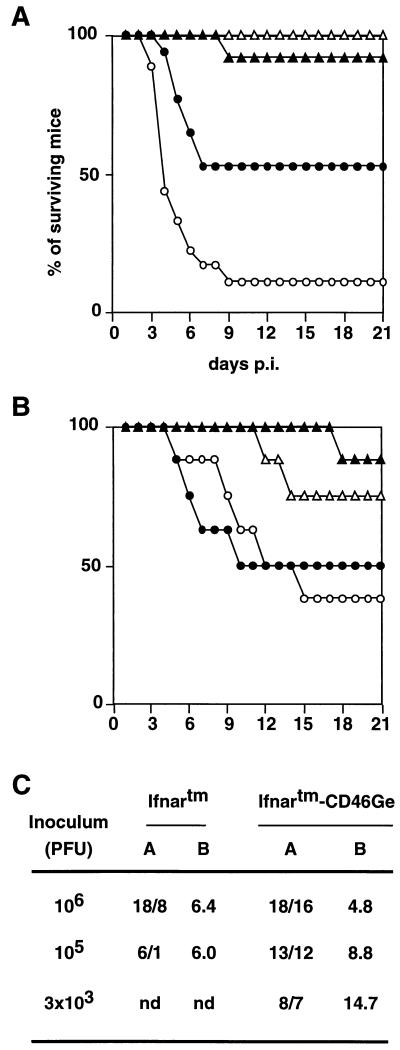
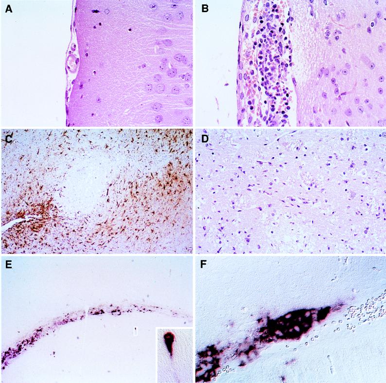
Attenuated Edmonston measles virus (MV-Edm) is not pathogenic in standard mice. We show here that MV-Edm inoculated via the natural respiratory route has a limited propagation in the lungs of mice with a targeted mutation inactivating the alpha/beta interferon receptor. A high dose of MV-Edm administered intracerebrally is lethal for about half of these mice. To study the consequences of the availability of a high-affinity receptor for MV propagation, we generated alpha/beta interferon-defective mice expressing human CD46 with human-like tissue specificity. Intranasal infection of these mice with MV-Edm resulted in enhanced spread to the lungs and more prominent inflammatory response. Virus replication was also detected in peripheral blood mononuclear cells, the spleen, and the liver. Moreover, intracerebral inoculation of adult animals with low MV-Edm doses caused encephalitis with almost inevitably lethal outcome. We conclude that in mice alpha/beta interferon controls MV infection and that a high-affinity receptor facilitates, but is not strictly required for, MV spread and pathogenesis.


Figures

FIG. 1
FIG. 1
Genome analysis (A) and protein expression (B) of CD46Ge transgenic mice. (A) Genomic analysis of transgenic and control mice. Tail DNAs were digested with EcoRI, separated on an agarose gel, blotted onto a nylon membrane, and hybridized with two probes, one recognizing two fragments (0.95 and 7.5 kb) in the human CD46 promoter region and one recognizing a 2-kb fragment of the endogenous mouse Prn-p gene. Lane 2n, CD46 homozygous mouse; lane 1n, CD46 hemizygous mouse; lane 0n, control mouse. (B) CD46 protein expression in four organs obtained at autopsies of three humans (h1, h2, and h3) compared to the same organs from a CD46 homozygous mouse (2n), a CD46 hemizygous mouse (1n), and a control mouse (0n). Thirty micrograms of protein from human tissue homogenates or 15 μg of protein from mouse tissue homogenates were separated by sodium dodecyl sulfate-polyacrylamide gel electrophoresis, transferred to a nylon membrane, and reacted against a polyclonal rabbit anti-CD46 serum. The molecular masses of marker proteins are indicated on the left in kilodaltons. The approximate positions of the two major isoforms of the CD46 proteins (termed BC and C) are indicated on the right. CD46 proteins produced in different organs have different molecular masses due to differential splicing and heterogeneous N- and O-glycosylation. Note that in humans different patterns of CD46 expression have been recognized (19). Individuals h1 and h2 expressed approximately equivalent amounts of BC and C isoforms, whereas individual h3 may have had predominantly BC expression. CD46Ge mice express equivalent amounts of the BC and C isoforms. The low quantity of protein detected in certain human autopsy samples, e.g., the brain of h1, may be because of partial protein degradation.
FIG. 2
FIG. 2
Northern blot analysis of MV RNA in lungs of Ifnartm and Ifnartm-CD46Ge mice intranasally inoculated with MV-P-CAT. Mice were sacrificed at the days p.i. indicated. Five micrograms of total lung RNA was separated on a 1% formaldehyde-agarose gel, blotted to a nylon membrane, and reacted with an antisense MV N RNA probe (top panel). The blot was stripped and rehybridized with an actin RNA probe (bottom panel). As a positive control, 6 ng of total RNA from MV-infected Vero cells was used (first lane) and, as a negative control, 10 μg of total RNA from mock-infected Vero cells (second lane) was used. As additional negative controls, total RNAs from two mock-infected mice (−) were examined. A synthetic MV N plus-strand standard RNA 851 bases in length (st 851) was added to the positive control. The positions of this standard RNA, the N mRNA (about 1.7 kb), and the actin mRNA are indicated on the right. About 30,000 copies of MV N mRNA are produced in MV-infected primate cells (first lane) (9). Considering that about 800 times less RNA from Vero cells than RNA from mouse lungs was loaded, the signal in the positive control corresponds to about 40 copies of N mRNA per cell, and the signals in the mouse lung tissues correspond to a few N mRNA copies per average cell.
FIG. 3
FIG. 3
Results of histological analysis (A and B) and MV RNA detection (C) in lung sections of intranasally infected Ifnartm-CD46Ge mice. After mock infection (A) or inoculation with MV-Edm (B and C), the mice were sacrificed. (C) A 2-μm-thick lung section hybridized with a MV N mRNA-specific probe.
FIG. 4
FIG. 4
MV spread in different organs of Ifnartm-CD46Ge mice. Mice intranasally inoculated (+) or mock infected (−) with MV-P-CAT were sacrificed at the days p.i. indicated and tissues were collected, homogenized, and tested for CAT activity. As a positive control, Vero cells infected with MV-P-CAT were used (first lane), and as a negative control, mock-infected Vero cells (second lane) were used. The lung extract of a mouse sacrificed 4 days after mock infection is shown in the fifth lane.
FIG. 5
FIG. 5
Survival of mice from four strains after intracerebral inoculation with the vaccine strain MV-Edm (A and C) or with the neurotropic strain CAM/RBH (B). (A and B) Six- to 8-week-old mice were injected intracerebrally with 1 million infectious units of MV-Edm or with 104 infectious units of the rodent brain-adapted neurotropic CAM/RBH strain. Open circles, Ifnartm-CD46Ge mice; dots, Ifnartm mice; open triangles, CD46Ge mice; filled triangles, control C57BL/6 mice. The numbers of animals injected were as follows: (A) 18 Ifnartm-CD46Ge and Ifnartm and 12 CD46Ge and C57BL/6 mice; (B) 8 from each mouse strain. (C) Susceptibilities of Ifnartm and Ifnartm-CD46Ge mice to different doses of MV-Edm. Columns: A, numbers of inoculated/dead mice; B, average day of death. nd, not determined.
FIG. 6
FIG. 6
Histological analysis of the consequences of MV infection and of the MV RNA distribution in the brains of Ifnartm-CD46Ge animals. A mouse inoculated with 1 million infectious units of MV-Edm (B to F) and a mock-infected mouse (A) were sacrificed 3 days p.i., their brains were prepared, and brain slices were stained as indicated below. (A, B, and D) HE-stained sections showing meningeal inflammation (B) and necrosis (B and D) of the brain tissue. (C) Immunohistochemical staining for GFAP demonstrating reactive astrocytes (brown) surrounding necrotic lesions. (E and F) In situ hybridization with a MV N-specific probe showing strong staining in the ventricular region and in scattered neurons. One infected hippocampal neuron is enlarged in the inset. In panel F, many contiguous ependymal cells are stained.

References

    1. Albrecht P, Lorenz D, Klutch M J, Vickers J H, Ennis F A. Fatal measles infection in marmosets: pathogenesis and prophylaxis. Infect Immun. 1980;27:969–978. - PMC - PubMed
    1. Buchholz C J, Gerlier D, Hu A, Cathomen T, Liszewski M K, Atkinson J P, Cattaneo R. Selective expression of a subset of measles virus receptor-competent CD46 isoforms in human brain. Virology. 1996;217:349–355. - PubMed
    1. Buchholz C J, Koller D, Devaux P, Mumenthaler C, Schneider-Schaulies J, Braun W, Gerlier D, Cattaneo R. Mapping of the primary binding site of measles virus to its receptor CD46. J Biol Chem. 1997;272:22072–22079. - PubMed
    1. Buchholz C J, Schneider U, Devaux P, Gerlier D, Cattaneo R. Cell entry by measles virus: long hybrid receptors uncouple binding from membrane fusion. J Virol. 1996;70:3716–3723. - PMC - PubMed
    1. Bueler H, Fischer M, Lang Y, Bluethmann H, Lipp H P, DeArmond S J, Prusiner S B, Aguet M, Weissmann C. Normal development and behaviour of mice lacking the neuronal cell-surface PrP protein. Nature. 1992;356:577–582. - PubMed

Publication types

MeSH terms

LinkOut - more resources