. 1998 Sep;72(9):7626-31.
doi: 10.1128/JVI.72.9.7626-7631.1998.

The role of influenza A virus hemagglutinin residues 226 and 228 in receptor specificity and host range restriction

Affiliations

The role of influenza A virus hemagglutinin residues 226 and 228 in receptor specificity and host range restriction

A Vines et al. J Virol. 1998 Sep.

Abstract

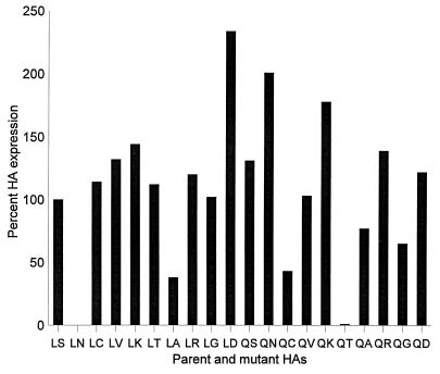
Influenza A viruses can be isolated from a variety of animals, but their range of hosts is restricted. For example, human influenza viruses do not replicate in duck intestine, the major replication site of avian viruses in ducks. Although amino acids at positions 226 and 228 of hemagglutinin (HA) of the H3 subtype are known to be important for this host range restriction, the contributions of specific amino acids at these positions to restriction were not known. Here, we address this issue by generating HAs with site-specific mutations of a human virus that contain different amino acid residues at these positions. We also let ducks select replication-competent viruses from a replication-incompetent virus containing a human virus HA by inoculating animals with 10(10.5) 50% egg infectious dose of the latter virus and identified a mutation in the HA. Our results showed that the Ser-to-Gly mutation at position 228, in addition to the Leu-to-Gln mutation at position 226 of the HA of the H3 subtype, is critical for human virus HA to support virus replication in duck intestine.


Figures

FIG. 1
FIG. 1
Cell surface expression of mutant HAs analyzed by FACS. Cos-1 cells (60% confluency) were transfected with 2 μg of purified plasmid DNA per well of a 6-well tissue culture plate with Lipofectamine (Gibco). The cells and transfection mixture were incubated for 5 h at 37°C, after which the transfection medium was replaced with 2 ml of Opti-Mem medium (Life Technologies, Inc.) containing 5% fetal calf serum (FCS). The cells were then incubated for 40 h at 37°C. Cells expressing HA were washed with PBS, treated with Vibrio cholerae sialidase (5.5 milliunits/ml; Life Technologies, Inc.) for 1 h at 37°C (which abolishes the susceptibility of cells to influenza virus infection) to remove sialic acid from the HA, which interferes with receptor recognition (17), and then maintained in suspension following trypsinization. Cells were spun down, washed twice with PBS containing 10% FCS, and then resuspended in PBS containing 10% FCS and an anti-H3 HA monoclonal antibody (S11/4, S28/1, S37/2, and 121/1) pool (diluted 1:400). After a 1-h incubation at 4°C, the cells were washed twice with PBS containing 10% FCS, and then incubated with fluorescein isothiocynate-labeled goat anti-mouse immunoglobulin (diluted 1:20 in PBS containing 10% FCS) (Boehringer Mannheim Biochemicals) for 30 min at 4°C. The cells were washed twice as before and then fixed in PBS containing 3.7% paraformaldehyde for 20 min at 4°C. Cells were centrifuged, resuspended in PBS containing 10% FCS, and stored at 4°C until analyzed by FACS with a FACScan (Becton Dickinson). The relative expression levels presented are based on mean values of fluorescence intensity. Experiments were repeated three times, and representative data are shown.
FIG. 2
FIG. 2
Effects of mutations at positions 226 and/or 228 on the hemadsorbing activity of human virus HA. Cos-1 cells were transfected with plasmid expressing HA. At 40 h posttransfection, they were washed twice with PBS containing 10% FCS and incubated with chilled 1% erythrocyte suspensions (chicken [in house], human [type O, St. Jude Children’s Research Hospital blood bank], or horse [Rockland]) in PBS. After a 1-h incubation at 4°C, the cells were washed at least five times with PBS and rinsed with methanol to remove erythrocytes that were nonspecifically bound. The cells were then air dried and stained with a 1:20 dilution of Giemsa stain (Sigma) for 15 min at 4°C. The numbers of hemadsorption-positive cells were recorded by examining five randomly selected microscopic fields that consisted of approximately 500 cells. Experiments were performed three times with similar results.
FIG. 2
FIG. 2
Effects of mutations at positions 226 and/or 228 on the hemadsorbing activity of human virus HA. Cos-1 cells were transfected with plasmid expressing HA. At 40 h posttransfection, they were washed twice with PBS containing 10% FCS and incubated with chilled 1% erythrocyte suspensions (chicken [in house], human [type O, St. Jude Children’s Research Hospital blood bank], or horse [Rockland]) in PBS. After a 1-h incubation at 4°C, the cells were washed at least five times with PBS and rinsed with methanol to remove erythrocytes that were nonspecifically bound. The cells were then air dried and stained with a 1:20 dilution of Giemsa stain (Sigma) for 15 min at 4°C. The numbers of hemadsorption-positive cells were recorded by examining five randomly selected microscopic fields that consisted of approximately 500 cells. Experiments were performed three times with similar results.
FIG. 3
FIG. 3
Positions of residues 226 and 228 in the RBS of influenza virus HA in relation to the Neu5Ac2,3Gal moiety. Only the relevant portion of the human virus X31 HA complexed with 3′ sialylactose (1HGG structure, Brookhaven Protein Databank [19]) and the Neu5Acα2-3Gal moiety of 3′ sialyl lactose (heavy atoms, stick presentation) are shown for clarity. Atoms of sialic acid (NAN) and galactose (GAL) that are thought to participate in hydrogen bonding with HA (19) are shown as small white balls; atoms of the protein that participate in these hydrogen bonds are shown as white on black background. Substitution 226S→G results in a loss of a hydrogen bond between the hydroxyl group of serine and the O-9 hydroxyl of sialic acid, whereas mutation 226L→Q results in new hydrogen bonds between the amide side chain of glutamine and the C-8 hydroxyl and carboxylic groups of Neu5Ac (30). This figure was generated with a WebLab viewer (Molecular Simulations, Inc., San Diego, Calif.).

References

    1. Almond J W. A single gene determines the host range of influenza virus. Nature. 1977;270:617–618. - PubMed
    1. Bean W J, Schell M, Katz J, Kawaoka Y, Naeve C, Gorman O, Webster R G. Evolution of the H3 influenza virus hemagglutinin from human and nonhuman hosts. J Virol. 1992;66:1129–1138. - PMC - PubMed
    1. Beare A S, Webster R G. Replication of avian influenza viruses in humans. Arch Virol. 1991;119:37–42. - PubMed
    1. Connor R J, Kawaoka Y, Webster R G, Paulson J C. Receptor specificity in human, avian, and equine H2 and H3 influenza virus isolates. Virology. 1994;205:17–23. - PubMed
    1. Enami M, Luytjes W, Krystal M, Palese P. Introduction of site-specific mutations into the genome of influenza virus. Proc Natl Acad Sci USA. 1990;87:3802–3805. - PMC - PubMed

Publication types

LinkOut - more resources