. 1998 Sep;72(9):7653-8.
doi: 10.1128/JVI.72.9.7653-7658.1998.

Lipofection of purified adeno-associated virus Rep68 protein: toward a chromosome-targeting nonviral particle

Affiliations

Lipofection of purified adeno-associated virus Rep68 protein: toward a chromosome-targeting nonviral particle

S Lamartina et al. J Virol. 1998 Sep.

Abstract

Adeno-associated virus (AAV) integrates very efficiently into a specific site (AAVS1) of human chromosome 19. Two elements of the AAV genome are sufficient: the inverted terminal repeats (ITRs) and the Rep78 or Rep68 protein. The incorporation of the AAV integration machinery in nonviral delivery systems is of great interest for gene therapy. We demonstrate that purified recombinant Rep68 protein is functionally active when directly delivered into human cells by using the polycationic liposome Lipofectamine, promoting the rescue-replication of a codelivered ITR-flanked cassette in adenovirus-infected cells and its site-specific integration in noninfected cells. The sequencing of cloned virus-host DNA junctions confirmed that lipofected Rep68 protein triggers site-specific integration at the same sites in chromosome 19 already characterized in cells latently infected with AAV.


Figures

FIG. 1
FIG. 1
Site-specific integration in HeLa cell clones. Transfected plasmids are schematically represented. Black boxes represent the AAV ITRs flanking the Hook and Neo expression cassettes. The Neo gene was derived from plasmid PRc/RSV, and the Hook gene was obtained from plasmid pHook-1 (both from Invitrogen). The individual cDNAs coding for Rep78 and Rep68 were obtained by site-directed mutagenesis of the Rep open reading frame (nucleotides 320 to 2252 of the AAV genome [33]) as described elsewhere (34). Integration at the AAVS1 site was assessed by DNA hybridization analysis with AAVS1 and neo-specific probes as described in the text.
FIG. 2
FIG. 2
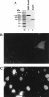
Lipofection of Rep68 protein. (A) Silver staining of a sodium dodecyl sulfate-polyacrylamide gel. Rep68 protein was expressed in E. coli as a fusion protein with maltose binding protein (MBP) as described elsewhere (4) and was partially purified by amylose affinity chromatography (lane 1), cleaved with Factor Xa to remove the maltose-binding moiety, and purified to homogeneity (lane 2) by fast protein liquid chromatography with MonoQ (anion exchange) and Superdex-75 (gel filtration) columns (both from Pharmacia). M, molecular size markers. (B and C) Intracellular localization of lipofected Rep68 protein. Two hours after transfection, 293 cells were washed, fixed in 3% formaldehyde, and permeabilized by treatment with 0.1% Triton X-100. The intracellular location of Rep68 was monitored by sequential incubation with the mouse monoclonal anti-Rep antibody 226.7 (Progen) and a rhodamine-conjugated anti-mouse immunoglobulin G goat polyclonal serum. Shown are results of staining of cells incubated for 2 h with Rep68 alone (B) or with the Rep68–Lipofectamine complex (C).
FIG. 3
FIG. 3
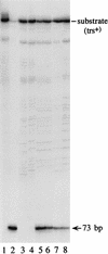
Endonuclease reaction with nuclear extracts of cells lipofected with Rep68 protein. Ten thousand counts per minute of trs+ hairpin substrate that had been 5′-end labelled with 32P, prepared as described elsewhere (32), was incubated for 1 h in endonuclease assay buffer (32) with 1 and 3 μg of nuclear extracts from untransfected cells (lanes 3 and 4, respectively), cells lipofected with the Rep68 expression vector pCMVrep68 (lanes 5 and 6), or cells lipofected with Rep68 protein (lanes 7 and 8). A standard endonuclease reaction was carried out (32). The reaction was terminated by treatment with proteinase K, phenol extraction, and ethanol precipitation; samples were then resolved on an 8% polyacrylamide sequencing gel. The position of the 73-base product of the nicking reaction is indicated on the right. In lane 1, the starting trs+ substrate was loaded. Lane 2 shows the results of a control experiment with 10 ng of purified Rep68. Nuclear extracts were prepared as described elsewhere (36).
FIG. 4
FIG. 4
Southern blot analysis of rescue-replication from plasmid ITR/Hook-Neo in Ad-infected cells lipofected with Rep68 protein. Rescue-replication assays were performed in HeLa (A) and HepG2 (B) cells. In both cases, cells were lipofected with 7.5 μg of plasmid ITR/Hook-Neo in combination with either an equivalent amount of pCMVrep68 expression vector (lanes 1) or increasing concentrations (0.5, 1, 2, 4, 8, 16, and 32 μg) of Rep68 protein (lanes 3, 4, 5, 6, 7, 8, and 9, respectively). As a control, DNA was extracted from cells transfected only with the ITR/Hook-Neo plasmid (lanes 2). Equivalent amounts of low-Mr DNA samples isolated at 68 h posttransfection were digested with DpnI, electrophoresed on agarose gels, and analyzed on Southern blots with a neo probe. The arrows on the left indicate the rescued monomer; dimeric forms were also observed after longer exposure (data not shown). Molecular sizes are shown in kilobases.
FIG. 5
FIG. 5
Lipofected Rep68 protein mediates site-specific integration into AAVS1. (A and B) Southern blot analysis of PCR amplification products generated from ITR-AAVS1 junctions. (A) Hybridization with the AAVS1 probe; (B) hybridization with the ITR probe. PCRs were carried out by using as a template the genomic DNA isolated from HeLa cells 48 h after lipofection with plasmid ITR/Hook-Neo alone (lanes 2) or in combination with the pCMVrep68 expression vector (lanes 1) or with Rep68 protein (lanes 3). Control reactions were performed with DNA extracted from untransfected cells (lanes 4). Amplification products were blotted onto nylon membranes; the same filter was probed first with a 32P-labelled AAVS1-specific probe (spanning nucleotides 210 to 1140 of the published AAVS1 sequence [17]) and then, after stripping, with an ITR-specific probe, excised as an MscI-PvuII fragment from plasmid pSub201 (28). (C) Sequence analysis of ITR-AAVS1 junctions. At the top is a schematic representation of the PCR assay. The sequence of one strand of the ITRs in plasmid ITR/Hook-Neo, in the “flop” orientation (3, 27), is shown; the nucleotide numbering is relative to the right end of the AAV genome (33). Letters (D, A, B, B′, C, C′, and A′) indicate palindromic sequences. Junctions obtained with the Rep68 proteins (r68-1 through -4) and with the pCMVrep68 expression vector (p68-1 through -6) are shown below the PCR assay representation. The numbers of the last evident cellular and viral nucleotides are given. AAVS1 breakpoints are based on the published AAVS1 sequence (17). Overlapping sequences between the ITR and AAVS1 are underlined. Insertions between the ITR and AAVS1 breakpoints are boldfaced.
FIG. 5
FIG. 5
Lipofected Rep68 protein mediates site-specific integration into AAVS1. (A and B) Southern blot analysis of PCR amplification products generated from ITR-AAVS1 junctions. (A) Hybridization with the AAVS1 probe; (B) hybridization with the ITR probe. PCRs were carried out by using as a template the genomic DNA isolated from HeLa cells 48 h after lipofection with plasmid ITR/Hook-Neo alone (lanes 2) or in combination with the pCMVrep68 expression vector (lanes 1) or with Rep68 protein (lanes 3). Control reactions were performed with DNA extracted from untransfected cells (lanes 4). Amplification products were blotted onto nylon membranes; the same filter was probed first with a 32P-labelled AAVS1-specific probe (spanning nucleotides 210 to 1140 of the published AAVS1 sequence [17]) and then, after stripping, with an ITR-specific probe, excised as an MscI-PvuII fragment from plasmid pSub201 (28). (C) Sequence analysis of ITR-AAVS1 junctions. At the top is a schematic representation of the PCR assay. The sequence of one strand of the ITRs in plasmid ITR/Hook-Neo, in the “flop” orientation (3, 27), is shown; the nucleotide numbering is relative to the right end of the AAV genome (33). Letters (D, A, B, B′, C, C′, and A′) indicate palindromic sequences. Junctions obtained with the Rep68 proteins (r68-1 through -4) and with the pCMVrep68 expression vector (p68-1 through -6) are shown below the PCR assay representation. The numbers of the last evident cellular and viral nucleotides are given. AAVS1 breakpoints are based on the published AAVS1 sequence (17). Overlapping sequences between the ITR and AAVS1 are underlined. Insertions between the ITR and AAVS1 breakpoints are boldfaced.

Similar articles

Cited by

References

    1. Balagué C, Kalla M, Zhang W-W. Adeno-associated virus Rep78 protein and terminal repeats enhance integration of DNA sequences into the cellular genome. J Virol. 1997;71:3299–3306. - PMC - PubMed
    1. Baubonis W, Sauer B. Genomic targeting with purified Cre recombinase. Nucleic Acids Res. 1993;21:2025–2029. - PMC - PubMed
    1. Berns K I, Linden R M. The cryptic life style of adeno-associated virus. Bioessays. 1995;17:237–245. - PubMed
    1. Chiorini J A, Weitzman M D, Owens R A, Urcelay E, Safer B, Kotin R M. Biologically active Rep proteins of adeno-associated virus type 2 produced as fusion proteins in Escherichia coli. J Virol. 1994;68:797–804. - PMC - PubMed
    1. Deshnukh H M, Huang L. Liposome and polylysine mediated gene transfer. New J Chem. 1997;21:113–124.

LinkOut - more resources