. 1998 Sep;72(9):7669-75.
doi: 10.1128/JVI.72.9.7669-7675.1998.

Caspase activation and specific cleavage of substrates after coxsackievirus B3-induced cytopathic effect in HeLa cells

Affiliations

Caspase activation and specific cleavage of substrates after coxsackievirus B3-induced cytopathic effect in HeLa cells

C M Carthy et al. J Virol. 1998 Sep.

Abstract

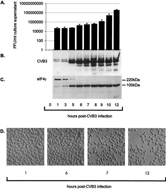
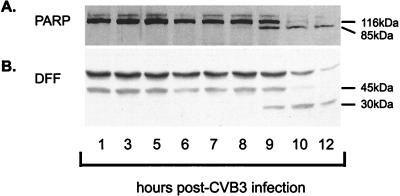
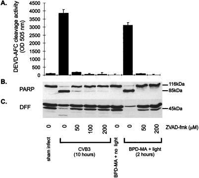
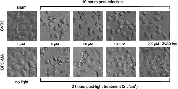
Coxsackievirus B3 (CVB3), an enterovirus in the family Picornaviridae, induces cytopathic changes in cell culture systems and directly injures multiple susceptible organs and tissues in vivo, including the myocardium, early after infection. Biochemical analysis of the cell death pathway in CVB3-infected HeLa cells demonstrated that the 32-kDa proform of caspase 3 is cleaved subsequent to the degenerative morphological changes seen in infected HeLa cells. Caspase activation assays confirm that the cleaved caspase 3 is proteolytically active. The caspase 3 substrates poly(ADP-ribose) polymerase, a DNA repair enzyme, and DNA fragmentation factor, a cytoplasmic inhibitor of an endonuclease responsible for DNA fragmentation, were degraded at 9 h following infection, yielding their characteristic cleavage fragments. Inhibition of caspase activation by benzyloxycarbonyl-Val-Ala-Asp-fluoromethylketone (ZVAD.fmk) did not inhibit the virus-induced cytopathic effect, while inhibition of caspase activation by ZVAD.fmk in control apoptotic cells induced by treatment with the porphyrin photosensitizer benzoporphyrin derivative monoacid ring A and visible light inhibited the apoptotic phenotype. Caspase activation and cleavage of substrates may not be responsible for the characteristic cytopathic effect produced by picornavirus infection yet may be related to late-stage alterations of cellular homeostatic processes and structural integrity.


Figures

FIG. 1
FIG. 1
Release of progeny CVB3 virus, host cell production of CVB3 viral protein, viral protease cleavage of host eIF4G, and cell morphology changes following infection with CVB3. (A) Culture medium was collected and assayed for infectious virus by the agar overlay plaque assay method. There was an increase in the amount of infectious virus (in PFU per milliliter) released over the 12-h experiment (B). Cellular lysate was collected from CVB3-infected HeLa cells, and immunoblot analysis with a CVB3 polyclonal antibody that recognizes major viral proteins was performed. (C) Cytosolic extract was then analyzed for the presence of the 220-kDa eIF4G component of the translation initiation complex. (D) Contrast microscopy of HeLa cells at 1, 6, 7, and 12 h postinfection was performed. Note the extensive cytopathic changes that occurred between 6 and 7 h postinfection.
FIG. 2
FIG. 2
Caspase 3 activation and cleavage of the 32-kDa proform following CVB3 infection of HeLa cells. (A) Ten micrograms of cell lysate was incubated in 150 μl of reaction buffer containing the caspase 3-specific substrate Ac-DEVD-AMC. After incubation at 37°C for 1 h, fluorescence levels were determined with an excitation wavelength of 380 nm and an emission wavelength of 460 nm. Note the increase in fluorescence, representing caspase activity, beginning after 7 h postinfection and increasing to maximum levels by 10 h postinfection. (B) HeLa cell lysates were separated by sodium dodecyl sulfate-polyacrylamide gel electrophoresis and transferred to nitrocellulose. Immunoblotting for the presence of the 32-kDa proform of caspase 3 demonstrates that this protein is processed between 7 and 12 h postinfection.
FIG. 3
FIG. 3
Specific cleavage of PARP and DFF substrates by caspase 3 following CVB3 infection. (A) Cellular lysate was collected from CVB3-infected HeLa cells, and immunoblot analysis was performed with an anti-PARP antibody which recognizes a 85-kDa cleavage fragment. Note that cleavage of PARP began at 9 h following infection, with a marked loss of the 116-kDa native protein occurring by 10 h postinfection. (B) Cellular lysate was similarly analyzed for DFF cleavage by immunoblotting following CVB3 infection. Note the change in DFF status beginning at 9 h postinfection, with the appearance of a 30-kDa fragment.
FIG. 4
FIG. 4
ZVAD-fmk inhibits caspase activation as well as cleavage of PARP and DFF following CVB3 infection or induction of apoptosis by treatment of HeLa cells with BPD-MA and light. (A) Cell lysate was incubated in reaction buffer containing the caspase 3-specific substrate Ac-DEVD-AFC. After incubation at 37°C for 1 h, fluorescence levels were determined with an excitation wavelength of 400 nm and an emission wavelength of 505 nm. Note the lack of caspase activation in ZVAD-fmk (50 to 200 μM)-treated HeLa cells at 10 h following CVB3 infection and at 2 h following treatment with BPD-MA and light. (B) Cellular lysate was collected from treated HeLa cells, and immunoblot analysis was performed with an anti-PARP antibody. Note the equivalent cleavage of PARP in the CVB3-infected HeLa cells without ZVAD.fmk and the BPD-MA- and light-treated HeLa cells without ZVAD.fmk. ZVAD.fmk treatment (50 to 200 μM) of HeLa cells prevented PARP processing in the BPD-MA- and light-treated HeLa cells, while in CVB3-treated HeLa cells the PARP processing was limited, but not completely inhibited, by treatment with ZVAD.fmk at 50 or 100 μM. (C) Immunoblot analysis of DFF cleavage at 10 h following CVB3 infection and at 2 h following treatment with BPD-MA and light showed that the cleavage pattern was similar to that of PARP. Sham-infected cultures were treated the same as infected cultures, without virus.
FIG. 5
FIG. 5
Effects of ZVAD.fmk treatment on cell morphological changes following CVB3 infection or BPD-MA and light treatment of HeLa cells. HeLa cells were treated with ZVAD.fmk at 0 to 200 μM and then infected with CVB3 or treated with BPD-MA and light. At 10 h postinfection, caspases were processed and substrates were cleaved in virus-infected cells, and at 2 h post-light treatment, caspases were processed and substrates were cleaved in BPD-MA-treated cells. Note the difference in the morphological appearances of the CVB3-infected and photodynamically treated HeLa cells. CVB3-infected HeLa cells appeared rounded, with smooth cell surfaces, while the HeLa cells treated with BPD-MA and light displayed extensive membrane blebbing and shrinkage, with cellular heterogeneity. At higher concentrations of ZVAD-fmk, the morphological changes produced by BPD-MA and light treatment were inhibited, and the cell appearance was similar to that of control HeLa cells (BPD-MA treated plus no light). In CVB3-infected HeLa cells, the morphological changes were not inhibited with increasing concentrations of ZVAD.fmk, suggesting that the morphological alterations were independent of caspase processing and cleavage of substrates.

References

    1. Aldabe R, Irurzun A, Carrasco L. Poliovirus protein 2BC increases cytosolic free calcium concentrations. J Virol. 1997;71:6214–6217. - PMC - PubMed
    1. Ambrosini G, Adida C, Altieri D C. A novel anti-apoptosis gene, survivin, expressed in cancer and lymphoma. Nat Med. 1997;3:917–921. - PubMed
    1. Anderson D R, Wilson J E, Carthy C M, Yang D, Kandolf R, McManus B M. Direct interactions of coxsackievirus B3 with immune cells in the splenic compartment of mice susceptible or resistant to myocarditis. J Virol. 1996;70:4632–4645. - PMC - PubMed
    1. Bablanian R, Eggers H, Tamm I. Studies on the mechanism of poliovirus-induced cell damage. I. The relation between poliovirus-induced metabolic and morphological alterations in cultured cells. Virology. 1965;26:100–113. - PubMed
    1. Bablanian R, Eggers H J, Tamm I. Studies on the mechanism of poliovirus-induced cell damage. II. The relation between poliovirus growth and virus-induced morphological changes in cells. Virology. 1965;26:114–121. - PubMed

Publication types

Substances