. 1998 Aug 15;18(16):6218-29.
doi: 10.1523/JNEUROSCI.18-16-06218.1998.

Tenascin-R is antiadhesive for activated microglia that induce downregulation of the protein after peripheral nerve injury: a new role in neuronal protection

Affiliations

Tenascin-R is antiadhesive for activated microglia that induce downregulation of the protein after peripheral nerve injury: a new role in neuronal protection

D N Angelov et al. J Neurosci. .

Abstract

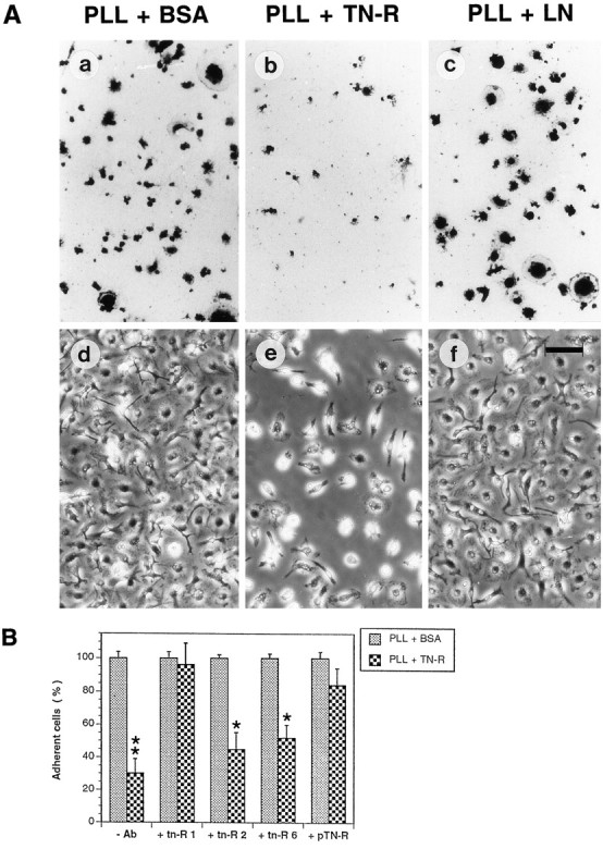
Microglial activation in response to pathological stimuli is characterized by increased migratory activity and potential cytotoxic action on injured neurons during later stages of neurodegeneration. The initial molecular changes in the CNS favoring neuronofugal migration of microglia remain, however, largely unknown. We report that the extracellular matrix protein tenascin-R (TN-R) present in the intact CNS is antiadhesive for activated microglia, and its downregulation after facial nerve axotomy may account for the loss of motoneuron protection and subsequent neurodegeneration. Studies on the protein expression in the facial and hypoglossal nucleus in rats demonstrate that TN-R is a constituent of the perineuronal net of motoneurons and 7 d after peripheral nerve injury becomes downregulated in the corresponding motor nucleus. This downregulation is reversible under regenerative (nerve suture) conditions and irreversible under degenerative (nerve resection) conditions. In short-term adhesion assays, the unlesioned side of brainstem cryosections from unilaterally operated animals is nonpermissive for activated microglia, and this nonpermissiveness is almost abolished by a monoclonal antibody to TN-R. Microglia-conditioned media and tumor necrosis factor-alpha downregulate TN-R protein and mRNA synthesis by cultured oligodendrocytes, which are one of the sources for TN-R in the brainstem. Our findings suggest a new role for TN-R in neuronal protection against activated microglia and the participation of the latter in perineuronal net destruction, e.g., downregulation of TN-R.


Figures

Fig. 1.
Fig. 1.
Expression pattern of TN-R (A, D, E) and TN-C (B) in the facial nucleus of the adult rat.A, B, Low-power photomicrographs of nuclei immunostained with Ab tn-R1 to TN-R and polyclonal Abs to TN-C. Note the dense (for TN-R) and faint (for TN-C) deposits of immunoreaction product outlining the cell bodies of motoneurons (arrowheads) and the diffuse immunostaining throughout the neuropil (asterisks). C,D, Double-immunofluorescent labeling of a nucleus with Vicia villosa agglutinin (C) and Ab tn-R4 to TN-R (D). Arrows indicate the overlapping staining pattern for TN-R and the lectin at perineuronal nets of motoneurons. Scale bar (in D):A–D, 50 μm. E, Immunoelectron micrograph of a peripheral portion of a facial motoneuron (MN). The immunoreaction product (arrowheads) outlines the neuronal cell membrane and fills homogeneously the perineuronal ECM. Note that the axosomatic synaptic clefts (arrows) are devoid of immunoreactivity.Inset shows an axosomatic synapse lacking TN-R at a higher magnification. Scale bar (in inset):E, 1 μm; inset, 0.3 μm.
Fig. 2.
Fig. 2.
Time course of TN-R and TN-C expression in the hypoglossal (TN-R) and facial nucleus (TN-C) after unilateral transection of the corresponding motor nerve. Brainstem sections derived from animals at PODs 3, 5, 7, and 14 and containing control (left) and axotomized (right) hypoglossal (A–D, I) or facial nuclei (E–H) were immunostained in parallel with Ab tn-R1 to TN-R (A–D) and polyclonal Abs to TN-C (E–H), respectively. I, Extended photomicrograph of the hypoglossal nucleus shown in C. Note the marked decrease in TN-R immunostaining in the axotomized hypoglossal nucleus with the exception of the ventromedial hypoglossal subnucleus (asterisk) whose axons have not been transected. Scale bar (in I):A–D, 100 μm; E–H, 50 μm;I, 80 μm.
Fig. 3.
Fig. 3.
Immunoelectron microscopic analysis of TN-R expression in the hypoglossal nucleus 7 d after unilateral transection of the hypoglossal nerve. A,B, Immunoelectron micrographs of the periphery of a hypoglossal motoneuron demonstrating that the axosomatic synapses have been replaced by activated microglial cells (Bcorresponds to inset in A). The contact side (large arrowheads) between a microglial cell (MG), containing an elongated nucleus (nc) and phagolysosome (asterisk), and a motoneuron (MN) is visible. Note the faint immunoreaction product for TN-R in the neuropil (small arrowheads) and the virtual lack of TN-R immunostaining in the tortuous cleft between the motoneuron and the microglial cell. Scale bar (in B): A, 0.9 μm;B, 0.5 μm.
Fig. 4.
Fig. 4.
Western blot analysis of the expression of TN-R and TN-C in the facial nucleus 7 d after unilateral resection of the facial nerve. TN-R and TN-C were isolated from tissue extracts derived from the operated (lanes 2 and 4) and unoperated (lanes 3 and 5) sides of the brainstem by sequential immunoprecipitation. Immunoprecipitates and TN-R 160 immunoaffinity purified from adult mouse brain (lane 1) were analyzed by Western blot using monoclonal Abs tn-R2 to TN-R (lanes 1–3) and J1/tn2 to TN-C (lanes 4 and 5). The apparent molecular weights (in kilodaltons) are shown at the left.
Fig. 5.
Fig. 5.
Effect of TN-R on microglial adhesion to PLL.A, Adhesion pattern of microglial cells on BSA-containing (PLL + BSA), TN-R-containing (PLL + TN-R), and LN-containing (PLL + LN) PLL substrates after 30 min (a–c) and 24 hr (d–f) in culture. a–c, Cells were stained with toluidine blue. Scale bar, 75 μm. B, Effect of monoclonal (tn-R1, tn-R2, andtn-R6) and polyclonal (pTN-R) Abs to TN-R on microglial adhesion to PLL + BSA and PLL + TN-R substrates. Protein substrates were incubated in the absence (-Ab) or presence of Abs to TN-R before plating the cells. The number of adherent cells after 30 min of incubation on PLL + BSA (control substrates) was set as 100%. Values represent the mean ± SD of one representative of three to five independent experiments performed in triplicate.Asterisks indicate statistically significant differences in the number of adherent cells. * p < 0.1; **p < 0.01.
Fig. 6.
Fig. 6.
TN-R present in the intact CNS is antiadhesive for activated microglial cells. A, Adhesion pattern of bisbenzimide-labeled microglial cells on brainstem cryosections containing lesioned (operated side) and control (unoperated side) hypoglossal nucleus (outlined) in the absence (-Ab) or presence of monoclonal Ab tn-R1 (+tn-R1). The central canal is marked by an asterisk. Scale bar, 75 μm.B, Effect of Abs to TN-R on microglial adhesion to brainstem cryosections. Microglial cells were plated onto cryosections preincubated in the absence (-Ab) or presence of Abs to TN-R, and the number of cells adherent on each hypoglossal nucleus (unoperated vs operated side) was estimated microscopically. Values for each Ab represent the mean ± SD of the cell number per 500 μm2 surface area from five equidistant sections. One representative of three independent experiments is shown.Asterisks indicate statistically significant differences in the number of adherent cells. * p < 0.1; **p < 0.01.
Fig. 7.
Fig. 7.
Activated microglia downregulate TN-R expression by cultured OLs. A, Effect of MCM and cytokines on TN-R protein expression. Media conditioned by OLs maintained for 2 d in MCM, TNF-α (+TNF), or IL-1β (+IL-1β) were analyzed by sandwich ELISA using monoclonal Ab tn-R1 as a linker. Values for the TN-R content represent mean ± SD of triplicate measurements of OL-conditioned media obtained from three (for MCM and TNF-α) and two (for IL-1β) independent experiments. In each experimental set, values for TN-R from OLs maintained in culture medium only (-MCM) were set as 1.0. B, Effect of MCM and TNF-α on TN-R mRNA expression by cultured OLs. Total RNA isolated from OLs maintained for 2 d in culture medium (lanes 1 and 4), MCM (lanes 2 and 5), or in culture medium containing TNF-α (lanes 3 and 6) was analyzed by RT-PCR using GAPDH-specific (lanes 1–3) and TN-R-specific primers (lanes 4–6). The apparent molecular weights of the DNA marker (in base pairs) are shown at theright.

Similar articles

Cited by

References

    1. Aldskogius H, Thomander L. Selective reinnervation of somatotopically appropriate muscles after facial nerve transection and regeneration in the neonatal rat. Brain Res. 1986;375:126–134. - PubMed
    1. Angelov DN, Neiss WF. Phagocytic microglia in the intracerebral presentation of antigen to the immune system. Morphological correlates. In: Ling EA, Tan CK, Tan CBC, editors. Topical issues in microglia research. Goh Brothers Enterprise Humanities; Singapore: 1996. pp. 165–188.
    1. Angelov DN, Gunkel A, Stennert E, Neiss WF. Phagocytic microglia during delayed neuronal loss in the facial nucleus of the rat: time course of the neuronofugal migration of brain macrophages. Glia. 1995;13:113–129. - PubMed
    1. Angelov DN, Neiss WF, Streppel M, Andermahr J, Mader K, Stennert E. Nimodipine accelerates axonal sprouting after surgical repair of rat facial nerve. J Neurosci. 1996a;16:1041–1048. - PMC - PubMed
    1. Angelov DN, Neiss WF, Streppel M, Walther M, Guntinas-Lichius O, Stennert E. ED2-positive perivascular cells act as neuronophages during delayed neuronal loss in the facial nucleus of the rat. Glia. 1996b;16:129–139. - PubMed

Publication types

LinkOut - more resources