. 1998 Aug 15;18(16):6241-53.
doi: 10.1523/JNEUROSCI.18-16-06241.1998.

Maturation-dependent vulnerability of oligodendrocytes to oxidative stress-induced death caused by glutathione depletion

Affiliations

Maturation-dependent vulnerability of oligodendrocytes to oxidative stress-induced death caused by glutathione depletion

S A Back et al. J Neurosci. .

Abstract

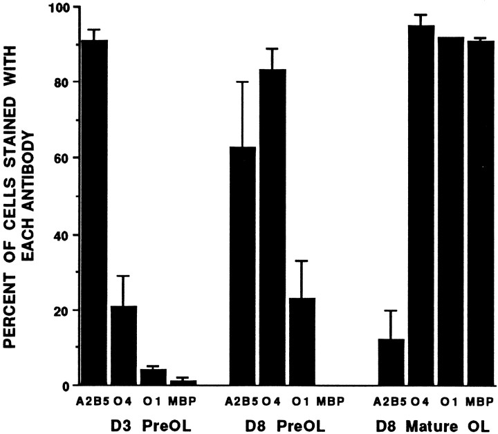
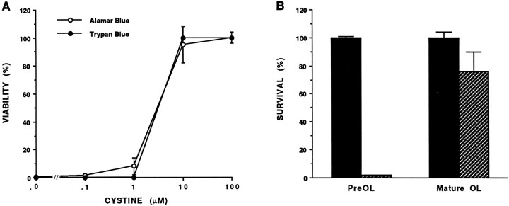
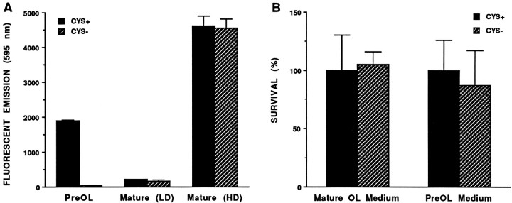
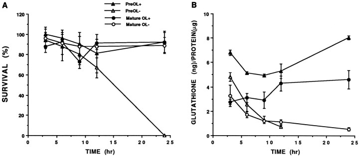
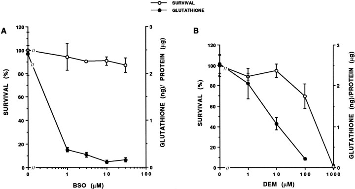
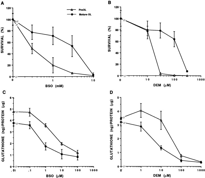
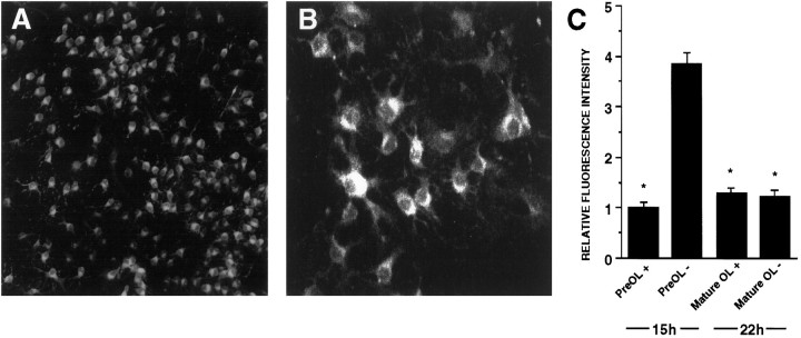
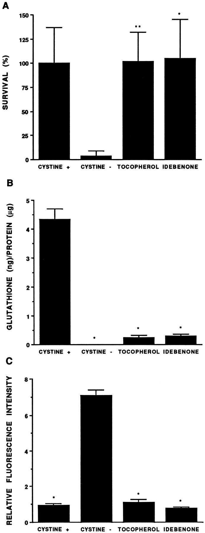
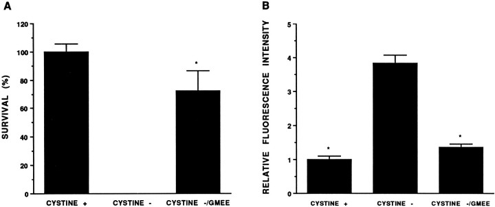
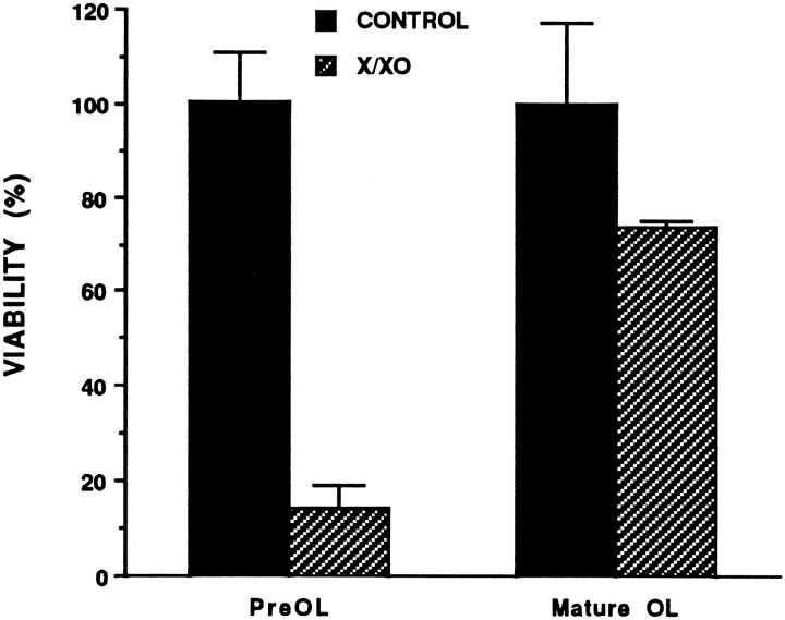
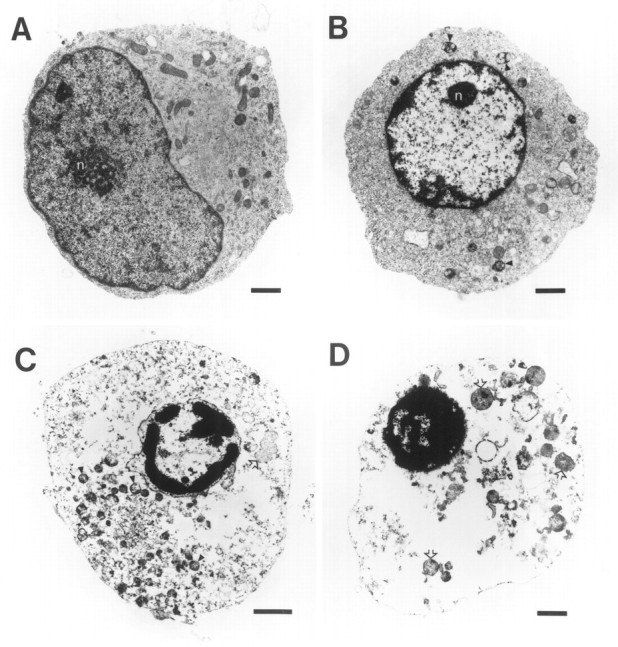
Death of oligodendrocyte (OL) precursors can be triggered in vitro by cystine deprivation, a form of oxidative stress that involves depletion of intracellular glutathione. We report here that OLs demonstrate maturation-dependent differences in survival when subjected to free radical-mediated injury induced by glutathione depletion. Using immunopanning to isolate rat preoligodendrocytes (preOLs), we generated highly enriched populations of preOLs and mature OLs under chemically defined conditions. Cystine deprivation caused a similar decrease in glutathione levels in OLs at both stages. However, preOLs were completely killed by cystine deprivation, whereas mature OLs remained viable. Although the glutathione-depleting agents buthionine sulfoximine and diethylmaleate were more potent in depleting glutathione in mature OLs, both agents were significantly more toxic to preOLs. Glutathione depletion markedly increased intracellular free radical generation in preOLs, but not in mature OLs, as indicated by oxidation of the redox-sensitive probe dihydrorhodamine 123. The antioxidants alpha-tocopherol, idebenone, and glutathione monoethylester prevented the oxidation of dihydrorhodamine in cystine-depleted preOLs and markedly protected against cell death. When the intracellular glutathione level was not manipulated, preOLs were also more vulnerable than mature OLs to exogenous free radical toxicity generated by a xanthine-xanthine oxidase system. Ultrastructural features of free radical-mediated injury in glutathione-depleted preOLs included nuclear condensation, margination of chromatin, and mitochondrial swelling. These observations indicate that preOLs are significantly more sensitive to the toxic effects of glutathione depletion and that oligodendroglial maturation is associated with decreased susceptibility to oxidative stress.


Figures

Fig. 1.
Fig. 1.
Quantitative immunocytochemical characterization of cultures generated when preOLs were maintained for 3 or 8 d in preOL medium or 8 d in mature OL medium. Immunocytochemical characterization of sister coverslips was done using the four monoclonal antibodies indicated. Values represent the mean ± SD from three separate experiments. Ten 20× fields (>1000 cells) were counted on one coverslip per condition in each experiment. Total cell number was determined by counting all cells labeled with Hoechst 33258 (see Materials and Methods).
Fig. 2.
Fig. 2.
A, AB is a direct measure of OL viability. Dose-response curves of preOL survival as a function of the concentration of cystine in the culture medium were compared using AB or cell counts with TB. In this experiment the EC50 for cystine was 3 μm, as determined by AB or TB.B, PreOLs are markedly more vulnerable to cystine deprivation than mature OLs. The survival of preOLs was 2 ± 0.3 versus 76 ± 14% for the mature OLs.
Fig. 3.
Fig. 3.
A, Cell density has no effect on the survival of mature OLs in cystine-depleted basal defined medium (CYS-). Survival of preOLs was also determined to confirm the relative toxicity of the cystine-depleted medium to the two OL stages. Mature OLs were generated at low density (LD) or high density (HD), as described in Materials and Methods. Values are expressed as the corrected fluorescent emission at 595 nm of the reaction product generated after a 2 hr incubation in AB. In this experiment, the survival of the preOLs was 2 ± 0.4%. The survival of the mature OLs at low density was 76 ± 14%, and survival was 99 ± 6% for the mature OLs at high density.B, Lack of effect of previous history of growth factor exposure on the survival of low-density mature OLs in cystine-depleted medium (CYS-). The survival of mature OLs generated by continuous exposure of preOLs to mature OL medium for 9 d (Mature OL Medium) did not differ from control. The survival of mature OLs, generated by a 7 d exposure of preOLs to mature OL medium, followed by a 2 d exposure to preOL medium (PreOL Medium), did not differ from control.
Fig. 4.
Fig. 4.
PreOLs are markedly more vulnerable to cystine deprivation than mature OLs, despite a similar time course for glutathione depletion. The time course of survival of preOLs and mature OLs in cystine-containing or cystine-depleted medium (A) was compared relative to the glutathione content of parallel cultures treated with the same conditions (B). After the indicated exposure times, viability was assayed with AB or glutathione was extracted and assayed.A, PreOLs were completely killed after a 24 hr exposure to cystine-depleted medium. There was no difference in viability at 24 hr for mature OLs in cystine-depleted medium relative to preOLs or mature OLs in cystine-containing medium. B, There was no difference in the decrease in glutathione content of preOLs and mature OLs after a 12 hr exposure to cystine-depleted medium. The glutathione content of the preOLs at 12 hr was 0.7 ± 0.1 ng/μg protein and did not differ significantly from that of mature OLs at 24 hr (0.5 ± 0.1 ng/μg protein). The glutathione content of preOLs at 24 hr is not shown, because it could not be determined due to complete cell death. Statistical comparisons were by factorial ANOVA.
Fig. 5.
Fig. 5.
Mature OLs are largely resistant to the toxicity of the glutathione-depleting agents BSO (A) or DEM (B). Dose-response curves compare the survival and glutathione content of mature OLs as a function of the concentration of BSO or DEM. Parallel cultures were exposed for 24 hr to BSO or DEM, after which they were assayed for viability with AB, or glutathione was extracted and assayed. A, There was no significant loss of viability at any concentration of BSO tested relative to control. The EC50 for the effect of BSO on the glutathione content of the mature OLs was 0.25 μm.B, The viability at 100 μm DEM was 70 ± 12% of control (NS), and the glutathione content was 0.2 ± 0.03 ng/μg protein. The glutathione content of the cells at 1 mm DEM could not be determined because of complete cell death. Statistical comparisons were by factorial ANOVA.
Fig. 6.
Fig. 6.
PreOLs are more vulnerable to the toxicity of BSO (A) or DEM (B), despite the fact that these agents more potently deplete glutathione in mature OLs (C, D). A, The EC50 for the toxicity of BSO to preOLs was 290 μm and to mature OLs was 3.3 mm.B, The EC50 for the toxicity of DEM to preOLs was 17 μm and to mature OLs was 103 μm. For the glutathione measurements, glutathione was extracted after an 8 hr exposure to BSO (C) or a 4 hr exposure to DEM (D), at which times there was no loss of cell viability, as described in Results. In this experiment, the EC50 for the effect of BSO on the glutathione content of the preOLs was 2.1 μm and on the mature OLs was 0.5 μm. The EC50 for the effect of DEM on the glutathione content of the preOLs was 33 μm and on the mature OLs was 5.2 μm. Similar results were obtained in one additional experiment.
Fig. 7.
Fig. 7.
Free radical generation, assessed by Rh 123 fluorescence, is greatly increased in cystine-deprived preOLs but not in mature OLs. A, Low-power fluorescent photomicrograph showing numerous Rh 123 fluorescent preOLs after a 15 hr exposure to cystine-depleted medium. B, Laser confocal digitalized image showing the cytoplasmic distribution of Rh 123 fluorescence in preOLs after a 15 hr exposure to cystine-depleted medium.C, Quantitation of the relative intensity of Rh 123 fluorescence (see Materials and Methods) in preOLs and mature OLs. There was a 3.9-fold increase in Rh 123 fluorescence in preOLs after 15 hr of cystine deprivation (PreOL-) compared with control (PreOL+). Statistical comparisons were among relative fluorescence intensity in control preOLs, control mature OLs, or treated mature OLs relative to treated preOLs. There was no significant increase in Rh 123 fluorescence in control versus treated mature OLs. *p < 0.001.
Fig. 8.
Fig. 8.
The free radical scavengers α-tocopherol and idebenone protect preOLs against cystine-deprivation (A) via a mechanism downstream of glutathione depletion (B) that suppresses free radical production (C). The relative effect of these free radical scavengers on the viability (A), intracellular glutathione content (B), and relative intensity of Rh 123 fluorescence (C) of preOLs in cystine-depleted medium is shown. Free radical scavengers were added to cystine-depleted medium, and after a 24 hr exposure, parallel cultures were either assayed for viability with AB (A) or glutathione was extracted and assayed (B). Rh 123 fluorescence was quantified after a 15 hr exposure to cystine-depleted medium (C) when preOLs in cystine-depleted medium remained viable. The α-tocopherol and idebenone were dissolved in DMSO, and an equivalent amount of DMSO (0.1% of final medium) was added to control cultures. A, The viability of the free radical scavenger-treated cultures differed significantly from cultures in cystine-depleted medium but did not differ significantly from control. *p < 0.05; **p < 0.05. B, Glutathione content was significantly decreased in cultures in cystine-depleted medium, with or without added free-radical scavengers, compared with control. *p< 0.001. C, The relative intensity of Rh 123 fluorescence of preOLs in cystine-depleted medium was increased significantly relative to control cultures or to those treated with either free radical scavenger. *p < 0.001. Statistical comparisons in A–C were by one-way ANOVA.
Fig. 9.
Fig. 9.
GMEE enhances the survival (A) of preOLs exposed to cystine-depleted medium (CYSTINE-) and suppresses free radical production (B) as measured by the relative intensity of Rh 123 fluorescence. Cells were exposed to 1 mm GMEE from the onset of the experiment. A, Survival in the presence of GMEE was 72 ± 14% relative to cultures in cystine-depleted medium. *p < 0.001. B, Treatment with GMEE suppressed Rh 123 fluorescence relative to preOLs exposed to cystine-depleted medium for 15 hr. Statistical comparisons were among relative fluorescence intensity in cystine-depleted medium versus cystine-containing medium (CYSTINE+) or cystine-depleted medium with GMEE. *p < 0.001. There was no significant difference between the GMEE-treated cultures and control (CYSTINE+). Statistical comparisons in Aand B were by one-way ANOVA.
Fig. 10.
Fig. 10.
PreOLs are more vulnerable than mature OLs to oxygen radicals generated by xanthine oxidase. OL viability was assessed 16 hr after exposure to xanthine (50 μm) and xanthine oxidase (5mU/ml), as described in Materials and Methods. The viability of preOLs was 14 ± 5%, whereas mature OLs had a significantly greater viability of 73 ± 1% (p < 0.001). There was no significant difference between mature OLs in the control or treated groups. The survival of the treated preOLs differed significantly from the control preOLs (p < 0.001). Statistical comparisons were by factorial ANOVA.
Fig. 11.
Fig. 11.
Ultrastructural analysis of the morphological changes observed in glutathione-depleted preOLs captured at various stages of degeneration (B–D) after a 14.5 hr exposure to cystine deprivation. A, Control preOL showing the eccentrically situated nucleus with a uniform distribution of chromatin adjacent to the nuclear membrane. The nucleolus (n) is localized at the center of the nucleus. Mitochondria are uniformly distributed and heterogeneous in shape, and the cristae are readily visualized. B, Note the margination and clumping of chromatin and the condensation of the nucleus and nucleolus (n). Mitochondria are uniformly spherical in shape, and the cristae are no longer visualized (arrowheads). C, In this very degenerated cell, the markedly electron-dense chromatin is condensed and marginated along the intact nuclear membrane, and several droplets of condensed chromatin are present within the nucleus. Most of the degenerated mitochondria (arrowheads) are localized to theleft of the nucleus, several swollen mitochondria are visible (open arrows), and the plasma membrane is intact. D, In this end-stage cell, the chromatin is extremely condensed within the markedly shrunken nucleus. Several extremely swollen mitochondria are visible (arrowheads). The plasma membrane is intact. Scale bars: A,B, D, 1 μm; C, 2 μm. .

Similar articles

Cited by

References

    1. Aizenman E. Modulation of N-methyl-d-aspartate receptors by hydroxyl radicals in rat cortical neurons in vitro. Neurosci Lett. 1995;189:57–59. - PubMed
    1. Anderson ME, Levy EJ, Meister A. Preparation and use of glutathione monoesters. In: Packer L, editor. Oxygen radicals in biological systems, Vol 234, Part D. Academic; San Diego: 1994. pp. 492–499. - PubMed
    1. Arends M, Morris R, Wyllie A. Apoptosis: the role of the endonuclease. Am J Pathol. 1990;136:593–608. - PMC - PubMed
    1. Back SA, Volpe JJ. Cellular and molecular pathogenesis of periventricular white matter injury. MRDD Research Reviews. 1997;3:96–107.
    1. Back SA, Yonezawa M, Gan X, Rosenberg PA, Volpe JJ. Oligodendrocyte death induced by cystine deprivation occurs by apoptosis. Soc Neurosci Abstr. 1995;21:41.

Publication types

MeSH terms

LinkOut - more resources