. 1998 Aug 15;18(16):6261-78.
doi: 10.1523/JNEUROSCI.18-16-06261.1998.

Potassium current development and its linkage to membrane expansion during growth of cultured embryonic mouse hippocampal neurons: sensitivity to inhibitors of phosphatidylinositol 3-kinase and other protein kinases

Affiliations

Potassium current development and its linkage to membrane expansion during growth of cultured embryonic mouse hippocampal neurons: sensitivity to inhibitors of phosphatidylinositol 3-kinase and other protein kinases

R L Wu et al. J Neurosci. .

Abstract

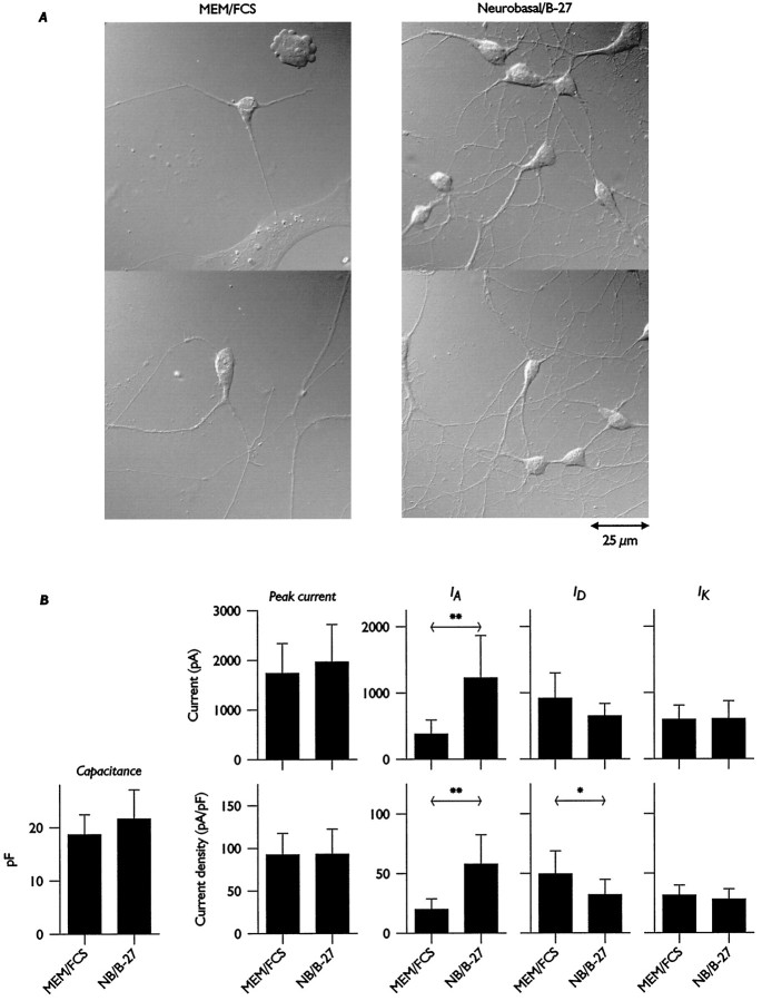
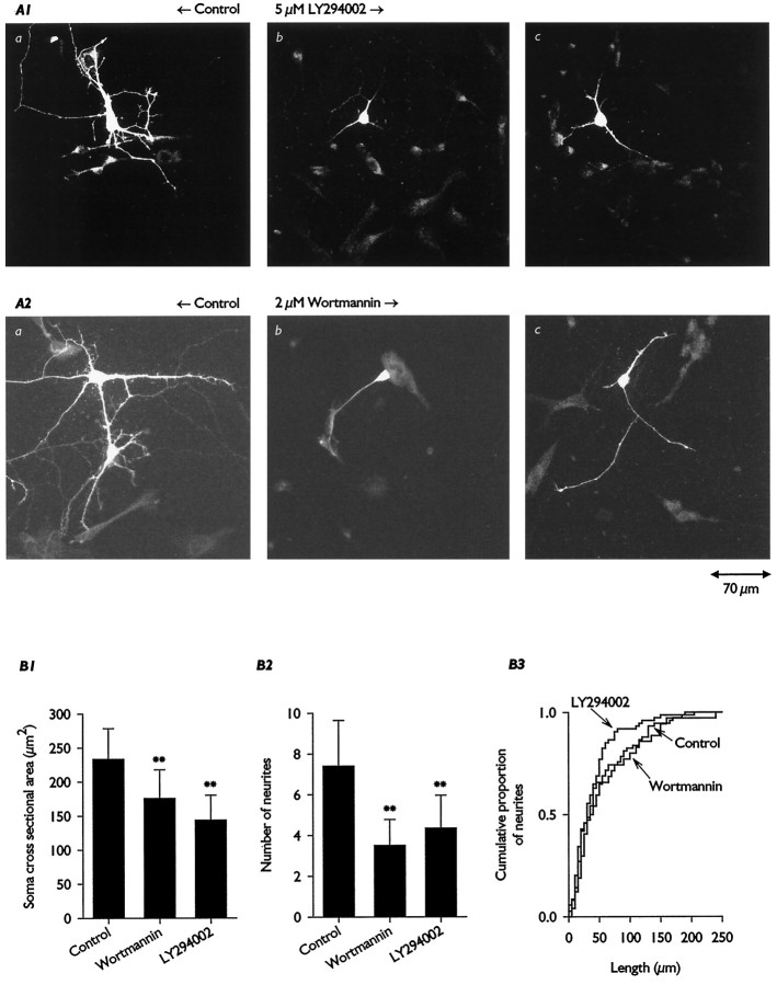
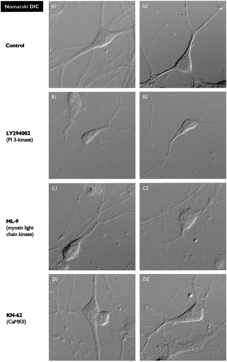
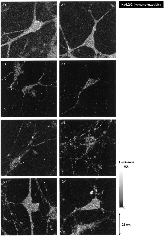
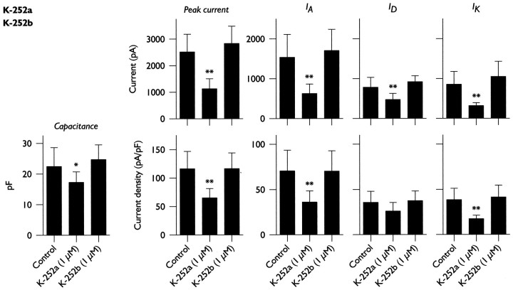
Hippocampal pyramidal neurons express three major voltage-dependent potassium currents, IA, ID, and IK. During hippocampal development, IA, the rapidly activating and inactivating transient potassium current, is detected soon after pyramidal neurons can be morphologically identified. Appearance of IA in developing pyramidal neurons is dependent on contact with cocultured astroglial cells; cultured pyramidal neurons not in contact with astroglial cells have reduced membrane area and IA (Wu and Barish, 1994). We have examined intracellular signaling pathways that could contribute to the regulation of IA development by probing developing pyramidal neurons with kinase inhibitors. We observed that exposure to LY294002 or wortmannin, inhibitors of phosphatidylinositol (PI) 3-kinase, reduced somatic cross-sectional area, neurite outgrowth, whole-cell capacitance, IA amplitude and density (amplitude normalized to membrane area), and immunoreactivity for Kv4.2 and/or Kv4.3 (potassium channel subunits likely to be present in the channels carrying IA). In contrast, exposure to ML-9 or KN-62, inhibitors of myosin light chain kinase or Ca2+-calmodulin-dependent protein kinase II (CaMKII), reduced membrane area and IA amplitude but did not affect IA density or Kv4. 2/3 immunoreactivity to the same extent as inhibitors of PI 3-kinase. Unexpectedly, exposure to bisindolymaleimide I or calphostin C, inhibitors of protein kinase C (PKC), did not affect membrane area or potassium current development. Our data suggest that PI 3-kinases regulate both A-type potassium channel synthesis and plasmalemmal insertion of vesicles bearing these potassium channels. CaMKII appears to regulate fusion of channel-bearing vesicles with the plasmalemma and myosin light chain kinase to regulate centripetal transport of channel-bearing vesicles from the Golgi. We further suggest that astroglial cells exert their influence on pyramidal neuron development through activation of PI 3-kinases.


Figures

Fig. 1.
Fig. 1.
A, Comparison of neuronal morphology after culture in serum-containing (MEM–FCS) and serum-free (Neurobasal–B-27) media. The images illustrate enhanced neurite outgrowth by neurons cultured in Neurobasal–B-27 medium. Neurons had been in culture for 5 d. B, Effects of the culture medium on potassium current development.IA was enhanced in neurons grown in Neurobasal–B-27 medium despite their lack of astroglial contact. Numbers of cells: 10 NB–B-27 and 8 MEM–FCS. In this and subsequent figures, * denotes p < 0.05; ** denotesp < 0.01; and *** denotes p < 0.001.
Fig. 2.
Fig. 2.
A, Morphology of neurons grown in the presence of PI 3-kinase inhibitors; exposure to LY294002 (A1) or wortmannin (A2) reduced soma size and neurite outgrowth. Neurons were labeled by placing a small drop of DiI dissolved in oil on a neuron soma, as described in Materials and Methods. Shown for each inhibitor are representative examples of a control image (note that two neurons are labeled in the wortmannin control) and two experimental images. B, Measurements of neurons imaged as above. Inhibition of PI 3-kinase reduced somatic cross-sectional area (B1) and numbers of neurites (B2). The lengths of shorter neurites (i.e., lengths <30–40 μm) were minimally effected, but exposure to LY294002 affected growth of longer neurites (B3, cumulative analysis). Numbers of cells analyzed: 10 control, 17LY294002, and 10 wortmannin.
Fig. 3.
Fig. 3.
Whole-cell potassium currents recorded at +40 mV from control and LY294002-treated neurons. The traces in the top row are currents recorded after conditioning prepulses to −120 and −40 mV (solid traces). Addition of 100 μm 4-AP to the bath reduced the current evoked after the prepulse to −120 mV (dotted trace). Thetraces in the bottom rows illustrate isolation of the individual currents, as described in Results. In this example, IA was almost completely eliminated in the cell grown for 4 d in the presence of LY294002 (5 μm). ID andIK were smaller in the LY294002-exposed neuron, as expected from the reduced membrane area of these cells.
Fig. 4.
Fig. 4.
Effects of growth in the presence of kinase inhibitors on whole-cell capacitance, determined as described in the Results. Shown are data for LY294002(A1), wortmannin (A2),ML-9 (B),KN-62 (C), andcalphostin C (D); putative targets of these inhibitors are indicated in parentheses. Membrane area was reduced by all inhibitors except calphostin C. Data presented in Figures 4-6 are drawn from the same population of neurons, and for all three figures numbers of cells are:wortmannin, 10 control, 11 experimental;LY294002, 3 control, 5 experimental;ML-9, 13 control, 10 experimental;KN-62, 8 control, 8 experimental; andcalphostin C, 14 control, 17 experimental.
Fig. 5.
Fig. 5.
Effects of growth in the presence of kinase inhibitors on amplitudes (as picoamperes measured at +40 mV) of voltage-gated potassium currents, determined as described in Results. Shown are data for LY294002(A1), wortmannin (A2),ML-9 (B),KN-62 (C), andcalphostin C (D); putative targets of these inhibitors are indicated in parentheses.IA,ID, and IKamplitudes were all reduced by all inhibitors except calphostin C; the largest and the most significant effects were seen for inhibitors of PI 3-kinases and for IA andIK (a clear trend was seen forID).
Fig. 6.
Fig. 6.
Effects of growth in the presence of kinase inhibitors on densities (as picoamperes per picofarad measured at +40 mV) of voltage-gated potassium currents, determined as described in Results. Shown are data for LY294002(A1), wortmannin (A2),ML-9 (B),KN-62 (C), andcalphostin C (D); putative targets of these inhibitors are indicated in parentheses.IA density was significantly reduced only by LY294002 and wortmannin; a trend that did not reach significance was evident for ML-9. In contrast, IA density was not altered by exposure to KN-62 or calphostin C, nor were densities of ID orIK affected by kinase inhibition.
Fig. 7.
Fig. 7.
Distributions of Kv4.2/3 immunoreactivity in control pyramidal neurons (A) and alterations induced by growth in the presence of the kinase inhibitors LY294002 (B), ML-9 (C), and KN-62 (D). In each case, Nomarski DIC and fluorescence images are shown for two representative neurons. Thearrows in the fluorescence images indicate areas enlarged in Figure 8. A, Control neurons demonstrating the disperse and granular character of the Kv4.2/3 immunoreactivity. Strong Kv4.2/3 immunoreactivity is found only in somata and dendrites; tubular axonal structures (A2, A4,arrowheads) are devoid of signal. B, In neurons exposed to LY294002, the density of Kv4.2/3-immunoreactive punctata was greatly reduced, but their distribution was not significantly affected. C, In neurons exposed to ML-9, the density of Kv4.2/3-immunoreactive punctata was reduced but not to the extent seen with LY294002. In somata, punctata were found in a perinuclear array surrounding a void volume occupied by the nucleus; this was much more evident in ML-9-treated neurons than in control neurons or neurons exposed to the other inhibitors. D, In neurons exposed to KN-62, the density of Kv4.2/3-immunoreactive punctata was only slightly reduced from the control, and, as in control neurons, punctata were distributed throughout the soma and major dendrites. (Figure and legend continue)The images in Figures 7 and 8 were all acquired from two sets of sister coverslips that were grown, processed for immunochemistry, and imaged in parallel. Numbers of neurons analyzed: 17 control, 10 LY294002, 24ML-9, and 10KN-62. In general, comparisons of Kv4.2/3 immunoreactivity were performed 2–4 times for each kinase inhibitor. The luminance scale applies to all fluorescence images. Control images, in which the primary antibody was omitted, showed only background luminance (as between punctata; images not shown).
Fig. 7.
Fig. 7.
Distributions of Kv4.2/3 immunoreactivity in control pyramidal neurons (A) and alterations induced by growth in the presence of the kinase inhibitors LY294002 (B), ML-9 (C), and KN-62 (D). In each case, Nomarski DIC and fluorescence images are shown for two representative neurons. Thearrows in the fluorescence images indicate areas enlarged in Figure 8. A, Control neurons demonstrating the disperse and granular character of the Kv4.2/3 immunoreactivity. Strong Kv4.2/3 immunoreactivity is found only in somata and dendrites; tubular axonal structures (A2, A4,arrowheads) are devoid of signal. B, In neurons exposed to LY294002, the density of Kv4.2/3-immunoreactive punctata was greatly reduced, but their distribution was not significantly affected. C, In neurons exposed to ML-9, the density of Kv4.2/3-immunoreactive punctata was reduced but not to the extent seen with LY294002. In somata, punctata were found in a perinuclear array surrounding a void volume occupied by the nucleus; this was much more evident in ML-9-treated neurons than in control neurons or neurons exposed to the other inhibitors. D, In neurons exposed to KN-62, the density of Kv4.2/3-immunoreactive punctata was only slightly reduced from the control, and, as in control neurons, punctata were distributed throughout the soma and major dendrites. (Figure and legend continue)The images in Figures 7 and 8 were all acquired from two sets of sister coverslips that were grown, processed for immunochemistry, and imaged in parallel. Numbers of neurons analyzed: 17 control, 10 LY294002, 24ML-9, and 10KN-62. In general, comparisons of Kv4.2/3 immunoreactivity were performed 2–4 times for each kinase inhibitor. The luminance scale applies to all fluorescence images. Control images, in which the primary antibody was omitted, showed only background luminance (as between punctata; images not shown).
Fig. 8.
Fig. 8.
Enlarged portions of fluorescence images near the regions indicated by the arrows in Figure 7, illustrating punctate areas of Kv4.2/3 immunoreactivity. Individual punctata had diameters of 0.5–0.75 μm, because each pixel represents ∼0.2 × 0.2 μm in the image plane. In neurons exposed toLY294002 (B),ML-9 (C), orKN-62 (D), the maximum luminance of individual punctata was comparable to that of control neurons (A), as indicated by the luminance scale; only punctata densities and distributions appeared to be affected by the kinase inhibitors.
Fig. 9.
Fig. 9.
Effects of growth in the presence of staurosporine on total cell capacitance and potassium current development when neurons were grown in normal (low serum) medium (A), or serum-free Neurobasal–B-27 medium (B). Comparable results were obtained under both conditions, a result indicating that neither serum nor astroglial contact were required for kinase-sensitive control ofIA development. A, Numbers of cells: 7 Control, 14 2 nm, and 7 20 nm.B, Numbers of cells: 4 Control, 42 nm, and 2 20 nm(neurons exposed to staurosporine in Neurobasal–B-27 medium were extremely fragile, and recordings sufficiently stable for acquisition of all data were difficult to maintain).
Fig. 10.
Fig. 10.
Effects of growth in the presence of the broad spectrum kinase inhibitor K-252a and its PKC-preferring variant K-252b on total cell capacitance and potassium current development. Exposure to K-252a affected membrane area, the amplitudes ofIA,ID, andIK, and densities ofIA and IK. In contrast, exposure to K252b did not affect membrane area or development of any of the potassium currents. Numbers of cells: 21control, 14 K252a, and 9K252b.

Similar articles

Cited by

References

    1. Audesirk G, Cablee L, Kern M. Modulation of neurite branching by protein phosphorylation in cultured rat hippocampal neurons. Dev Brain Res. 1997;102:247–260. - PubMed
    1. Bailey CH, Chen M. Time course of structural changes at identified sensory neuron synapses during long-term sensitization in Aplysia. J Neurosci. 1989;9:1774–1780. - PMC - PubMed
    1. Banker GA, Cowan WM. Rat hippocampal neurons in dispersed cell culture. Brain Res. 1977;126:397–425. - PubMed
    1. Banker GA, Cowan WM. Further observations on hippocampal neurons in dispersed cell culture. J Comp Neurol. 1979;187:469–494. - PubMed
    1. Barish ME. Modulation of the electrical differentiation of neurons by interactions with glia and other non-neuronal cells. Perspect Dev Neurobiol. 1995;2:357–370. - PubMed

Publication types