. 1998 Aug 15;18(16):6319-30.
doi: 10.1523/JNEUROSCI.18-16-06319.1998.

Localization of Ca2+ channel subtypes on rat spinal motor neurons, interneurons, and nerve terminals

Affiliations

Localization of Ca2+ channel subtypes on rat spinal motor neurons, interneurons, and nerve terminals

R E Westenbroek et al. J Neurosci. .

Abstract

Ca2+ channels in distinct subcellular compartments of neurons mediate voltage-dependent Ca2+ influx, which integrates synaptic responses, regulates gene expression, and initiates synaptic transmission. Antibodies that specifically recognize the alpha1 subunits of class A, B, C, D, and E Ca2+ channels have been used to investigate the localization of these voltage-gated ion channels on spinal motor neurons, interneurons, and nerve terminals of the adult rat. Class A P/Q-type Ca2+ channels were present mainly in a punctate pattern in nerve terminals located along the cell bodies and dendrites of motor neurons. Both smooth and punctate staining patterns were observed over the surface of the cell bodies and dendrites with antibodies to class B N-type Ca2+ channels, indicating the presence of these channels in the cell surface membrane and in nerve terminals. Class C and D L-type and class E R-type Ca2+ channels were distributed mainly over the cell soma and proximal dendrites. Class A P/Q-type Ca2+ channels were present predominantly in the presynaptic terminals of motor neurons at the neuromuscular junction. Occasional nerve terminals innervating skeletal muscles from the hindlimb were labeled with antibodies against class B N-type Ca2+ channels. Staining of the dorsal laminae of the rat spinal cord revealed a complementary distribution of class A and class B Ca2+ channels in nerve terminals in the deeper versus the superficial laminae. Many of the nerve terminals immunoreactive for class B N-type Ca2+ channels also contained substance P, an important neuropeptide in pain pathways, suggesting that N-type Ca2+ channels are predominant at synapses that carry nociceptive information into the spinal cord.


Figures
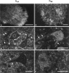
Fig. 1.
Fig. 1.
Distribution of class A and class B Ca2+ channels in the ventral horn.A–C, Tissue sections incubated with anti-CNA1 antibodies to the class A Ca2+ channels illustrating staining of terminals throughout the ventral horn and along the cell bodies and dendrites of motor neurons. Arrows point to dendritic regions. D–F, Tissue sections incubated with anti-CNB2 antibodies in the ventral horn of the spinal cord demonstrating both smooth and punctate staining along the cell soma and dendrites of motor neurons. Arrows point to regions of dendritic staining. Scale bars: A, 250 μm; B, C, E, F, 25 μm; D, 50 μm.
Fig. 2.
Fig. 2.
Localization of class C–ECa2+ channels in the ventral horn. A, B, Tissue sections labeled with anti-CNC1 antibodies illustrating the punctate pattern of staining along the cell surface and the proximal dendrites. C, D, Tissue sections labeled with anti-CND1 antibodies demonstrating the presence of class D channels mainly on the cell body and proximal dendrites.Arrows indicate regions of dendritic staining. E, F, Sections stained with anti-CNE2 antibodies showing their presence mainly along the cell bodies of spinal motor neurons.G, Control section incubated with normal rabbit serum to illustrate the lack of specific staining. Scale bars: A, D, E, 50 μm; B, F, G, 25 μm; C, 250 μm.
Fig. 3.
Fig. 3.
Ca2+ channels in nerve terminals. Diaphragm muscle stained with anti-CNA1 (A) or with anti-CNA5 (B). Tibialis anterior muscle stained with anti-CNA6 (C), anti-CNB2 (D), or anti-synaptotagmin (E) illustrating the presence of both class A and class B Ca2+ channels at the NMJ. Scale bar, 10 μm.
Fig. 4.
Fig. 4.
Localization of Ca2+ channels in the dorsal horn. A, B, Sections stained with anti-CNA1 illustrating the labeling of terminals in the superficial layers of the cord. Arrows point to the dorsal surface of the spinal cord. C, D, Tissue sections incubated with anti-CNB2 antibodies showing labeling of terminals and cell bodies in the superficial laminae of the spinal cord. E, F, Sections labeled with anti-CNC1 antibodies demonstrating punctate immunoreactivity associated with cell body in the dorsal horn. Scale bars: A, C, E, 100 μm; B, D, 10 μm;F, 50 μm.
Fig. 5.
Fig. 5.
Localization of class D and class E Ca2+ channels in the dorsal horn. A, Tissue section labeled with anti-CND1 antibodies illustrating localization in the cell bodies found throughout the dorsal horn.B, Tissue section stained with anti-CNE2 antibodies demonstrating the presence of class E channels along the cell bodies of neurons. Arrows point to the dorsal surface of the spinal cord. Scale bar, 100 μm.
Fig. 6.
Fig. 6.
Colocalization studies at the NMJ and in spinal cord. A, Muscle tissue section incubated with anti-CNA1 (green) and anti-synaptotagmin (red) antibodies illustrating the presence of class A channels at the NMJ. Regions of yellow represent colocalization. B, Muscle tissue section labeled with both anti-CNB2 (green) and anti-synaptotagmin (red) antibodies demonstrating the presence of class B Ca2+ channels at the NMJ. Regions ofyellow represent areas of colocalization.C, Tissue section from ventral horn double-labeled with anti-CNA1 (green) and anti-syntaxin (red) demonstrating colocalization (yellow) of these two proteins in terminals.D, Tissue sections from ventral horn were double-labeled with anti-CNB2 (green) and anti-syntaxin (red) to show colocalization in terminals (yellow). E, Section labeled with anti-CNA1 (green) and anti-CNB2 (red) antibodies illustrating the distribution of these terminals in the superficial layers of the dorsal horn. Areas ofyellow represent colocalization of these two Ca2+ channels in terminals. The topof the section is dorsal. Laminae 2 and 3 are illustrated. Scale bars:A, B, 10 μm; C–E, 25 μm.
Fig. 7.
Fig. 7.
Colocalization studies in the spinal cord.A, Dorsal horn of spinal cord labeled with anti-CNA1 (green) antibodies. B, Same section in A that was also labeled with anti-substance P (red) antibodies. C, Merged image ofA and B illustrating that in the dorsal horn colocalization between class A Ca2+ channels and substance P in terminals (yellow regions) is limited. D, Tissue section from the spinal cord labeled with anti-CNB2 (green). E, Same section as in C, double-labeled with anti-substance P (red) antibodies. F, Merged image ofC and D illustrating that some terminals that are labeled with class B Ca2+ channels are also labeled with substance P (yellow regions).G, Higher magnification of merged image shown inC illustrating distribution of class A channels in the superficial portions of the dorsal horn (red), the distribution of substance P in this area (green), and regions of colocalization (yellow) of these two antibodies. H, Higher magnification of the image inF showing double-labeling with anti-CNB2 antibodies (green), anti-substance P antibodies (red), and terminals containing both proteins (yellow). Scale bars: A–C, 50 μm; D–F, 50 μm; G, H, 10 μm.

Similar articles

Cited by

References

    1. Appel SH, Stefani E. Amyotrophic lateral sclerosis: etiology and pathogenesis. In: Appel SH, editor. Current neurology, Vol 11. Mosby; Chicago: 1991. pp. 287–310.
    1. Appel SH, Engelhardt JI, Barcia J, Stefani E. Immunoglobulins from animal models of motor neuron disease and from human amyotrophic lateral sclerosis patients passively transfer physiological abnormalities to the neuromuscular junction. Proc Natl Acad Sci USA. 1991;88:647–651. - PMC - PubMed
    1. Appel SH, Smith RG, Engelhardt JI, Stefani E. Evidence for autoimmunity in amyotrophic lateral sclerosis. J Neurol Sci. 1993;118:169–174. - PubMed
    1. Bading H, Ginty DD, Greenberg ME. Regulation of gene expression in hippocampal neurons by distinct Ca2+ signaling pathways. Science. 1993;260:181–186. - PubMed
    1. Basbaum AI, Fields HL. Endogenous pain control system: brainstem spinal pathways and endorphin circuitry. Annu Rev Neurosci. 1984;7:309–338. - PubMed

Publication types

Substances

LinkOut - more resources