. 1998 Aug 15;18(16):6370-7.
doi: 10.1523/JNEUROSCI.18-16-06370.1998.

Cyclin-dependent kinase 5-deficient mice demonstrate novel developmental arrest in cerebral cortex

Affiliations

Cyclin-dependent kinase 5-deficient mice demonstrate novel developmental arrest in cerebral cortex

E C Gilmore et al. J Neurosci. .

Abstract

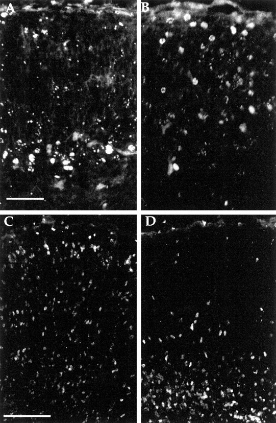
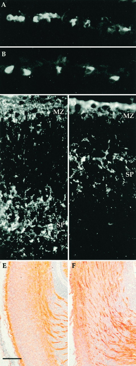
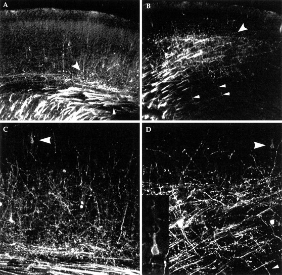
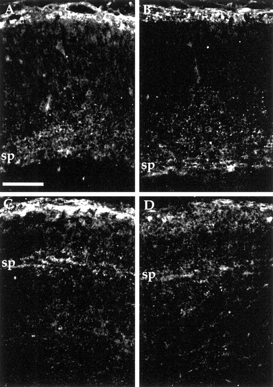
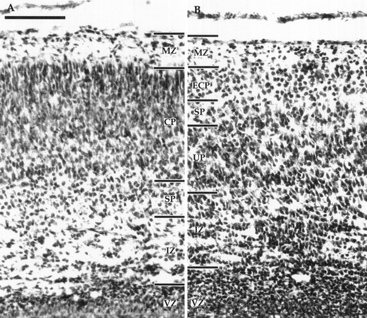
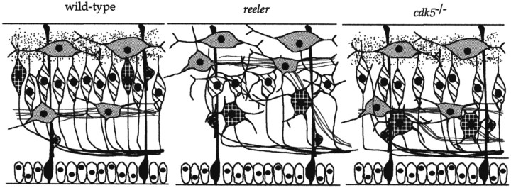
The cerebral cortex of mice with a targeted disruption in the gene for cyclin-dependent kinase 5 (cdk5) is abnormal in its structure. Bromodeoxyuridine labeling reveals that the normal inside-out neurogenic gradient is inverted in the mutants; earlier born neurons are most often found superficial to those born later. Despite this, the early preplate layer separates correctly and neurons with a normal, pyramidal morphology can be found between true marginal zone and subplate. Consistent with their identity as layer VI corticothalamic neurons, they can be labeled by DiI injections into thalamus. The DiI injections also reveal that the trajectories of the cdk5(-/-) thalamocortical axons are oblique and cut across the entire cortical plate, instead of being oriented tangentially in the subcortical white matter. We propose a model in which the cdk5(-/-) defect blocks cortical development at a heretofore undescribed intermediate stage, after the splitting of the preplate, but before the migration of the full complement of cortical neurons.


Figures

Fig. 1.
Fig. 1.
Coronal sections of cerebral cortex from E18.5 animals injected with BrdU at various gestational ages and stained with anti-BrdU antibody. A, B, Embryos from dams injected with BrdU at E12.5. Wild-type (A) cortex demonstrates expected pattern of labeling with BrdU-positive cells found in deeper portions of cerebral cortex. Thecdk5−/− (B) animals show BrdU-positive cells with a wider distribution including cells in positions near the pial surface. C, D, Embryos from dams injected with BrdU at E15.5. Cells in the wild type (C) were consistently found near the pial surface (and at lower positions, still in the process of migration). By contrast, in the cdk5−/− brain (D) very few cells were found near the pial surface. Indeed most appeared stalled in a layer below subplate. Scale bars: A, B, 50 μm; C, D, 100 μm.
Fig. 2.
Fig. 2.
Anti-reelin antibody (G10) stains Cajal-Retzius cells in both wild-type (A) andcdk5−/− (B) E18.5 cerebral cortices. Anti-chondroitin sulfate antibody stains preplate derivatives in the marginal zone (MZ) and subplate (SP). Subplate is seen separated from marginal zone in both wild-type (C) andcdk5−/− (D) animals at E15.5. Note, however, the difference in the absolute distance of subplate separation. For full discussion see Results and Table 1. E, F, L1 staining of axons in wild-type (E) andcdk5−/− (F) embryonic cerebral cortex. Coronal sections of E18.5 embryos were stained with L1CD, an antibody to the cytoplasmic domain of L1 (a generous gift of Vance P. Lemon). Dorsal is toward thetop; the pial surface is to the left; the internal capsule can be seen in the bottom right. The wild-type section (E) shows the expected pattern of stained fascicles deep within cerebral cortex. In thecdk5−/− mutant (F) some fibers run deep, but many are found coursing obliquely through the cerebral cortex toward the pial surface. L1-positive bundles run parallel and near but not adjacent to the pial surface. Scale bars: E, F, 100 μm.
Fig. 3.
Fig. 3.
Thalamic injections of DiI reveal corticothalamic and thalamocortical axons in E18.5 wild-type (A, C) andcdk5−/− mutant (B, D) cerebral cortex. In A and B, the heavily labeled internal capsule is seen in the bottom left corner. The small arrowhead in Aindicates the location of the tract of cortical efferents (running in the presumptive subcortical white matter). In mutant (B), note the sparse horizontal stained fibers, near arrowheads, which are the corticothalamic tract. The large arrowheads in A andB mark the plexus of afferent fibers (the endings of the thalamocortical tract axons). In both genotypes, this plexus is found within the subplate. Above this, small fibers ascend into the developing cortical plate in both wild type and mutant. In wild type, these fibers can often be traced to a cell body of origin, superficial to the subplate, with a clear pyramidal morphology. In thecdk5−/− cerebral cortex (B), a number of thin fibers (B, D, small arrowheads), nearly perpendicular to the larger mass of fibers, can be traced to cell bodies deep to subplate, indicating they too are cortical efferents (connections not seen because of limitations in plane of focus). In the cdk5−/−mutant no labeled cell bodies located above the subplate had an abnormal morphology, whereas no cells in or below the subplate had the normal pyramidal morphology. Thalamic DiI injections also backfill neuronal cell bodies in or below the afferent plexus. Many of these are likely to be subplate cells but may also include aberrantly located and shaped layer VI neurons. The inset in Dshows a higher magnification of the DiI-filled cell, indicated by thelarge arrowhead.
Fig. 4.
Fig. 4.
Anti-calretinin immunostaining of wild-type andCdk5−/− cerebral cortex at E18.5. Coronal sections of medial (A, C) and lateral (B, D) cerebral cortex indicate location of subplate cells and fibers. Subplate (sp) is located deep within cerebral cortex of wild-type (A, B) but near the pial surface ofCdk5−/− mice (C, D). Because A and B and C andD are from the same coronal section, direct comparisons can be made concerning their degree of development. Separation of subplate away from pial surface was used as a marker for development of the cortex and quantified in Table 1. Scale bar, 100 μm.
Fig. 5.
Fig. 5.
Comparison of the cytoarchitectonics of wild-type (A) and mutant (B) E18.5 cerebral cortex. In the wild type, the marginal zone (MZ) is adjacent to the pial surface. The layer of differentiating cells marking the cortical plate (CP) is easily distinguished from the cells of the subplate (SP), which in turn are found above the relatively cell-sparse intermediate zone (IZ). The ventricular zone (VZ) is still present at this age. In thecdk5−/− mutant (B) the MZ is present as is a thin layer with some cells. Deep to this lie the layer of neurons of the early cortical plate (ECP). Below these cells is a layer resembling subplate, and deep to these cells is a thick tier of cells, the underplate (UP), that contains the later-generated cortical plate neurons. The IZ is present in its normal position but appears to have a higher density of cells, as if the backup in the underplate has extended to this depth. Scale bar, 100 μm.
Fig. 6.
Fig. 6.
Models of the proposed phenotypic differences of the early cortical plate stage in wild-type, reeler, andcdk5−/− mouse cerebral cortices. Phenotypic differences between wild-type and reelerdeveloping cortical plate have been reviewed elsewhere (Caviness and Rakic, 1978; Goffinet, 1984; Gilmore and Herrup, 1997). Ventricular zone cells (open ovals) lie at the bottom of the cerebral wall with radial glia (darkly stippled) running the length of the wall. Cells derived from the preplate, Cajal–Retzius and subplate cells, are lightly stippled, whereas reelin protein is represented by the speckles around the Cajal–Retzius cells (absent in reeler). Cells of the early cortical plate are cross-hatched, whereas later born cells are crisscrossed. Cortical efferents are represented by dark lines, whereas cortical afferents are light gray. Shape of cortical plate neurons is a representation of final phenotype rather than morphology at the stage depicted. See Discussion for details.

References

    1. Allendoerfer KL, Shatz CJ. The subplate, a transient neocortical structure: its role in the development of connections between thalamus and cortex. Annu Rev Neurosci. 1994;17:185–218. - PubMed
    1. Angevine JB, Sidman RL. Autoradiographic study of cell migration during histogenesis of cerebral cortex in mice. Nature. 1961;192:766–768. - PubMed
    1. Bicknese AR, Sheppard AM, O’Leary DDM, Pearlman AL. Thalamocortical axons extend along a chondroitin sulfate proteoglycan-enriched pathway coincident with the neocortical subplate and distinct from the efferent path. J Neurosci. 1994;14:3500–3510. - PMC - PubMed
    1. Boulder-Committee. Embryonic vertebrate CNS: revised terminology. Anat Rec. 1970;166:257–261. - PubMed
    1. Brittis P, Meiri K, Dent E, Silver J. The earliest patterns of neuronal differentiation and migration in the mammalian central nervous system. Exp Neurol. 1995;134:1–12. - PubMed

Publication types

MeSH terms