. 1998 Aug 15;18(16):6378-87.
doi: 10.1523/JNEUROSCI.18-16-06378.1998.

Differential response of cortical plate and ventricular zone cells to GABA as a migration stimulus

Affiliations

Differential response of cortical plate and ventricular zone cells to GABA as a migration stimulus

T N Behar et al. J Neurosci. .

Abstract

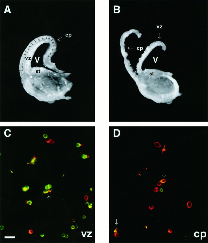
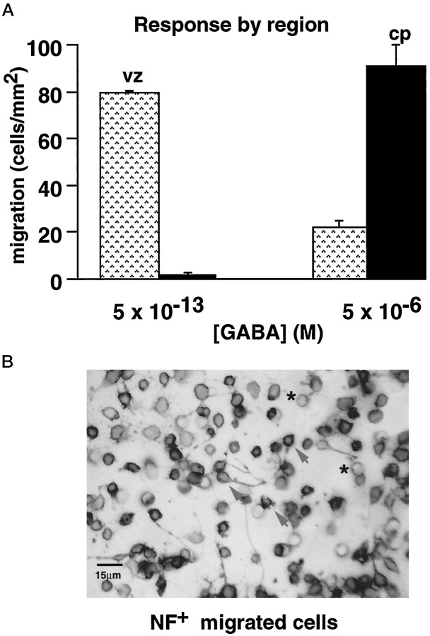
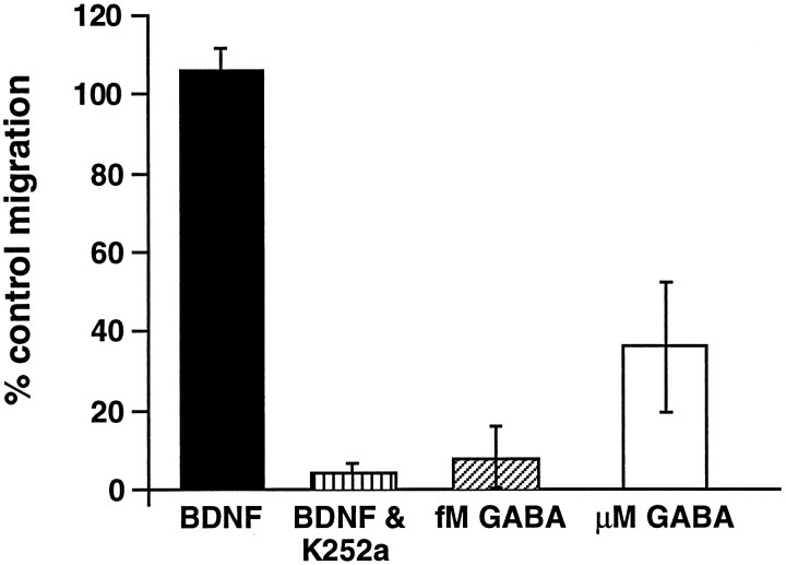
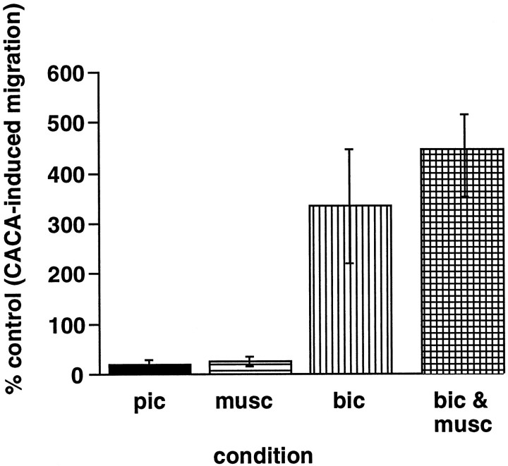
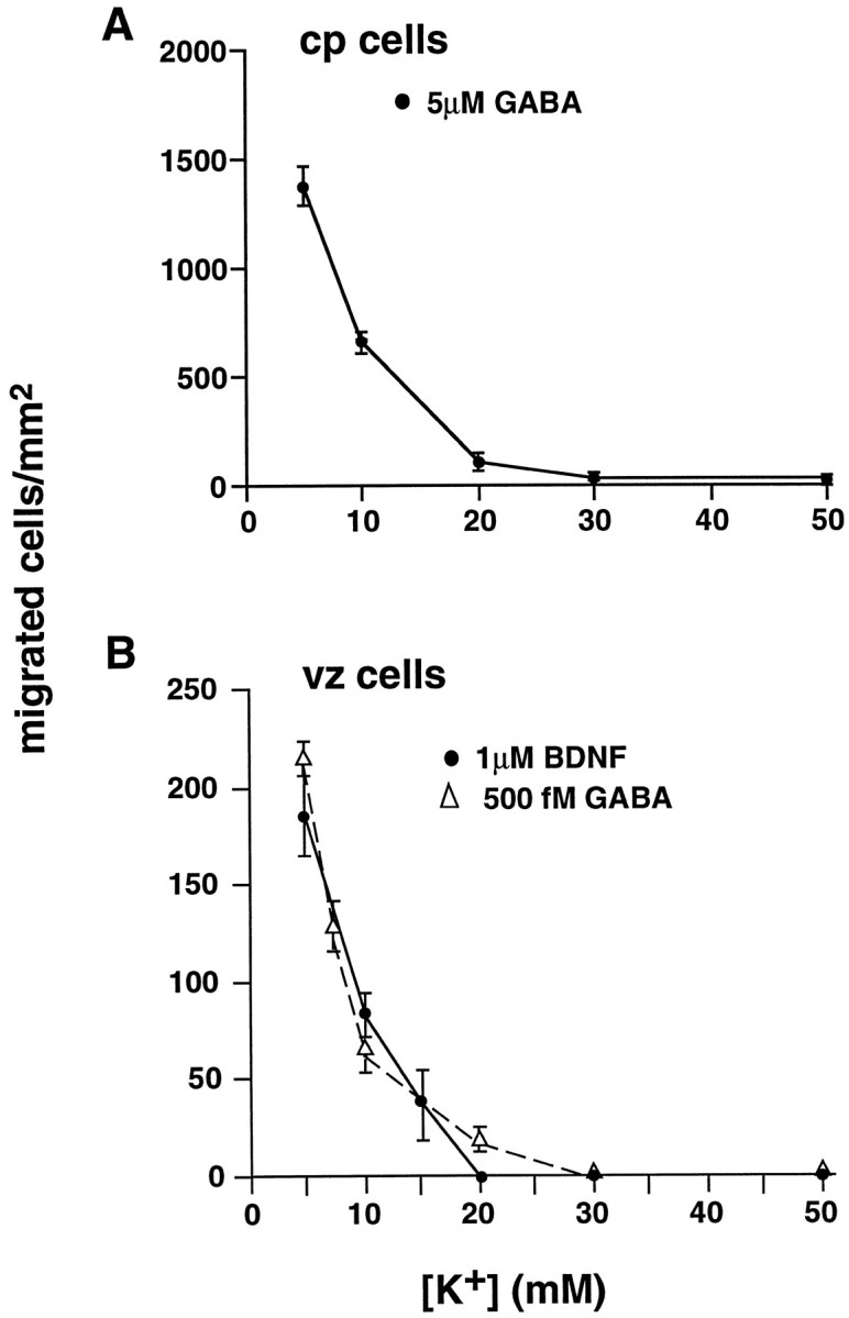
A microdissection technique was used to separate differentiated cortical plate (cp) cells from immature ventricular zone cells (vz) in the rat embryonic cortex. The cp population contained >85% neurons (TUJ1(+)), whereas the vz population contained approximately 60% precursors (nestin+ only). The chemotropic response of each population was analyzed in vitro, using an established microchemotaxis assay. Micromolar GABA (1-5 microM) stimulated the motility of cp neurons expressing glutamic acid decarboxylase (GAD), the rate-limiting enzyme in GABA synthesis. In contrast, femtomolar GABA (500 fM) directed a subset of GAD- vz neurons to migrate. Thus, the two GABA concentrations evoked the motility of phenotypically distinct populations derived from different anatomical regions. Pertussis toxin (PTX) blocked GABA-induced migration, indicating that chemotropic signals involve G-protein activation. Depolarization by micromolar muscimol, elevated [K+]o, or micromolar glutamate arrested migration to GABA or GABA mimetics, indicating that migration is inhibited in the presence of excitatory stimuli. These results suggest that GABA, a single ligand, can promote motility via G-protein activation and arrest attractant-induced migration via GABAA receptor-mediated depolarization.


Figures

Fig. 1.
Fig. 1.
Chemotropically active concentration ranges of GABA form bell-shaped dose–response curves. Shown is GABA-induced migration of E18 cells dissociated from the whole cortex. At E18 the dose–response curve of cells isolated from the whole cortex is bimodal. Femtomolar GABA and micromolar GABA induce similar numbers of cells to migrate. Error bars indicate SEM.
Fig. 2.
Fig. 2.
The cortical plate can be microdissected from the ventricular zone in coronal sections of the rat cortex.A, A 350 μm cross section of one hemisphere from an E18 brain. The intermediate zone (iz,arrows) lies between the cortical plate (cp) and the ventricular zone (vz).B, The cross section has been dissected along theiz, resulting in the separation of the cpfrom the vz. Cells in preparations from both regions contain iz cells. C, D, Photomicrographs of cells from each dissociate are double-labeled for nestin (green) and TUJ1 (red).C, The majority of cells in the vz preparation labels with anti-nestin antibody. Other TUJ1+ cells are scattered throughout the field. D, Most cells derived from the cp label with TUJ1, indicating that they are neurons. A minority of cells express nestin only. Arrows inC and D denote newly generated neurons that appear yellow/orange, labeling for both TUJ1 and nestin. st, Striatum; V, ventricle. Scale bar in C, 20 μm.
Fig. 3.
Fig. 3.
Vz and cp cells migrate to distinct concentration ranges of GABA. A, The vz dissociate (░) responds to the femtomolar range of GABA, whereas the majority of cells in the cp dissociate (▪) migrates in response to higher, micromolar levels of GABA. Error bars indicate SEM. B, Photomicrograph of E18 vz cells that were immunoperoxidase-labeled for neurofilament protein after migration to femtomolar GABA. After migration in the chemotaxis assay, the cells were stained with cresyl violet and then immunolabeled for neurofilament protein, using peroxidase-labeled second antibodies, as described in Materials and Methods. All vz cells that migrate to femtomolar GABA in vitro express neurofilament protein (arrows), indicating that they are neurons.Asterisks denote 8 μm pores in the membrane. Scale bar, 15 μm.
Fig. 4.
Fig. 4.
GAD expression in vz and cp cells. A, B, Photomicrographs of the vz and cp starting populations immunostained for GAD67. A, Although many cells in the vz dissociate express GAD, unlabeled vz cells are abundant (arrows). B, In contrast, nearly all of the cells in the cp dissociate express GAD protein. Anarrow highlights an unlabeled cp cell. C, Histogram depicting the percentage of GAD+ cells in the starting (▪) versus migrated (■) populations. Nearly 55% of the starting vz dissociate expresses GAD; however, the majority of vz cells that migrate to femtomolar GABA (∼82%) is GAD. Most cp cells in the starting population are GAD+ (>90%), and all cp cells that migrate to micromolar GABA express GAD. Error bars indicate SEM. D, Photomicrograph of migrated vz cells immunostained for GAD67. After migration to femtomolar GABA, vz cells were stained with cresyl violet and then immunolabeled for GAD67 protein, using peroxidase-labeled secondary antibodies. A few migrated vz cells express GAD protein (arrows); most migrated vz cells are unlabeled.Asterisks denote 8 μm pores in the membrane. Scale bars in B (also applies to A),D: 20 μm.
Fig. 5.
Fig. 5.
Pertussis toxin treatment selectively blocksin vitro migration to GABA. Treatment with PTX inhibits vz cell migration to GABA ∼90% as compared with control levels. Cp cell responses to micromolar GABA also are inhibited (>50%) in the presence of PTX. In contrast, BDNF-induced motility of vz cells is not blocked by PTX treatment; however, K252a, an alkaloid kinase inhibitor, significantly attenuates vz cell migration to BDNF. Data represent the percentage of the control level of migration averaged from three separate trials. Error bars indicate SEM.
Fig. 6.
Fig. 6.
GABAA receptor activation arrestsin vitro migration. Migration induced by 1 μm CACA is inhibited by the activation of GABAA receptors with a 10 μm concentration of the agonist muscimol (▤). Direct inhibition of GABAAreceptors with a 10 μm concentration of the antagonist bicuculline (▥) results in a potentiation of migration to CACA. In the presence of a 10 μm concentration of both muscimol and bicuculline (▩), levels of CACA-induced migration are comparable to migration in the presence of bicuculline only. CACA-induced migration is inhibited in the presence of picrotoxin (▪), which blocks GABAC receptors. Data represent the percentage of the control level of migration averaged from multiple trials (muscimol,n = 5; bicuculline, n = 5; picrotoxin, n = 5; muscimol plus bicuculline,n = 3). Error bars indicate SEM.
Fig. 7.
Fig. 7.
GABA-induced motility is arrested by increased [K+]o. A, Increasing concentrations of [K+]o attenuate cp cell migration to micromolar GABA. At 10 mm[K+]o, motility to GABA is inhibited 50%. At 20 mm[K+]o, migration to GABA is blocked completely. B, Femtomolar GABA-induced chemotaxis (▵) and BDNF-induced chemotaxis (•) of vz cells are blocked by elevated [K+]o. An increase to 10 mm [K+]o results in significant inhibition of motility. Higher levels completely block migration. Error bars indicate SEM.

References

    1. Amatruda TT, Gerard NP, Gerard C, Simon MI. Specific interactions of chemoattractant factor receptors with G-proteins. J Biol Chem. 1993;268:10139–10144. - PubMed
    1. Armstrong RC, Harvath L, Dubois-Dalcq M. Type 1 astrocytes and oligodendrocyte-type 2 astrocyte glial progenitors migrate toward distinct molecules. J Neurosci Res. 1990;27:400–407. - PubMed
    1. Bayer SA. Development of the lateral and medial limbic cortices in the rat in relation to cortical phylogeny. Exp Neurol. 1990;107:118–131. - PubMed
    1. Behar TN, Schaffner AE, Colton CA, Somogyi R, Olah Z, Lehel C, Barker JL. GABA-induced chemokinesis and NGF-induced chemotaxis of embryonic spinal cord neurons. J Neurosci. 1994;14:29–38. - PMC - PubMed
    1. Behar TN, Schaffner A, Tran H, Barker J. GABA-induced motility of spinal neuroblasts develops along a ventrodorsal gradient and can be mimicked by agonists of GABAA and GABAB receptors. J Neurosci Res. 1995;42:97–108. - PubMed

MeSH terms

LinkOut - more resources