. 1998 Aug 15;18(16):6558-67.
doi: 10.1523/JNEUROSCI.18-16-06558.1998.

NMDA EPSCs at glutamatergic synapses in the spinal cord dorsal horn of the postnatal rat

Affiliations

NMDA EPSCs at glutamatergic synapses in the spinal cord dorsal horn of the postnatal rat

R Bardoni et al. J Neurosci. .

Abstract

In rat dorsal horn, little is known about the properties of synaptic NMDA receptors during the first two postnatal weeks, a period of intense synaptogenesis. Using transverse spinal cord slices from postnatal day 0-15 rats, we show that 20% of glutamatergic synapses tested at low-stimulation intensity in spinal cord laminae I and II were mediated exclusively by NMDA receptors. Essentially all of the remaining glutamatergic EPSCs studied were attributable to the activation of both NMDA and AMPA receptors. Synaptic NMDA receptors at pure and mixed synapses showed similar sensitivity to membrane potential, independent of age, indicating similar Mg2+ sensitivity. Kinetic properties of NMDA EPSCs from pure and mixed synapses were measured at +50 mV. The 10-90% rise times of the pure NMDA EPSCs were slower (16 vs 10 msec), and the decay tau values were faster (tau1, 24 vs 42 msec; tau2, 267 vs 357 msec) than NMDA EPSCs at mixed synapses. Our results indicate that NMDA receptors are expressed at glutamatergic synapses at a high frequency, either alone or together with AMPA receptors, consistent with the prominent role of NMDA receptors in central sensitization (McMahon et al., 1993).


Figures
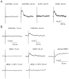
Fig. 1.
Fig. 1.
Some glutamatergic synapses evoked in lamina II neurons are mediated by NMDA receptors only. A, EPSCs were recorded from a P10 lamina II neuron in 10 μmbicuculline and 5 μm strychnine at different holding potentials to test for the presence of pure NMDA EPSCs. Stimulation of the neuron held at −70 mV produced no detectable AMPA EPSC. EPSCs recorded at +50 mV were completely blocked by 50 μmd-APV. After washout of d-APV for 5 min, the NMDA EPSCs tested at +50 mV recovered. All currents are averages of three traces. B, EPSCs were recorded from a P3 lamina II neuron in 10 μm bicuculline and 5 μmstrychnine. Application of 10 μm NBQX did not affect the EPSC recorded at +50 mV. When the rising phase of the EPSCs recorded in control (solid line) and in NBQX (dashed line) are superimposed, no effect of NBQX is detectable. Addingd-APV to the NBQX containing extracellular solution blocks the EPSC. All currents are averages of five traces.
Fig. 2.
Fig. 2.
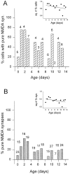
Analysis of the proportion of pure NMDA synapses during the first 2 weeks of postnatal development. A,Bars at each postnatal day represent the percentage of lamina II neurons in which pure NMDA EPSCs were observed over the total number of tested cells. The number over each bar is the number of cells tested at that age. Inset, Linear regression fit to the square root of the percentage of cells at each age (r = 0.33; p > 0.05).B, Bars represent the percentage of pure NMDA synapses over the total number of glutamatergic (i.e., NMDA plus AMPA) EPSCs for each age evoked by focally stimulating lamina II neurons. The number over each bar is the number of synapses tested at that age. Inset, Regression fit to the square root of the percentage of synapses at each age (r = 0.39; p > 0.05).
Fig. 3.
Fig. 3.
Kinetic analysis of pure NMDA EPSCs.A, EPSCs were recorded from a P1 lamina II neuron held at +50 mV, averaged, and fit with a double-exponential function. The current is the average of five traces. The double-exponential curve is superimposed on the EPSC decay phase (solid line), whereas the two components of the double-exponential function are represented by dashed lines (τ1, 21.4; τ2, 209 msec). B, EPSCs were recorded from a P10 lamina II neuron held at +50 mV, averaged, and fit with a double-exponential function (τ1, 25.3; τ2, 292 msec). The current is the average of five traces.
Fig. 4.
Fig. 4.
Pure NMDA EPSCs are strongly voltage-dependent.A, I–V relationships obtained by plotting the peak amplitude of averaged pure NMDA EPSCs recorded in bicuculline and strychnine from lamina II neurons as a function of holding potential at different ages (P1, P4, and P13). Each point was determined by averaging five EPSC traces. B, Voltage dependence of NMDA receptors at pure NMDA synapses was assessed by calculating the ratio of peak synaptic current amplitudes recorded at −70 mV and +50 mV in bicuculline and strychnine from lamina II neurons. Average ratio values and SDs are plotted in 4 d bins and compared as a function of age. One-way ANOVA revealed no relationship between voltage dependence and age (F = 0.91;p = 0.457).
Fig. 5.
Fig. 5.
NMDA EPSCs isolated from mixed glutamatergic synapses in laminae I and II over the first 2 postnatal weeks.A, EPSCs were recorded from a P4 lamina II neuron held at −70 mV and assessed for sensitivity to different antagonists. Glutamatergic EPSCs evoked in bicuculline and strychnine are mediated by AMPA receptors, blocked by 10 μm CNQX and NMDA receptors, blocked by 50 μmd-APV. The antagonist effects were reversible after a 5 min wash.B, EPSCs were recorded from the same neuron under the same conditions as in A, except the membrane potential was held at +50 mV. All currents are the average of five traces.
Fig. 6.
Fig. 6.
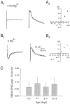
NMDA receptors at mixed glutamatergic synapses on lamina I and II neurons are strongly blocked by Mg2+during the first 2 postnatal weeks. A, Voltage dependence of the NMDA EPSC was determined for a P3 lamina II neuron in the presence of 1 mm Mg2+. A1, NMDA EPSCs recorded at −70 and +50 mV. Almost no synaptic current was apparent in 10 μm CNQX when membrane potential was held at −70 mV (average of 5 traces), whereas under the same conditions, an EPSC was evoked at +50 mV.A2, I–V curve representing the peak amplitudes of the average of five consecutive traces at different membrane potentials. B, Voltage dependence of the NMDA EPSCs for the same lamina II neuron was determined in a bath with no added Mg2+. B1, In low Mg2+, EPSCs evoked at −70 mV in CNQX were readily apparent. EPSCs are averages of five traces. B2,I–V curve was obtained by averaging five consecutive traces recorded at different potentials in 0 Mg2+.C, Mg2+ sensitivity of NMDA receptors at mixed synapses in lamina I and II neurons was assessed by calculating the ratio of NMDA current peak amplitudes recorded at −70 and +50. Ratio values are binned over 4 d and plotted as function of age. Error bars represent SD. One-way ANOVA revealed no dependence of Mg2+ sensitivity on age (F = 0.8; p = 0.457).
Fig. 7.
Fig. 7.
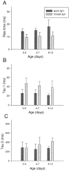
Kinetic analysis of NMDA EPSCs at mixed synapses and comparison with pure NMDA EPSCs. A, Ten to 90% rise time values determined from NMDA EPSCs at mixed and pure NMDA synapses are binned and plotted as a function of age. The first two bins are 4 d, and the third bin includes data from 5 d. Two-way ANOVA indicates no dependence of rise time on age for either type of synapse (F = 1.43; p = 0.254) and a highly significant difference in rise time of pure and mixed NMDA synapses (F = 27.57; p≪ 0.01). B, The decay phase of NMDA EPSCs at mixed and pure synapses was fit with a double-exponential function. τ1 values are plotted in bins as a function of age. The decay τ1 values of the two types of NMDA EPSCs are significantly different (two-way ANOVA, F = 25.44; p ≪ 0.01), although neither changes significantly as a function of age (F = 1.032; p = 0.37).C, τ2 values determined from NMDA EPSCs at mixed and pure synapses are binned and plotted against age. τ2 values for mixed and pure synapses are not significantly different (two-way ANOVA,F = 3.4; p = 0.074) nor are they dependent from age (F = 0.661;p = 0.524). All kinetic parameters were determined from averaged EPSCs (5–15 traces) recorded at +50 mV. Error bars indicate SD.

References

    1. Bardoni R, Magherini PC, MacDermott AB. Magnesium sensitivity of synaptic NMDA receptors in rat spinal cord lamina I and II neurons during postnatal development. Soc Neurosci Abstr. 1996;22:797.
    1. Bardoni R, Magherini PC, MacDermott AB. NMDA receptor mediated synaptic transmission in rat spinal cord lamina I and II during postnatal development. Pflügers Arch. 1997a;434:R59.
    1. Bardoni R, Magherini PC, MacDermott AB. Some glutamatergic synapses on neurons in rat spinal cord lamina II are mediated by NMDA receptors alone. Soc Neurosci Abstr. 1997b;23:2284.
    1. Bessou P, Perl ER. Response of cutaneous sensory units with unmyelinated fibers to noxious stimuli. J Neurophysiol. 1969;32:1025–1043. - PubMed
    1. Carmignoto G, Vicini S. Activity-dependent decrease in NMDA receptor responses during development of the visual cortex. Science. 1992;258:1007–1011. - PubMed

Publication types

LinkOut - more resources