

Object recognition and location memory in monkeys with excitotoxic lesions of the amygdala and hippocampus
- PMID: 9698344
- PMCID: PMC6793180
- DOI: 10.1523/JNEUROSCI.18-16-06568.1998
Object recognition and location memory in monkeys with excitotoxic lesions of the amygdala and hippocampus
Abstract
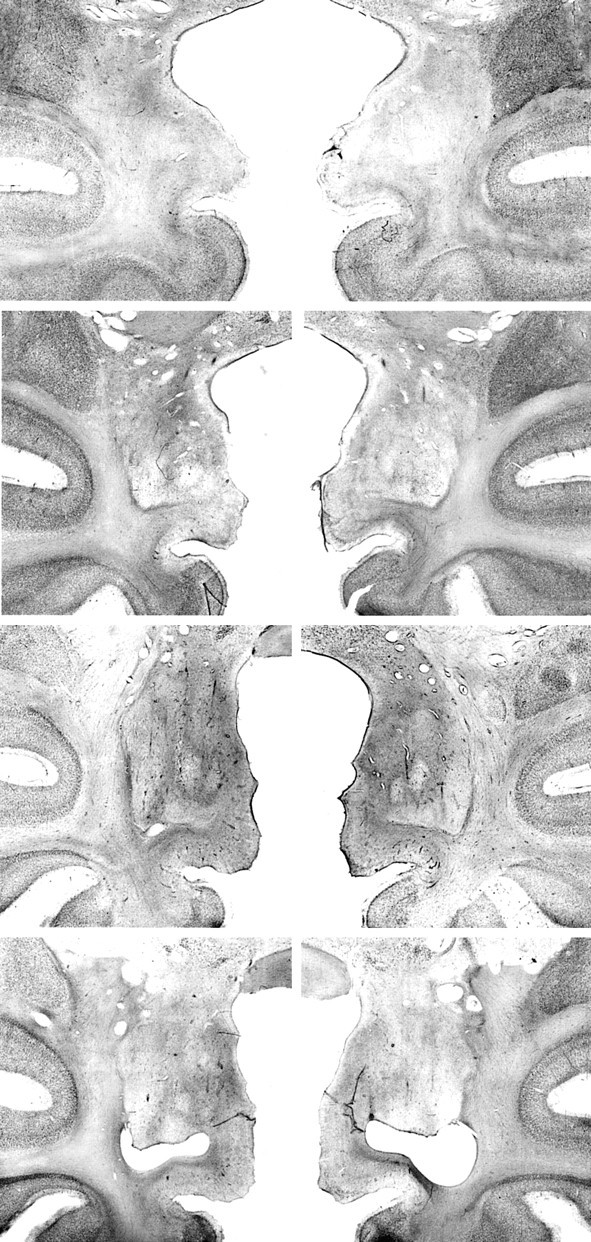
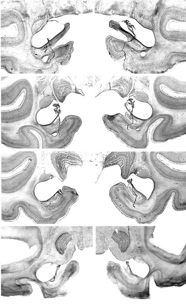
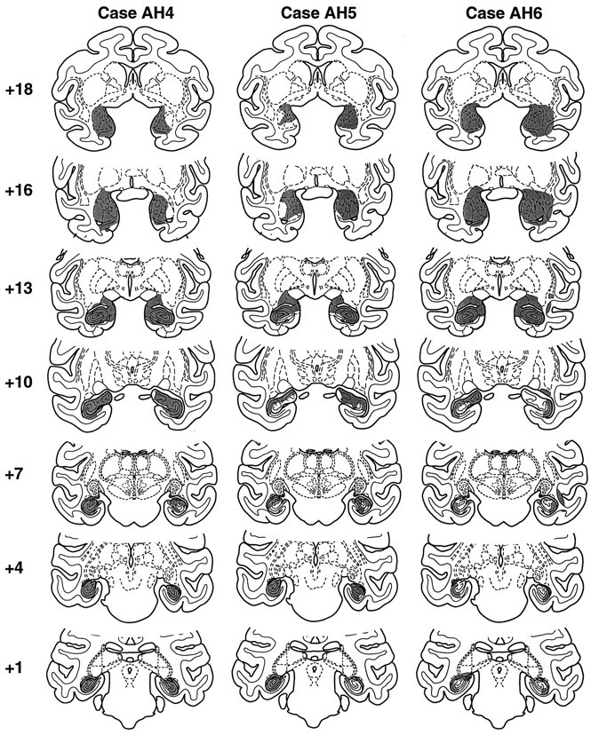
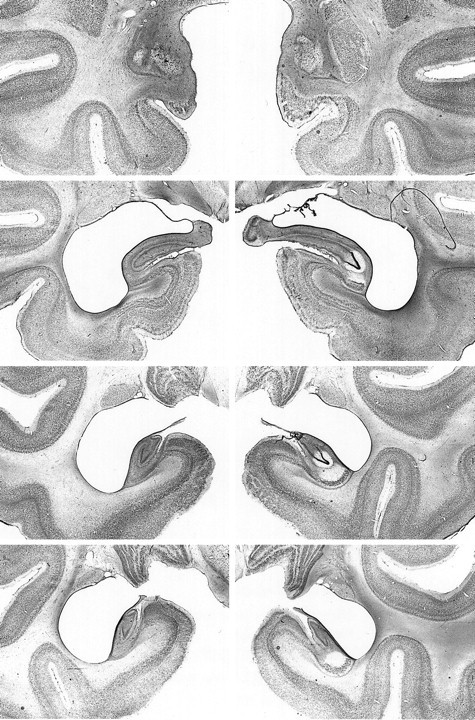
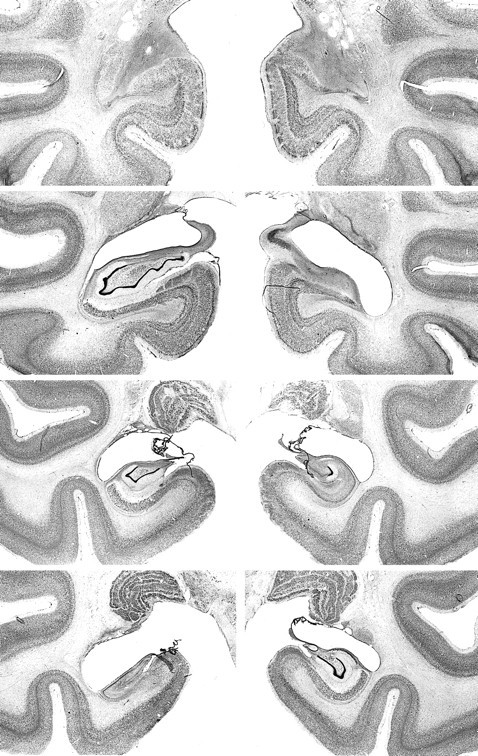
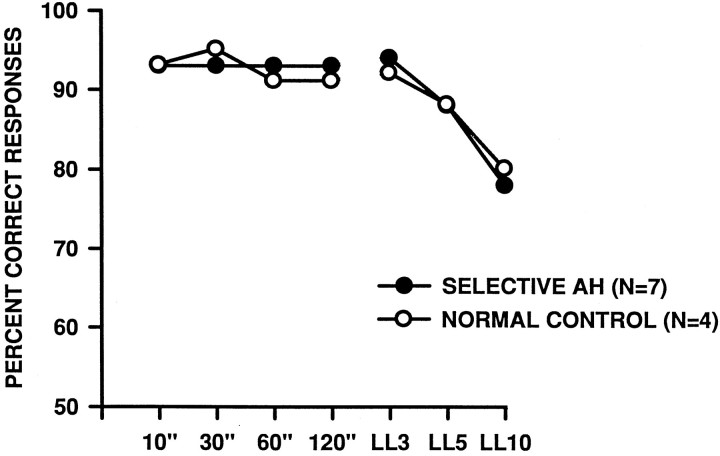
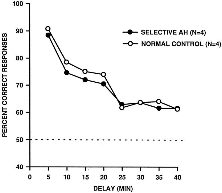
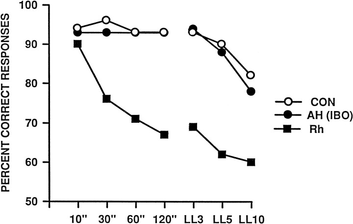
Earlier work indicated that combined but not separate removal of the amygdala and hippocampus, together with the cortex underlying these structures, leads to a severe impairment in visual recognition. More recent work, however, has shown that removal of the rhinal cortex, a region subjacent to the amygdala and rostral hippocampus, yields nearly the same impairment as the original removal. This raises the possibility that the earlier results were attributable to combined damage to the rostral and caudal portions of the rhinal cortex rather than to the combined amygdala and hippocampal removal. To test this possibility, we trained rhesus monkeys on delayed nonmatching-to-sample, a measure of visual recognition, gave them selective lesions of the amygdala and hippocampus made with the excitotoxin ibotenic acid, and then assessed their recognition abilities by using increasingly longer delays and list lengths, including delays as long as 40 min. Postoperatively, monkeys with the combined amygdala and hippocampal lesions performed as well as intact controls at every stage of testing. The same monkeys also were unimpaired relative to controls on an analogous test of spatial memory, delayed nonmatching-to-location. It is unlikely that unintended sparing of target structures can account for the lack of impairment; there was a significant positive correlation between the percentage of damage to the hippocampus and scores on portions of the recognition performance test, suggesting that, paradoxically, the greater the hippocampal damage, the better the recognition. The results show that, within the medial temporal lobe, the rhinal cortex is both necessary and sufficient for visual recognition.
Figures
Similar articles
-
Effects of rhinal cortex lesions combined with hippocampectomy on visual recognition memory in rhesus monkeys.J Neurophysiol. 1996 Mar;75(3):1190-205. doi: 10.1152/jn.1996.75.3.1190. J Neurophysiol. 1996. PMID: 8867128
-
The hippocampal/parahippocampal regions and recognition memory: insights from visual paired comparison versus object-delayed nonmatching in monkeys.J Neurosci. 2004 Feb 25;24(8):2013-26. doi: 10.1523/JNEUROSCI.3763-03.2004. J Neurosci. 2004. PMID: 14985444 Free PMC article.
-
Visual recognition in monkeys following rhinal cortical ablations combined with either amygdalectomy or hippocampectomy.J Neurosci. 1986 Jul;6(7):1991-2003. doi: 10.1523/JNEUROSCI.06-07-01991.1986. J Neurosci. 1986. PMID: 3734871 Free PMC article.
-
Effects of aging on visual recognition memory in the rhesus monkey.Neurobiol Aging. 1988 Sep-Dec;9(5-6):495-502. doi: 10.1016/s0197-4580(88)80103-9. Neurobiol Aging. 1988. PMID: 3062461 Review.
-
The primate hippocampus: ontogeny, early insult and memory.Curr Opin Neurobiol. 2005 Apr;15(2):168-74. doi: 10.1016/j.conb.2005.03.015. Curr Opin Neurobiol. 2005. PMID: 15831398 Review.
Cited by
-
Neonatal hippocampal damage impairs specific food/place associations in adult macaques.Behav Neurosci. 2013 Feb;127(1):9-22. doi: 10.1037/a0031498. Behav Neurosci. 2013. PMID: 23398438 Free PMC article.
-
Spatial object recognition enables endogenous LTD that curtails LTP in the mouse hippocampus.Cereb Cortex. 2013 May;23(5):1118-25. doi: 10.1093/cercor/bhs089. Epub 2012 Apr 17. Cereb Cortex. 2013. PMID: 22510536 Free PMC article.
-
Predictive feedback and conscious visual experience.Front Psychol. 2013 Jan 21;3:620. doi: 10.3389/fpsyg.2012.00620. eCollection 2012. Front Psychol. 2013. PMID: 23346068 Free PMC article.
-
Effects of pharmacologically induced changes in NMDA-receptor activity on long-term memory in humans.Learn Mem. 2001 Jan-Feb;8(1):20-5. doi: 10.1101/lm.33701. Learn Mem. 2001. PMID: 11160760 Free PMC article.
-
Transient inactivation of perirhinal cortex disrupts encoding, retrieval, and consolidation of object recognition memory.J Neurosci. 2005 Jan 5;25(1):52-61. doi: 10.1523/JNEUROSCI.3827-04.2005. J Neurosci. 2005. PMID: 15634766 Free PMC article.
References
-
- Aggleton JP, Burton MJ, Passingham RE. Cortical and subcortical afferents to the amygdala of the rhesus monkey (Macaca mulatta). Brain Res. 1980;36:243–248. - PubMed
-
- Aguirre GK, Detre JA, Alsop DC, D’Esposito M. The parahippocampus subserves topographic learning in man. Cereb Cortex. 1996;6:823–829. - PubMed
-
- Angeli SJ, Murray EA, Mishkin M. Hippocampectomized monkeys can remember one place but not two. Neuropsychologia. 1993;31:1021–1030. - PubMed
Publication types
MeSH terms
Substances
LinkOut - more resources
Full Text Sources
Medical