Comparative Study
. 1998;7(2):115-29.

Cell-specific transcription of the smooth muscle gamma-actin gene requires both positive- and negative-acting cis elements

Affiliations
Comparative Study

Cell-specific transcription of the smooth muscle gamma-actin gene requires both positive- and negative-acting cis elements

A M Kovacs et al. Gene Expr. 1998.

Abstract

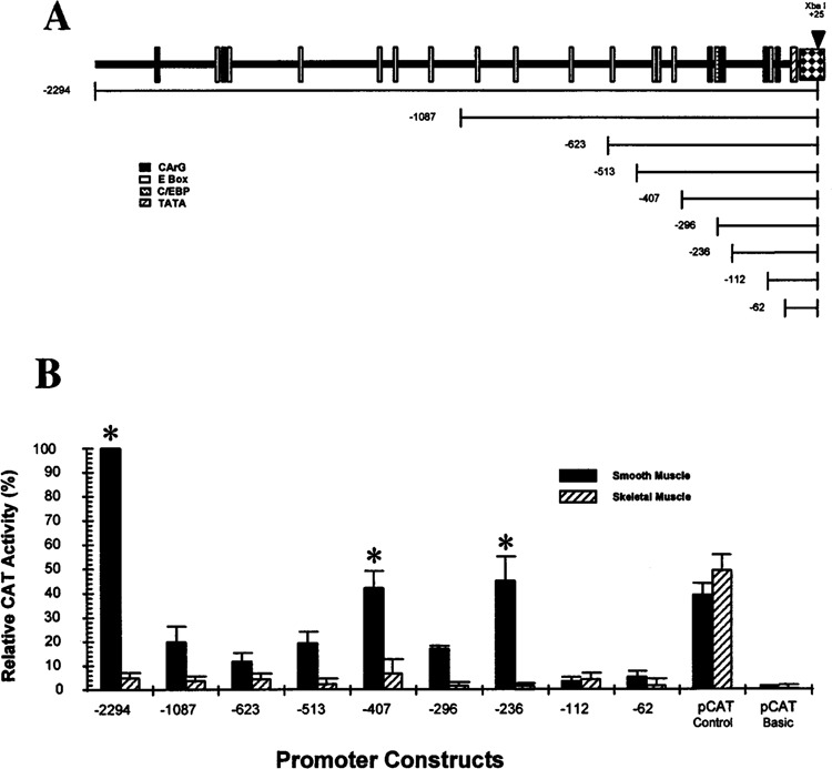
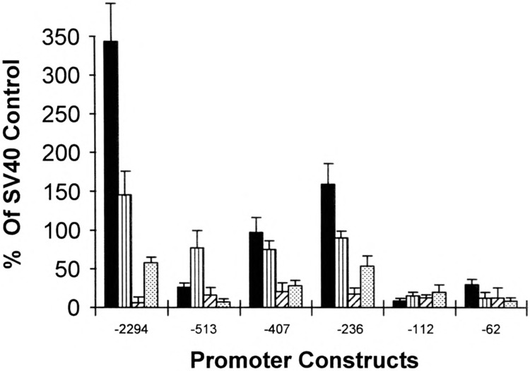
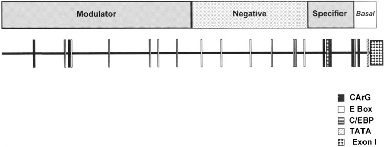
We have characterized the function of putative regulatory sequences upon the smooth muscle transcription of the SMGA gene, using promoter deletion analyses. We demonstrate that the SMGA promoter contains four domains: a basal promoter (-1 to -100), a smooth muscle specifier sequence (-100 to -400), a negative regulator (-400 to -1000), and a smooth muscle-specific modulator (-1000 to -2000). The basal or core promoter supports equivalent transcription in both smooth and skeletal muscle cells. Addition of sequences containing a CArG motif juxtaposed to an E-box element stimulates smooth muscle transcription by five- to sixfold compared to skeletal muscle. This smooth muscle-specific segment is maintained for about 200 bp, after which is a segment of DNA that appears to inhibit the transcriptional capacity of the SMGA promoter in smooth muscle cells. Within the boundary between the smooth muscle specifier and negative regulatory sequences (-400 to -500) are three E-box elements. The smooth muscle modulator domain contains two CArG elements and multiple E-boxes. When added to the SMGA promoter it causes an additional three- to fivefold increase in smooth muscle-specific transcription over that stimulated by the smooth muscle specifier domain. Thus, our studies show that the appropriate cell-specific transcription of the SMGA gene involves complex interactions directed by multiple cis-acting elements. Moreover, our characterization of a cell culture system employing embryonic gizzard smooth muscle cells lays the foundation for further molecular analyses of factors that regulate or control SMGA and other smooth muscle genes during differentiation.


Figures

FIG. 1
FIG. 1
Localization of the SMGA transcriptional initiation site. The 5′ boundary of exon 1 was located using primer extension (A) and RNase protection analyses (B). (A) A primer made to complement sequences of exon 2 beginning at the ATG translation codon (nucleotides 770 to 787, Fig. 2) was labeled with 32P and annealed with 10 μg of yeast tRNA (lane 3) or 10 μg of total RNA isolated from embryonic day 18 gizzard tissue (lane 2). Primer extension was then performed as detailed in Materials and Methods. Lane 1 shows the results of primer extension reactions performed with no added RNA. The sequence ladder (shown a, g, c, t) was derived from single-stranded M13mpl8 and run on the same gel as a molecular weight standard. The arrow shows the position of the major extension product (∼92 bases) with the asterisk showing a minor product (∼95 bases). (B) Uniformly labeled sense and antisense RNA probes to the region spanning exon 1 (+25 to −68, Fig. 1) were annealed to 10 μg yeast tRNA (lanes 2) or 10 μg embryonic gizzard total RNA (lane 3) and the annealed products subjected to RNase protection analyses as outlined in Materials and Methods. Lanes 1 show the position of the probes (sense or anti-sense) that received no RNase treatment. The arrow denotes the position (∼27 bases) of the protected fragment corresponding to exon 1, which is only observed with the antisense probe in gizzard RNAs.
FIG. 2
FIG. 2
Characterization of embryonic gizzard smooth muscle cell primary cultures. Cultures of cells from 7–8 day embryonic gizzard tissue were established as outlined in Materials and Methods, and the cultured cells examined for the expression of SMGA (C, D) and smooth muscle myosin heavy chain (E, F) expression by immunofluorescence microscopy. (C) and (E) represent γ-actin and myosin heavy chain expression, respectively, in cells just prior to switching media to the differentiation media (DMEM:F12 plus 2 mM l-glutamine, 10−6 units insulin, 5 ml/ml Apo-transferrin, and 0.2 mM L-ascorbic acid), and (D) and (F) represent staining of cells after 48-h incubation in this media. (C), (D′), (E′), and (F) show the staining of nuclei in the same cultures using Hoechst 33258. (A)/(A′) and (B)/(B′) show staining obtained from replicating (A) and differentiated (B) cells using an unrelated, control monoclonal antibody (antispectrin, rat astrocyte-specific, monoclonal antibody, kind gift of Dr. S. Goodman, University of South Alabama). (I) and (J) show replicating and differentiated smooth muscle cells stained with a polyclonal actin antibody that is not isotype specific. (G) and (H) show staining in similar cultures using a preimmune rabbit control antibody.
FIG. 3
FIG. 3
Comparison of SMGA gene proximal promoter segments. Sequences from the 5′ flanking regions of the human (44), mouse (61), and chicken SMGA genes were compared using the Bestfit and Pileup programs of the GCG package (University of Wisconsin). The top line shows the human sequence, the middle shows the mouse sequence, and the bottom line, in lowercase letters, represents the chicken sequence. The 5′ boundaries of the SMGA gene exon 1 segment are denoted by +1 and underlining. The sequences have been aligned to preserve maximal homology. Sequences conforming to CArG and C/EBP DNA elements that are conserved among the SMGA proximal promoters are illustrated with the motif identified above the sequence. The distal two CArG sequences deviate by one nucleotide from the CC(A/T)6GG motif. All the motifs were identified by the GCG program, patterns, with a threshold of 85%.
FIG. 4
FIG. 4
Analysis of the chicken SMGA promoter. Sequences flanking the 5′ region of the chicken SMGA gene were cloned in front of the CAT gene and the hybrid SMGA promoter/CAT genes then placed into cultured smooth muscle or skeletal muscle myoblast cells. (A) The top portion shows a diagram illustrating the position of CArG, E-box, C/EBP, and TATA consensus sequences in front of the chicken SMGA gene. The position of unique restriction sites (EcoRI: −2294, Nar I: −1087, Apa I: −623, Xho I: −513, Sma I: −407, Nde I: −112, and BamHI: −62) or synthetic oligonucleotides (−293 and −233) used to form SMGA promoter deletions are shown. The bottom segment shows the relative position of the promoter mutations tested, each having a common 3′ boundary within exon 1 of the SMGA gene. (B) The constructs shown in (A) were placed into either smooth muscle or skeletal muscle myoblast cells and the resultant CAT activity within the transfected cells assayed 48 h later as described in Materials and Methods. A constant amount of CMV-βgal plasmid was included in each experiment and the amount of CAT activity was evaluated relative to the activity measured from the CMV-βgal control in the same lysate. The data are reported as relative CAT activity, normalizing the −2294 construct to a value of 100%. Each construct was evaluated in a minimum of 20 separate assays (duplicate plates in three different cultures with three separate plasmid preparations) with the SEM shown by the error bars. *Statistical differences between the transcription observed in the smooth muscle and skeletal muscle cells (p < 0.05).
FIG. 5
FIG. 5
Cellular specificity of SMGA transcription. Selected SMGA promoter/reporter gene constructs were placed into parallel cultures of differentiated smooth muscle cells (filled bars), replicating smooth muscle mesenchyme (vertical striped bars), skeletal muscle myoblasts (striped bars), and chicken embryonic fibroblasts (dotted bars). The resultant reporter activity was assayed as described in Materials and Methods and this activity was normalized to the activity obtained by a constant amount of CMV-βgal plasmid as an internal control for transfection efficiency. Each cell type also recieved a reporter plasmid that contained the SV40 promoter and enhancer sequence (pCAT-Control), and the data are shown as the amount of activity obtained with the SMGA promoter fragment compared with the amount of activity derived for the SV40 control vector (% of SV40 control).
FIG. 6
FIG. 6
Summary of DNA transfection experiments. This is a model of the SMGA promoter activities in smooth muscle cells as determined by promoter deletions. The placement of potential, muscle-specific DNA elements is shown by the boxes along the ∼2.3 kb of DNA 5′ to the chicken SMGA gene. The promoter is divided into four segments or domains, with the boundaries shown above the promoter segment. The sequences extending to −100 bp 5′ to the gene confer basal promoter activity, exhibiting similar transcriptional capacity in smooth and skeletal muscle cells. The DNA segment between −100 and −400 has been termed a smooth muscle specifier because this DNA shows enhanced transcriptional capability in visceral smooth muscle compared to skeletal muscle myoblasts. The segment from −400 to −1087 imparts a diminished transcriptional activity and is referred to as a negative regulator. Finally, the sequences between −1087 and −2294 show enhanced smooth muscle transcriptional activation in visceral smooth muscle cells. We therefore have denoted this region as the smooth muscle modulator activity, as discussed in the text.

Similar articles

Cited by

References

    1. Al-Molish M. I.; Dubes G. R. Enhanced DNA transfer with dextrans. J. Gen. Virol. 18:189–193; 1973. - PubMed
    1. Balczon R.; West K. The identification of mammalian centrosomal antigens using human autoimmune anti-centrosome antisera. Cell Motil. Cytoskeleton 20:121–135; 1991. - PubMed
    1. Blank R. S.; McQuinn T. C.; Yin K. C.; Thompson M. M.; Takeyasu K.; Schwartz R. J.; Owens G. K. Elements of the smooth muscle α-actin promoter required in cis for transcriptional activation in smooth muscle. J. Biol. Chem. 267:984–989; 1992. - PubMed
    1. Boxer L. M.; Prywes R.; Roeder R. G.; Kedes L. The sarcomeric actin CArG-binding factor is indistinguishable from the c-fos serum response factor. Mol. Cell. Biol. 9:515–522; 1989. - PMC - PubMed
    1. Breathnach R.; Chambon P. Organization and expression of eukaryotic split genes coding for proteins. Annu. Rev. Biochem. 50:349–383; 1981. - PubMed

Publication types

MeSH terms

LinkOut - more resources