. 1998 Aug 10;142(3):613-23.
doi: 10.1083/jcb.142.3.613.

The sorting of mitochondrial DNA and mitochondrial proteins in zygotes: preferential transmission of mitochondrial DNA to the medial bud

Affiliations

The sorting of mitochondrial DNA and mitochondrial proteins in zygotes: preferential transmission of mitochondrial DNA to the medial bud

K Okamoto et al. J Cell Biol. .

Abstract

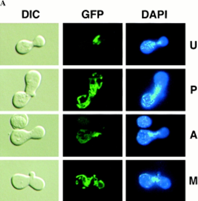
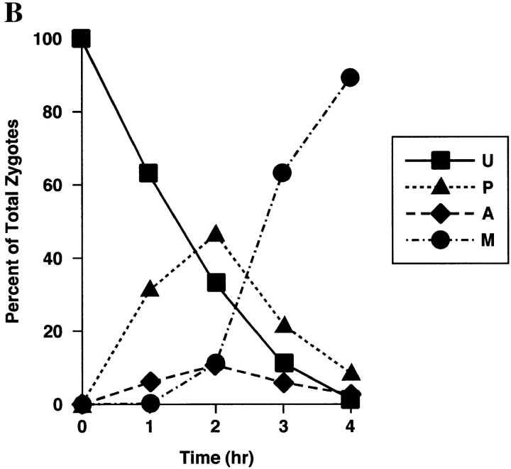
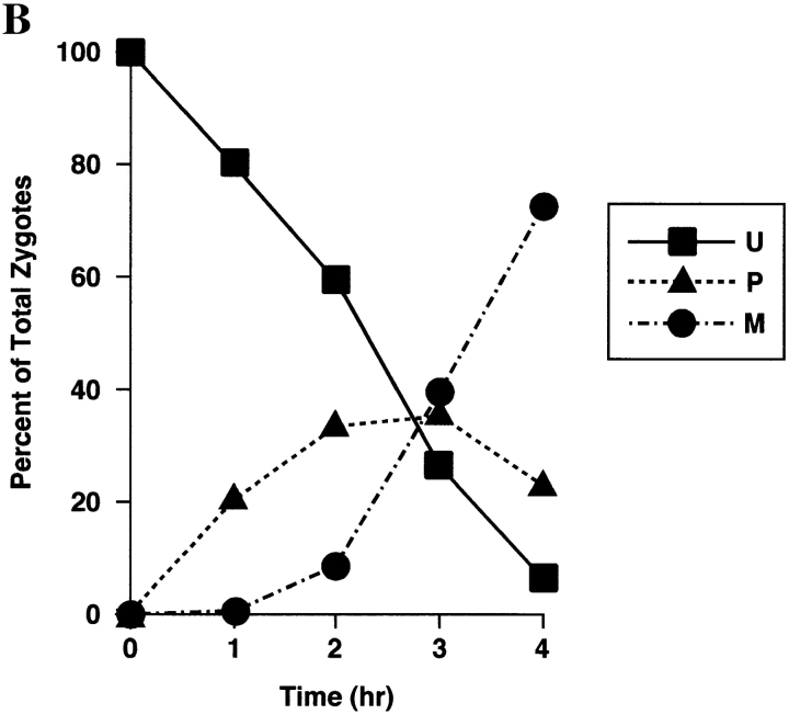
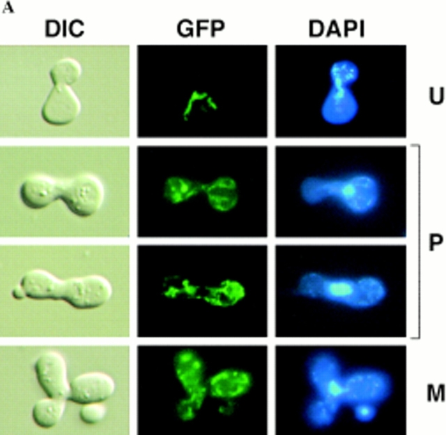
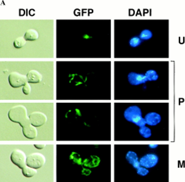
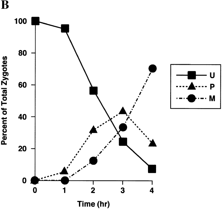
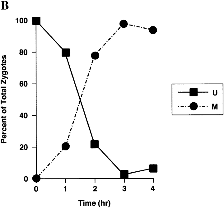
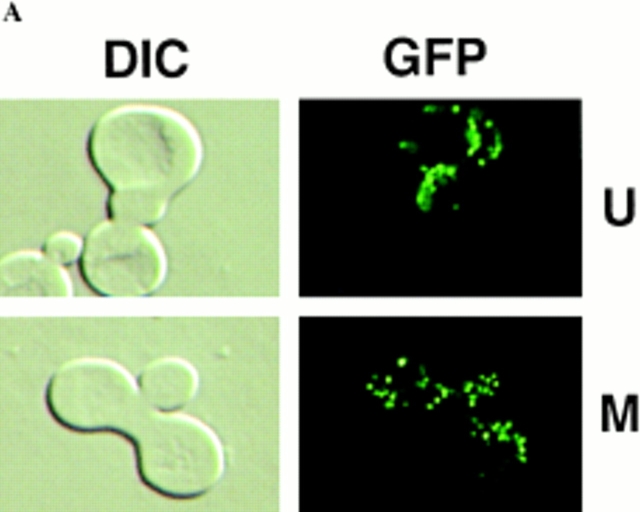
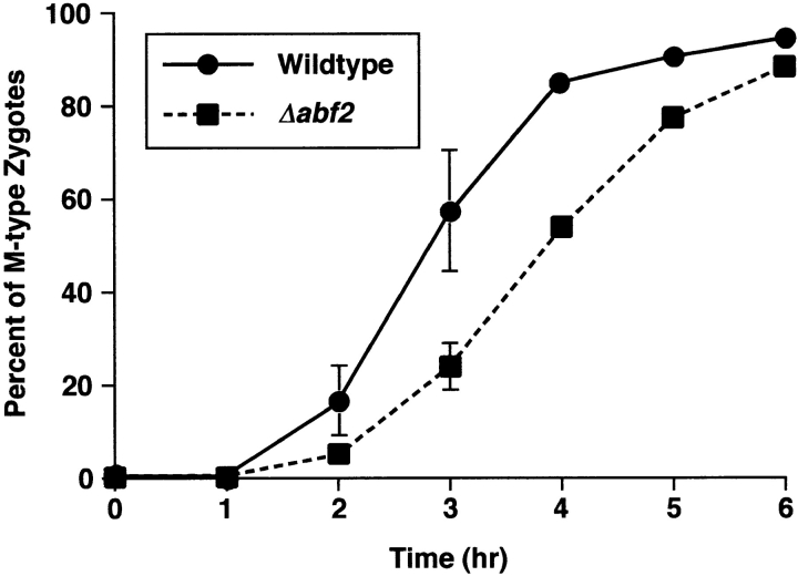
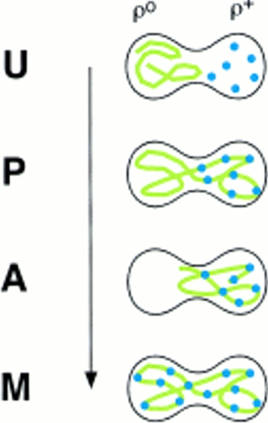
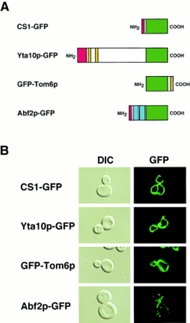
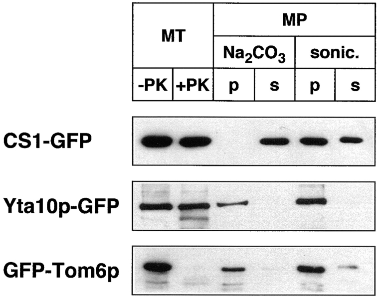
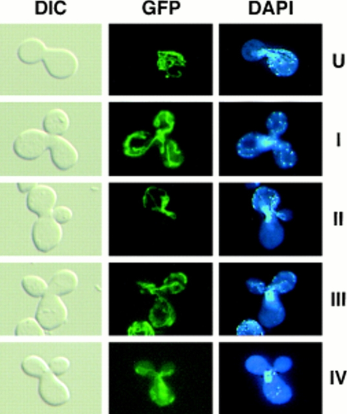
Green fluorescent protein (GFP) was used to tag proteins of the mitochondrial matrix, inner, and outer membranes to examine their sorting patterns relative to mtDNA in zygotes of synchronously mated yeast cells in rho+ x rho0 crosses. When transiently expressed in one of the haploid parents, each of the marker proteins distributes throughout the fused mitochondrial reticulum of the zygote before equilibration of mtDNA, although the membrane markers equilibrate slower than the matrix marker. A GFP-tagged form of Abf2p, a mtDNA binding protein required for faithful transmission of rho+ mtDNA in vegetatively growing cells, colocalizes with mtDNA in situ. In zygotes of a rho+ x rho+ cross, in which there is little mixing of parental mtDNAs, Abf2p-GFP prelabeled in one parent rapidly equilibrates to most or all of the mtDNA, showing that the mtDNA compartment is accessible to exchange of proteins. In rho+ x rho0 crosses, mtDNA is preferentially transmitted to the medial diploid bud, whereas mitochondrial GFP marker proteins distribute throughout the zygote and the bud. In zygotes lacking Abf2p, mtDNA sorting is delayed and preferential sorting is reduced. These findings argue for the existence of a segregation apparatus that directs mtDNA to the emerging bud.


Figures

Figure 4
Figure 4
Sorting of CS1–GFP in a PSY142 ρ0 [CS1–GFP] × S150-2B ρ+ cross. (A) Shown are representative examples of the different zygote forms, indicated diagrammatically in Fig. 3, detected in the cross based on the distribution of GFP and DAPI (mtDNA) fluorescence. (B) Kinetics of appearance and disappearance of the different zygote forms.
Figure 4
Figure 4
Sorting of CS1–GFP in a PSY142 ρ0 [CS1–GFP] × S150-2B ρ+ cross. (A) Shown are representative examples of the different zygote forms, indicated diagrammatically in Fig. 3, detected in the cross based on the distribution of GFP and DAPI (mtDNA) fluorescence. (B) Kinetics of appearance and disappearance of the different zygote forms.
Figure 5
Figure 5
Sorting of Yta10p–GFP in a PSY142 ρ0 [Yta10p–GFP] × S150-2B ρ+ cross. (A) Representative examples of the different zygote forms generated in the cross. (B) Kinetics of appearance and disappearance of the different zygote forms.
Figure 5
Figure 5
Sorting of Yta10p–GFP in a PSY142 ρ0 [Yta10p–GFP] × S150-2B ρ+ cross. (A) Representative examples of the different zygote forms generated in the cross. (B) Kinetics of appearance and disappearance of the different zygote forms.
Figure 6
Figure 6
Sorting of GFP–Tom6p in a PSY142 ρ0 [GFP–Tom6p] × S150-2B ρ+ cross. (A) Representative examples of the different zygote forms generated in the cross. (B) Kinetics of appearance and disappearance of the different zygote forms.
Figure 6
Figure 6
Sorting of GFP–Tom6p in a PSY142 ρ0 [GFP–Tom6p] × S150-2B ρ+ cross. (A) Representative examples of the different zygote forms generated in the cross. (B) Kinetics of appearance and disappearance of the different zygote forms.
Figure 7
Figure 7
Sorting of Abf2p–GFP in a PSY142 ρ+ [Abf2p–GFP] × S150-2B ρ+ cross. (A) Representative examples of the U and M form zygotes generated in the cross. In this experiment, PSY142 ρ+ cells were prelabled with Abf2p–GFP before synchronous mating with the S150-2B ρ+ strain. (B) Kinetics of sorting of Abf2p–GFP.
Figure 7
Figure 7
Sorting of Abf2p–GFP in a PSY142 ρ+ [Abf2p–GFP] × S150-2B ρ+ cross. (A) Representative examples of the U and M form zygotes generated in the cross. In this experiment, PSY142 ρ+ cells were prelabled with Abf2p–GFP before synchronous mating with the S150-2B ρ+ strain. (B) Kinetics of sorting of Abf2p–GFP.
Figure 9
Figure 9
The sorting of CS1–GFP is delayed in a S150-2B Δabf2 ρ+ [CS1–GFP] × PSY142 Δabf2 ρ0 cross. Wild-type and Δabf2 derivatives of S150-2B ρ+ cells transiently expressing CS1–GFP and PSY142 ρ0 wild-type and Δabf2 cells were synchronously mated and the kinetics of appearance of M form zygotes was determined by direct microscopic analysis.
Figure 3
Figure 3
Diagrammatic representation of the different zygote forms detected in synchronous ρ0 × ρ+ crosses with a matrix marker protein prelabeled in the ρ0 parent. Green lines, marker protein in mitochondria; blue dots, mtDNA. U, unmixed; P, partially mixed; A, asymmetric; M, mixed.
Figure 1
Figure 1
GFP fusion proteins targeted to mitochondria. (A) Diagrammatic representation of fusion proteins between GFP and all or a portion of the indicated mitochondrial proteins. Green, GFP; red, mitochondrial presequence; yellow, putative transmembrane domains; blue, HMG boxes in Abf2p. (B) Fluorescence pattern of the indicated GFP fusion proteins (right panels) in cultures of PSY142 ρ+ cells grown on YNBGalR + cas medium. Cells were transformed with plasmids encoding either the matrix (CS1-GFP), inner membrane (Yta10p-GFP), outer membrane (GFP-Tom6p), or mtDNA (Abf2p-GFP) marker proteins (refer to Materials and Methods). Left panels, cells visualized by DIC.
Figure 2
Figure 2
Localization of GFP fusion proteins in mitochondria. Mitochondria were isolated from PSY142 ρ+ cells grown on YNBGalR + cas medium that were transformed with the plasmid encoding the indicated GFP fusion protein. As described in Materials and Methods, mitochondria (MT) were incubated with or without proteinase K (PK). Crude mitoplasts (MP) were treated with Na2CO3 or sonicated and separated into a pellet (p) and soluble (s) fraction. Proteins were fractionated on a 12% SDS polyacrylamide gel and detected by Western blotting with anti-GFP antiserum.
Figure 8
Figure 8
Differential sorting of CS1–GFP and mtDNA in ρ+ [CS1–GFP] × ρ0 crosses. The top four panel sets are representative micrographs of the different zygote types observed in the cross between S150-2B ρ+ [CS1–GFP] and PSY142 ρ0 cells. The U form zygotes appear immediately after mating. Types I–III are the different zygote forms detected in populations of zygotes with medial buds. The type IV zygote is from the cross S150-2B Δabf2 ρ+ [CS1–GFP] × PSY142 Δabf2 ρ0.

References

    1. Alconada A, Kübrich M, Moczko M, Hönlinger A, Pfanner N. The mitochondrial receptor complex: the small subunit Mom8b/Isp6 supports association of receptors with the general insertion pore and transfer of preproteins. Mol Cell Biol. 1995;15:6196–6205. - PMC - PubMed
    1. Arlt H, Tauer R, Feldmann H, Neupert W, Langer T. The YTA10-12 complex, an AAA protease with chaperone-like activity in the inner membrane of mitochondria. Cell. 1996;85:875–885. - PubMed
    1. Azpiroz R, Butow RA. Patterns of mitochondrial sorting in yeast zygotes. Mol Biol Cell. 1993;4:21–36. - PMC - PubMed
    1. Azpiroz R, Butow RA. Mitochondrial inheritance in yeast. Methods Enzymol. 1995;260:453–466. - PubMed
    1. Berger KH, Yaffe MP. Mitochondrial distribution and inheritance. Experentia. 1996;52:1111–1116. - PubMed

Publication types

MeSH terms