. 1998 Aug 10;142(3):635-49.
doi: 10.1083/jcb.142.3.635.

Dual roles for Ste24p in yeast a-factor maturation: NH2-terminal proteolysis and COOH-terminal CAAX processing

Affiliations

Dual roles for Ste24p in yeast a-factor maturation: NH2-terminal proteolysis and COOH-terminal CAAX processing

A Tam et al. J Cell Biol. .

Abstract

Maturation of the Saccharomyces cerevisiae a-factor precursor involves COOH-terminal CAAX processing (prenylation, AAX tripeptide proteolysis, and carboxyl methylation) followed by cleavage of an NH2-terminal extension (two sequential proteolytic processing steps). The aim of this study is to clarify the precise role of Ste24p, a membrane-spanning zinc metalloprotease, in the proteolytic processing of the a-factor precursor. We demonstrated previously that Ste24p is necessary for the first NH2-terminal processing step by analysis of radiolabeled a-factor intermediates in vivo (Fujimura-Kamada, K., F.J. Nouvet, and S. Michaelis. 1997. J. Cell Biol. 136:271-285). In contrast, using an in vitro protease assay, others showed that Ste24p (Afc1p) and another gene product, Rce1p, share partial overlapping function as COOH-terminal CAAX proteases (Boyartchuk, V.L., M.N. Ashby, and J. Rine. 1997. Science. 275:1796-1800). Here we resolve these apparently conflicting results and provide compelling in vivo evidence that Ste24p indeed functions at two steps of a-factor maturation using two methods. First, direct analysis of a-factor biosynthetic intermediates in the double mutant (ste24Delta rce1Delta) reveals a previously undetected species (P0*) that fails to be COOH terminally processed, consistent with redundant roles for Ste24p and Rce1p in COOH-terminal CAAX processing. Whereas a-factor maturation appears relatively normal in the rce1Delta single mutant, the ste24Delta single mutant accumulates an intermediate that is correctly COOH terminally processed but is defective in cleavage of the NH2-terminal extension, demonstrating that Ste24p is also involved in NH2-terminal processing. Together, these data indicate dual roles for Ste24p and a single role for Rce1p in a-factor processing. Second, by using a novel set of ubiquitin-a-factor fusions to separate the NH2- and COOH-terminal processing events of a-factor maturation, we provide independent evidence for the dual roles of Ste24p. We also report here the isolation of the human (Hs) Ste24p homologue, representing the first human CAAX protease to be cloned. We show that Hs Ste24p complements the mating defect of the yeast double mutant (ste24Delta rce1Delta) strain, implying that like yeast Ste24p, Hs Ste24p can mediate multiple types of proteolytic events.


Figures
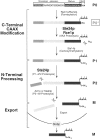
Figure 1
Figure 1
Pathway of a-factor biogenesis. The a-factor precursor, biosynthetic intermediates, and components of the biogenesis machinery are shown. The biogenesis of a-factor is comprised of three stages, indicated at left: (a) COOH-terminal CAAX processing, (b) two sequential NH2-terminal proteolytic cleavages, and (c) export. The a-factor precursor (P0, top) consists of the mature, bioactive portion (black) flanked by an NH2-terminal extension (two shades of gray) and a COOH-terminal CAAX motif. Mature a-factor is a prenylated and carboxyl methylated dodecapeptide (M, bottom). The known a-factor biosynthetic intermediates that can be directly visualized by SDS-PAGE are designated P0, P1, P2, and M (Chen et al., 1997b ). In this study, we present the previously undetected intermediate P0*. The cellular components that mediate COOH-terminal CAAX modification events (prenylation, AAX proteolysis, carboxyl methylation) are the Ram1p/Ram2p complex, Rce1p and Ste24p, and Ste14p, respectively (refer to text for more details). NH2-terminal processing involves two successive proteolytic steps. The first NH2-terminal cleavage step (P1→ P2) is mediated by Ste24p (as confirmed by this study), and the second step (P2→ M) requires Axl1p or Ste23p. The dual roles of Ste24p in COOH-terminal AAXing and the first NH2-terminal (P1→ P2) processing step is the subject of this study.
Figure 2
Figure 2
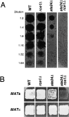
STE24 and RCE1 are required for the efficient production of a-factor by MAT a cells. (A) a-Factor spot dilution assay. The a-factor– induced growth arrest of a MATα supersensitive (sst2) tester lawn results in a clear zone or spot. Dilutions of concentrated, extracellular a-factor produced by a wild-type strain, the single (rce1Δ and ste24Δ) and double (ste24Δ rce1Δ) mutants were prepared as described in the Materials and Methods and spotted onto a lawn of the MATα halo tester (SM1086) on YPD. The highest dilution yielding a clear spot is the titer of a-factor for that strain. The papillae arising in the clear spots are revertants arising from the sst2 lawn (Chan and Otte, 1982). (B) Patch mating assay. Strains bearing the single and double rce1Δ ste24Δ mutations were tested for the ability to mate with cells of the opposite mating type. Patches of the MAT a or MATα strains to be tested for mating were replica-plated from a patch master plate onto a lawn of auxotrophic mating tester cells spread on minimal SD media. The growth of prototrophic diploids is indicative of mating. MATα strains do not exhibit mating defects even under stringent mating conditions (5% YPD) (data not shown). MAT a strains tested are wild-type (SM1058), rce1Δ (SM3613), ste24Δ (SM3103), and rce1Δ ste24Δ (SM3614); the isogenic MATα strains tested are wild-type (SM1059), rce1Δ (SM3811), ste24Δ (SM3102), and rce1Δ ste24Δ (SM3812). The mating tester lawns are MATα (SM1068) and MAT a (SM1067) and were resuspended in 5% YPD and 50% YPD, respectively, as described in the Materials and Methods.
Figure 3
Figure 3
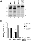
The rce1Δ ste24Δ double mutant produces a novel a-factor intermediate, P0*, that is unmethylated. (A) To examine the a-factor biogenesis profile in wild-type and mutant strains, cells were metabolically labeled with [35S]cysteine for 5 min. Intracellular (I) and extracellular (E) fractions were separated by centrifugation and extracts were prepared as described in the Materials and Methods. Protein extracts were immunoprecipitated with anti–a-factor antiserum no. 9-137, and subjected to SDS-PAGE and PhosphorImager analysis. The previously characterized precursor species (P1, P2) and mature a-factor (M) are shown. The band migrating slightly faster than M is the a-factor– relabeled peptide (AFRP) and is discussed elsewhere (Chen et al., 1997a ). The asterisk (*) marks the new a-factor intermediate P0* (refer to Fig. 1). (B) Carboxyl methylation levels of a-factor were used as an indirect measure of AAXing activity in wild-type and mutant strains. Cells were double-labeled with S-adenosyl-l- [3H-methyl]methionine and [35S]cysteine for 6 min. Total cell extracts were prepared, immunoprecipitated with anti–a-factor antiserum 9-137, and then subjected to SDS-PAGE and autoradiography. Bands representing each a-factor species (P0* or P1, P2, and M) were excised from the dried gel. The degree of methylation for each band was measured by determining the [3H]/[35S] cpm ratio. The relative methylation level was then calculated as a fraction of the corresponding species from the wild-type strain (100%). The ste14Δ strain is a control that lacks carboxyl methylation activity. NP, no protein was detected by autoradiography. The data are averaged from three independent experiments. Strains are wild-type (SM3310), rce1Δ (SM3644), ste24Δ (SM3286) rce1Δ ste24Δ (SM3651), and ste14Δ (SM1871).
Figure 4
Figure 4
P0* is metabolically unstable. (A) Pulse-chase analysis of a-factor in wild-type and mutant strains. Cells were pulse labeled with [35S]cysteine for 5 min and chased with excess cold cysteine for the indicated times (min). Protein extracts were prepared, immunoprecipitated with anti–a-factor antiserum 9-137, and then analyzed by SDS-PAGE. Strains are wild-type (SM3310), rce1Δ (SM3644), ste24Δ (SM3286), and rce1Δ ste24Δ (SM3651). (B) Graph of a-factor precursor stability versus time. The amount of the single a-factor species (either P1 or P0*) present at each time point in the single (ste24Δ) and double (rce1Δ ste24Δ) mutants, respectively, was quantitated by PhosphorImager analysis and graphed as shown.
Figure 5
Figure 5
Processing of Ubi–a-factor fusion proteins generate the expected a-factor species. (A) Ubi–a-factor fusions. To generate Ubi– a-factor fusions, the sequence encoding a single copy of ubiquitin (Ubi) was fused precisely to the full-length a-factor gene MFA1 or to truncated MFA1 corresponding to P2 or mature a-factor, yielding Ubi-P1, Ubi-P2, and Ubi-M, respectively. In all cases, the CAAX motif was retained in the fusion construct. The site of cleavage by the ubiquitin-specific proteases (Ubp) is indicated. The shading of regions within a-factor correspond to that shown in Fig. 1. (B) Pulse-chase analysis of a-factor derived from wild-type MFA1 and Ubi– a-factor fusions. The pulse-chase experiment was carried out as described in Fig. 4, except that the anti–a-factor antiserum 9-497 was used. Chase times (min) are indicated. It should be noted that we do not detect the full-length forms of the Ubi–a-factor fusions, presumably because processing by the Ubps is cotranslational. We have observed the expected full-length Ubi–a-factor fusion upon introduction of a COOH-terminal G76→ V mutation in ubiquitin that abolishes the normal cut site for the Ubps (Nouvet, F. and S. Michaelis, unpublished data). Strains bear deletions of the chromosomal a-factor genes (mfa1Δ mfa2Δ) and contain plasmids harboring wild-type MFA1 (SM3683), Ubi-P1 (SM3685), Ubi-P2 (SM3686), or Ubi-M (SM3684). (C) a-Factor spot dilution assay. The amount of extracellular active a-factor produced by the Ubi–a-factor fusions was measured as described in the Fig. 2 legend. (D) Patch mating assay of Ubi– a-factor fusions. For permissive and stringent mating conditions, the mating tester lawn is MATα (SM1068) resuspended in 100% YPD and 1% YPD, respectively, as described in the Materials and Methods.
Figure 5
Figure 5
Processing of Ubi–a-factor fusion proteins generate the expected a-factor species. (A) Ubi–a-factor fusions. To generate Ubi– a-factor fusions, the sequence encoding a single copy of ubiquitin (Ubi) was fused precisely to the full-length a-factor gene MFA1 or to truncated MFA1 corresponding to P2 or mature a-factor, yielding Ubi-P1, Ubi-P2, and Ubi-M, respectively. In all cases, the CAAX motif was retained in the fusion construct. The site of cleavage by the ubiquitin-specific proteases (Ubp) is indicated. The shading of regions within a-factor correspond to that shown in Fig. 1. (B) Pulse-chase analysis of a-factor derived from wild-type MFA1 and Ubi– a-factor fusions. The pulse-chase experiment was carried out as described in Fig. 4, except that the anti–a-factor antiserum 9-497 was used. Chase times (min) are indicated. It should be noted that we do not detect the full-length forms of the Ubi–a-factor fusions, presumably because processing by the Ubps is cotranslational. We have observed the expected full-length Ubi–a-factor fusion upon introduction of a COOH-terminal G76→ V mutation in ubiquitin that abolishes the normal cut site for the Ubps (Nouvet, F. and S. Michaelis, unpublished data). Strains bear deletions of the chromosomal a-factor genes (mfa1Δ mfa2Δ) and contain plasmids harboring wild-type MFA1 (SM3683), Ubi-P1 (SM3685), Ubi-P2 (SM3686), or Ubi-M (SM3684). (C) a-Factor spot dilution assay. The amount of extracellular active a-factor produced by the Ubi–a-factor fusions was measured as described in the Fig. 2 legend. (D) Patch mating assay of Ubi– a-factor fusions. For permissive and stringent mating conditions, the mating tester lawn is MATα (SM1068) resuspended in 100% YPD and 1% YPD, respectively, as described in the Materials and Methods.
Figure 6
Figure 6
Ubi–a-factor fusions provide independent evidence that Ste24p plays a critical role in NH2-terminal processing of a-factor. (A) Schematic representation of wild-type a-factor, Ubi-P1, and Ubi-P2, with the expected cleavages are shown. (B) Processing of Ubi–a-factor fusions is compared in the wild-type and ste24Δ mutant strains, as indicated. Radiolabeling and immunoprecipitation with anti–a-factor antiserum 9-497 was carried out as described in Fig. 3. Strains are wild-type and ste24Δ with MFA1 (SM3683 and SM3714), Ubi-P1 (SM3685 and SM3715), and Ubi-P2 (SM3686 and SM3716), respectively.
Figure 7
Figure 7
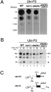
Ubi-P2 confirms overlapping roles for Ste24p and Rce1p in COOH-terminal AAXing of a-factor; similar extracellular a-factor levels are produced from Ubi-P2 in the single ste24Δ and rce1Δ mutants. (A) The a-factor spot dilution assay was carried out as described in Fig. 2. The amount of a-factor derived from Ubi-P2 is roughly equivalent when the endpoint dilutions of the single rce1Δ and ste24Δ strains are compared. It is notable that the amount of a-factor derived from MFA1 differs markedly in the single rce1Δ and ste24Δ mutants (compare with Fig. 2). (B) Pulse labeling and immunoprecipitation of a-factor expressed from Ubi-P2 in wild-type and mutant strains. Cells were pulse labeled with [35S]cysteine for 2 min and the label was chased with excess cold cysteine for the indicated times (min). Immunoprecipitation of a-factor was carried out with anti–a-factor antiserum 9-497. Strains contain a plasmid harboring Ubi-P2 in the wild-type (SM3686), rce1Δ (SM3721), ste24Δ (SM3716), or rce1Δ ste24Δ (SM3726) backgrounds. (C) The diagram illustrates the predicted structure of the Ubi-P2 and Ubi-P2* species.
Figure 8
Figure 8
Relative methylation levels of Ubi-P2 in wild-type and mutant strains. Carboxyl methylation of Ubi-P2–derived a-factor was used as an indirect measure of AAXing activity in wild-type and mutant strains, as described in Fig. 3 B, except that the radiolabel was chased for 0 and 30 min and immunoprecipitation of a-factor was carried out with anti–a-factor antiserum no. 9-497. The P2 or P2* bands were excised from dried gel. The data are averaged from two independent experiments. Strains contain a plasmid harboring Ubi-P2 in the wild-type (SM3686), rce1Δ (SM3721), ste24Δ (SM3716), or rce1Δ ste24Δ (SM3726) backgrounds.
Figure 9
Figure 9
The human homologue of Ste24p complements mating defects of the yeast ste24Δ rce1Δ strain. (A) Amino acid alignment of Ste24p from S. cerevisiae, S. pombe, and H. sapiens. The S. pombe homologue, called Sp Ste24p here, is known as YAN5 (GenBank/EMBL/DDBJ accession no. Q10071). Residues shaded in black and gray, regions of identity and similarity, respectively. The zinc metalloprotease motif (HEXXH) is marked by asterisks (*). I, II, and III, conserved regions unique to members of the Ste24p zinc metalloprotease subfamily. Dashed line (---) within Region I, an additional region found solely within Hs Ste24p (GenBank/EMBL/DDBJ accession no. AF064867). Solid circles (•), a degenerate COOH-terminal dilysine motif. (B) Kyte and Doolittle hydropathy analysis of Sc Ste24p and Hs Ste24p. (C) The human STE24 was tested for the ability to complement the mating defect of the double rce1Δ ste24Δ mutant strain. The patch mating assay was carried out as described in Fig. 2. The lawn of mating tester cells (SM1068) was resuspended in 5% YPD diluted in sterile water. Strains are ste24Δ rce1Δ containing the vector only (SM3650), Sc STE24 (SM3653), or Hs STE24 (SM3814). Hs STE24 has also been recently cloned independently by H. Kumagai, K. Yanagisawa, and H. Komano (all from National Institute for Longevity Sciences, Aichi, Japan) (personal communication).
Figure 9
Figure 9
The human homologue of Ste24p complements mating defects of the yeast ste24Δ rce1Δ strain. (A) Amino acid alignment of Ste24p from S. cerevisiae, S. pombe, and H. sapiens. The S. pombe homologue, called Sp Ste24p here, is known as YAN5 (GenBank/EMBL/DDBJ accession no. Q10071). Residues shaded in black and gray, regions of identity and similarity, respectively. The zinc metalloprotease motif (HEXXH) is marked by asterisks (*). I, II, and III, conserved regions unique to members of the Ste24p zinc metalloprotease subfamily. Dashed line (---) within Region I, an additional region found solely within Hs Ste24p (GenBank/EMBL/DDBJ accession no. AF064867). Solid circles (•), a degenerate COOH-terminal dilysine motif. (B) Kyte and Doolittle hydropathy analysis of Sc Ste24p and Hs Ste24p. (C) The human STE24 was tested for the ability to complement the mating defect of the double rce1Δ ste24Δ mutant strain. The patch mating assay was carried out as described in Fig. 2. The lawn of mating tester cells (SM1068) was resuspended in 5% YPD diluted in sterile water. Strains are ste24Δ rce1Δ containing the vector only (SM3650), Sc STE24 (SM3653), or Hs STE24 (SM3814). Hs STE24 has also been recently cloned independently by H. Kumagai, K. Yanagisawa, and H. Komano (all from National Institute for Longevity Sciences, Aichi, Japan) (personal communication).

References

    1. Adames N, Blundell K, Ashby MN, Boone C. Role of yeast insulin-degrading enzyme homologs in propheromone processing and bud site selection. Science. 1995;270:464–467. - PubMed
    1. Altschul SF, Gish W, Miller W, Myers EW, Lipman DJ. Basic local alignment search tool. J Mol Biol. 1990;215:403–410. - PubMed
    1. Andres DA, Milatovich A, Ozcelik T, Wenzlau JM, Brown MS, Goldstein JL, Francke U. cDNA cloning of the two subunits of human CAAX farnesyltransferase and chromosomal mapping of FNTA and FNTB loci and related sequences. Genomics. 1993;18:105–112. - PubMed
    1. Ashby MN, Rine J. Ras and a-factor converting enzyme. Methods Enzymol. 1995;250:235–250. - PubMed
    1. Ausubel, F.M., R. Brent, R.E. Kingston, D.D. Moore, J.G. Seidman, J.A. Smith, and K. Struhl. 1987. Current Protocols in Molecular Biology. Vol. 1. Greene Publishing Associates, New York.

Publication types

MeSH terms

Associated data