. 1998 Aug 10;142(3):651-63.
doi: 10.1083/jcb.142.3.651.

Retrograde traffic out of the yeast vacuole to the TGN occurs via the prevacuolar/endosomal compartment

Affiliations

Retrograde traffic out of the yeast vacuole to the TGN occurs via the prevacuolar/endosomal compartment

N J Bryant et al. J Cell Biol. .

Abstract

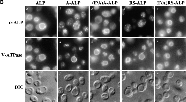
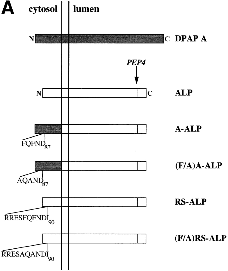
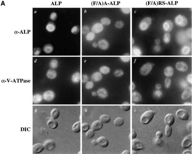
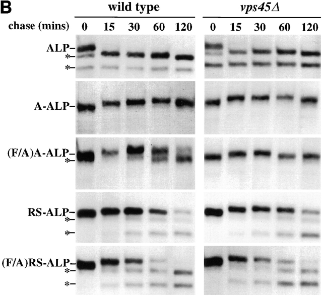
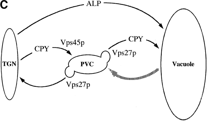
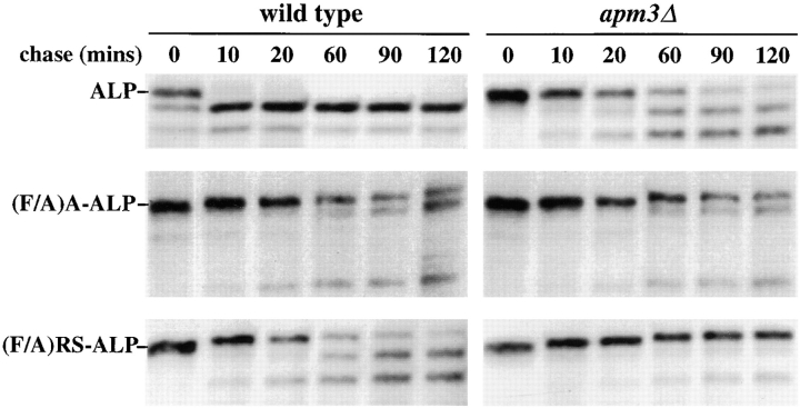
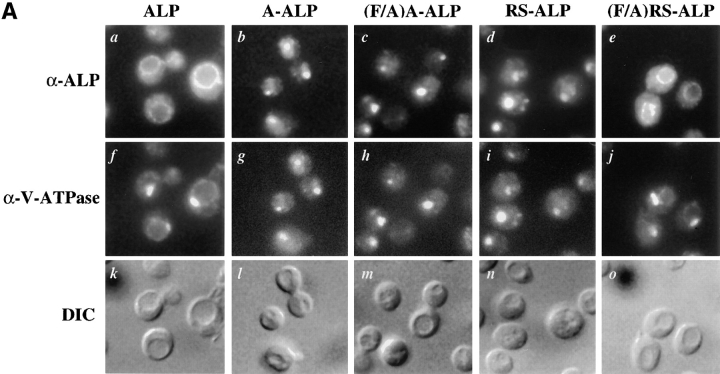
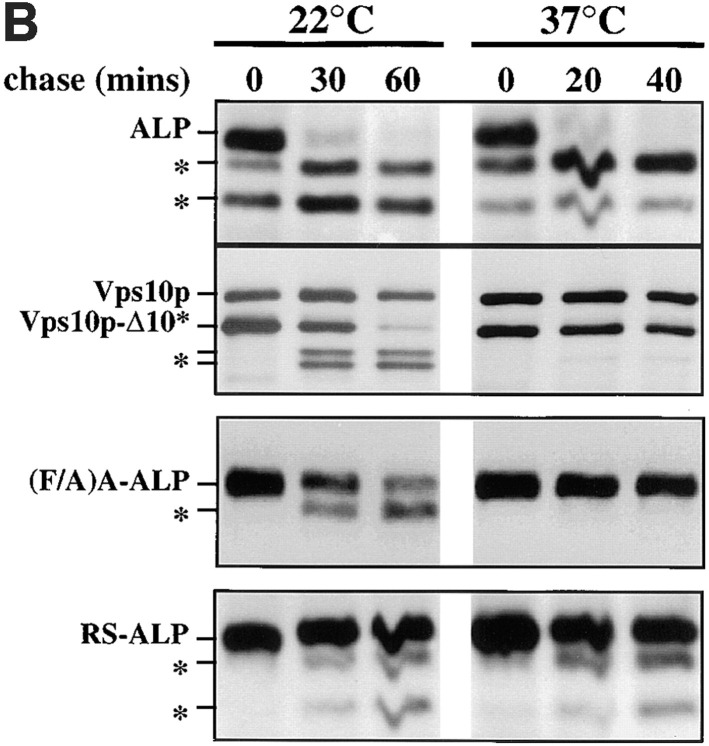
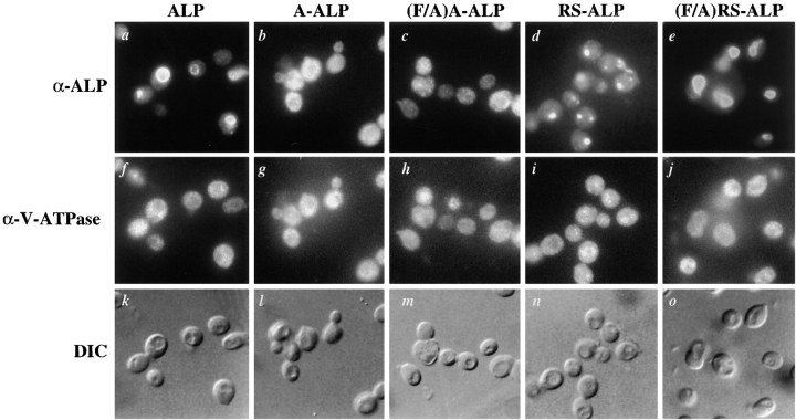
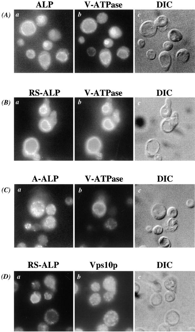
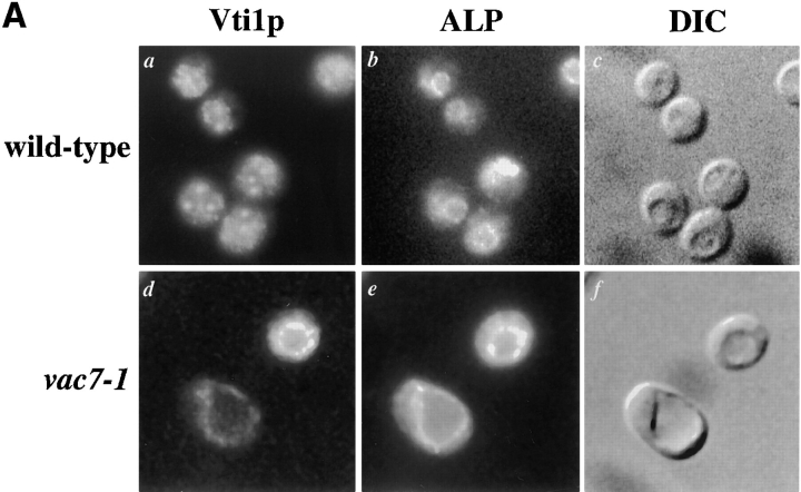
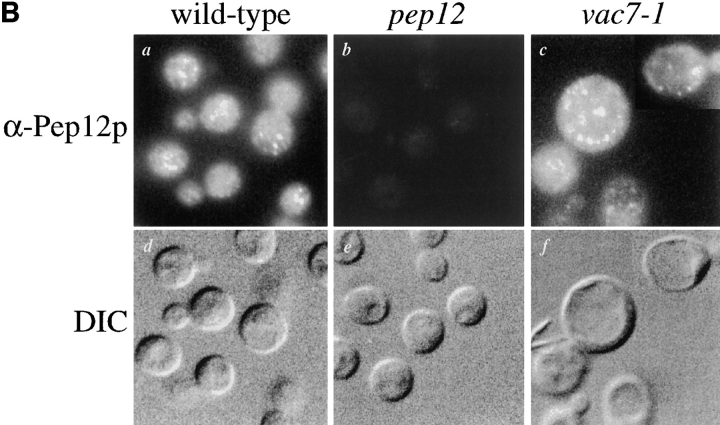
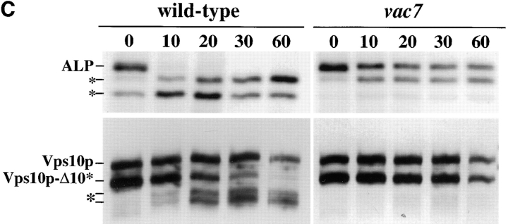
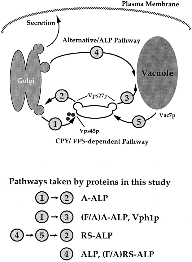
A large number of trafficking steps occur between the last compartment of the Golgi apparatus (TGN) and the vacuole of the yeast Saccharomyces cerevisiae. To date, two intracellular routes from the TGN to the vacuole have been identified. Carboxypeptidase Y (CPY) travels through a prevacuolar/endosomal compartment (PVC), and subsequently on to the vacuole, while alkaline phosphatase (ALP) bypasses this compartment to reach the same organelle. Proteins resident to the TGN achieve their localization despite a continuous flux of traffic by continually being retrieved from the distal PVC by virtue of an aromatic amino acid-containing sorting motif. In this study we report that a hybrid protein based on ALP and containing this retrieval motif reaches the PVC not by following the CPY sorting pathway, but instead by signal-dependent retrograde transport from the vacuole, an organelle previously thought of as a terminal compartment. In addition, we show that a mutation in VAC7, a gene previously identified as being required for vacuolar inheritance, blocks this trafficking step. Finally we show that Vti1p, a v-SNARE required for the delivery of both CPY and ALP to the vacuole, uses retrograde transport out of the vacuole as part of its normal cellular itinerary.


Figures

Figure 1
Figure 1
(A) Schematic representation of proteins used in this study. Sequences derived from DPAP A are shown shaded and those from ALP are unshaded. (B) Localization of ALP, A-ALP, (F/A)A-ALP, RS-ALP, and (F/A)RS-ALP in wild-type cells. NBY72 (pho8Δ-X pep4-3) cells harboring pSN92 (ALP; a, f, and k), pSN55 (A-ALP; b, g, and l), pSN100 ((F/A)A-ALP; c, h, and m), pSN97 (RS-ALP; d, i, and n), or pSN123 ((F/A)RS-ALP; e, j, and o) were prepared for double labeling indirect immunofluorescence using the α-ALP mAb, 1D3-A10 (a–e) and affinity-purified antibodies against the 100-kD subunit of the V-ATPase, Vph1p, to show the localization pattern of the V-ATPase (f–j) as described in Materials and Methods. Cells were also visualized using DIC microscopy (k–o).
Figure 1
Figure 1
(A) Schematic representation of proteins used in this study. Sequences derived from DPAP A are shown shaded and those from ALP are unshaded. (B) Localization of ALP, A-ALP, (F/A)A-ALP, RS-ALP, and (F/A)RS-ALP in wild-type cells. NBY72 (pho8Δ-X pep4-3) cells harboring pSN92 (ALP; a, f, and k), pSN55 (A-ALP; b, g, and l), pSN100 ((F/A)A-ALP; c, h, and m), pSN97 (RS-ALP; d, i, and n), or pSN123 ((F/A)RS-ALP; e, j, and o) were prepared for double labeling indirect immunofluorescence using the α-ALP mAb, 1D3-A10 (a–e) and affinity-purified antibodies against the 100-kD subunit of the V-ATPase, Vph1p, to show the localization pattern of the V-ATPase (f–j) as described in Materials and Methods. Cells were also visualized using DIC microscopy (k–o).
Figure 2
Figure 2
(A) Localization of ALP, (F/A)A-ALP, and (F/A)RS-ALP in vps45Δ cells. NBY83 (vps45Δ-X pho8Δ-X pep4Δ-X) harboring pSN92 (ALP; a, d, and g), pSN100 ((F/A)A-ALP; b, e, and h) or pSN123 ((F/A)RS-ALP; c, f, and i) were prepared for double labeling indirect immunofluorescence using the α-ALP mAb, 1D3-A10 (a–c) and affinity-purified antibodies against the 100-kD subunit of the V-ATPase, Vph1p (d–f), as described in Materials and Methods. Cells were also visualized using DIC microscopy (g–i). (B) Kinetics of processing of ALP, A-ALP, (F/A)A-ALP, RS-ALP, and (F/A)RS-ALP in wild-type and vps45Δ cells. AACY28 (wild type) and NBY68 (vps45Δ-X) pho8Δ-X PEP4 cells harboring pSN92 (ALP), pSN55 (A-ALP), pSN100 ((F/A)A-ALP), pSN97 (RS-ALP), or pSN123 ((F/A)RS-ALP) were labeled with [35S]Met for 10 min and chased by adding unlabeled methionine and cysteine, each to a final concentration of 50 μg/ ml. At the indicated times, proteins were immunoprecipitated from cell extracts using polyclonal antibodies against ALP. The resulting immunoprecipitates were subjected to SDS-PAGE and fluorography. The products of PEP4-dependent proteolysis are indicated using asterisks. (C) Model depicting the pathways taken by CPY and ALP to the vacuole. CPY reaches the vacuole by firstly transiting through a prevacuolar/endosomal compartment (PVC). Entry of proteins into this compartment requires the product of VPS45. Exit of proteins from the PVC, both back to the TGN and on to the vacuole requires the product of VPS27. ALP follows an alternative pathway to the vacuole bypassing the trafficking intermediates defined by mutations in VPS45 and VPS27. The large shaded arrow depicts the retrograde membrane trafficking pathway proposed to be taken by RS-ALP out of the vacuole to reach the PVC.
Figure 2
Figure 2
(A) Localization of ALP, (F/A)A-ALP, and (F/A)RS-ALP in vps45Δ cells. NBY83 (vps45Δ-X pho8Δ-X pep4Δ-X) harboring pSN92 (ALP; a, d, and g), pSN100 ((F/A)A-ALP; b, e, and h) or pSN123 ((F/A)RS-ALP; c, f, and i) were prepared for double labeling indirect immunofluorescence using the α-ALP mAb, 1D3-A10 (a–c) and affinity-purified antibodies against the 100-kD subunit of the V-ATPase, Vph1p (d–f), as described in Materials and Methods. Cells were also visualized using DIC microscopy (g–i). (B) Kinetics of processing of ALP, A-ALP, (F/A)A-ALP, RS-ALP, and (F/A)RS-ALP in wild-type and vps45Δ cells. AACY28 (wild type) and NBY68 (vps45Δ-X) pho8Δ-X PEP4 cells harboring pSN92 (ALP), pSN55 (A-ALP), pSN100 ((F/A)A-ALP), pSN97 (RS-ALP), or pSN123 ((F/A)RS-ALP) were labeled with [35S]Met for 10 min and chased by adding unlabeled methionine and cysteine, each to a final concentration of 50 μg/ ml. At the indicated times, proteins were immunoprecipitated from cell extracts using polyclonal antibodies against ALP. The resulting immunoprecipitates were subjected to SDS-PAGE and fluorography. The products of PEP4-dependent proteolysis are indicated using asterisks. (C) Model depicting the pathways taken by CPY and ALP to the vacuole. CPY reaches the vacuole by firstly transiting through a prevacuolar/endosomal compartment (PVC). Entry of proteins into this compartment requires the product of VPS45. Exit of proteins from the PVC, both back to the TGN and on to the vacuole requires the product of VPS27. ALP follows an alternative pathway to the vacuole bypassing the trafficking intermediates defined by mutations in VPS45 and VPS27. The large shaded arrow depicts the retrograde membrane trafficking pathway proposed to be taken by RS-ALP out of the vacuole to reach the PVC.
Figure 3
Figure 3
Kinetics of processing of ALP, (F/A)A-ALP, and (F/A) RS-ALP in wild-type and apm3Δ cells. SNY17 (wild-type) and NBY100 (apm3::HIS3) pho8Δ-X PEP4 cells harboring pSN92 (ALP), pSN55 (A-ALP), or pSN123 ((F/A)RS-ALP) were labeled with [35S]Met for 10 min and chased by adding unlabeled methionine and cysteine each to a final concentration of 50 μg/ml. At the indicated times, proteins were immunoprecipitated from cell extracts using polyclonal antibodies against ALP. The resulting immunoprecipitates were subjected to SDS-PAGE and fluorography.
Figure 4
Figure 4
(A) Localization of ALP, A-ALP, (F/A)A-ALP, RS-ALP, and (F/A)RS-ALP in vps27Δ cells. NBY60 (vps27Δ pho8Δ-X pep4-3) cells harboring pSN92 (ALP; a, f, and k), pSN55 (A-ALP; b, g, and l), pSN100 ((F/A)A-ALP; c, h, and m), pSN97 (RS-ALP; d, i, and n), or pSN123 ((F/A)RS-ALP; e, j, and o) were prepared for double-labeling indirect immunofluorescence using the α-ALP mAb, 1D3-A10 (a–e) and affinity-purified antibodies against the 100-kD subunit of the V-ATPase, Vph1p (f–j) as described in Materials and Methods. Cells were also visualized using DIC microscopy (k–o). (B) Rate of PEP4-dependent cleavage of ALP, (F/A)A-ALP, RS-ALP, and Vps10p-Δ10* in vps27-ts cells. RPY103 (VPS10::LEU2::vps10-10* pep4-3 vps27-ts) cells and NBY73 (pho8Δ-X pep4-3 vps27-ts) carrying the GAL1-PEP4 plasmid, pRCP39 and either pNB8 ((F/A)A-ALP) or pNB7 (RS-ALP). Cells were grown in galactose-containing media for 24 h at 22°C, and then shifted to glucose-containing media for 24 h. Before labeling with [35S]Met for 10 min, cells were either maintained at 22°C or shifted to 37°C for 10 min. Chase times used are indicated, after which aliquots were removed from which proteins were immunoprecipitated using α-ALP and α-Vps10p antibodies.
Figure 4
Figure 4
(A) Localization of ALP, A-ALP, (F/A)A-ALP, RS-ALP, and (F/A)RS-ALP in vps27Δ cells. NBY60 (vps27Δ pho8Δ-X pep4-3) cells harboring pSN92 (ALP; a, f, and k), pSN55 (A-ALP; b, g, and l), pSN100 ((F/A)A-ALP; c, h, and m), pSN97 (RS-ALP; d, i, and n), or pSN123 ((F/A)RS-ALP; e, j, and o) were prepared for double-labeling indirect immunofluorescence using the α-ALP mAb, 1D3-A10 (a–e) and affinity-purified antibodies against the 100-kD subunit of the V-ATPase, Vph1p (f–j) as described in Materials and Methods. Cells were also visualized using DIC microscopy (k–o). (B) Rate of PEP4-dependent cleavage of ALP, (F/A)A-ALP, RS-ALP, and Vps10p-Δ10* in vps27-ts cells. RPY103 (VPS10::LEU2::vps10-10* pep4-3 vps27-ts) cells and NBY73 (pho8Δ-X pep4-3 vps27-ts) carrying the GAL1-PEP4 plasmid, pRCP39 and either pNB8 ((F/A)A-ALP) or pNB7 (RS-ALP). Cells were grown in galactose-containing media for 24 h at 22°C, and then shifted to glucose-containing media for 24 h. Before labeling with [35S]Met for 10 min, cells were either maintained at 22°C or shifted to 37°C for 10 min. Chase times used are indicated, after which aliquots were removed from which proteins were immunoprecipitated using α-ALP and α-Vps10p antibodies.
Figure 5
Figure 5
Localization of ALP, A-ALP, (F/A)A-ALP, RS-ALP, and (F/A)RS-ALP in vps27Δ vps45Δ double mutant cells. NBY84 (vps27Δ vps45Δ pho8Δ-X pep4-3) cells harboring pSN92 (ALP; a, f, and k), pSN55 (A-ALP; b, g, and l), pSN100 ((F/A)A-ALP; c, h, and m), pSN97 (RS-ALP; d, i, and n), or pSN123 ((F/A)RS-ALP; e, j, and o) were prepared for double labeling indirect immunofluorescence using the α-ALP mAb 1D3-A10 (a–e) and affinity-purified antibodies against the 100-kD subunit of the V-ATPase, Vph1p (f–j), as described in Materials and Methods. Cells were also visualized using DIC microscopy (k–o).
Figure 6
Figure 6
RS-ALP is mislocalized to the vacuolar membrane of vac7 mutant cells. (A) NBY85 (vac7-1 pho8Δ-X pep4Δ-X) cells harboring pSN92 (ALP), (B) pSN97 (RS-ALP), and (C) pSN55 (A-ALP) were prepared for double labeling indirect immunofluorescence using the α-ALP mAb 1D3-A10, and affinity-purified antibodies against the 100-kD subunit of the V-ATPase, Vph1p, as described in Materials and Methods. Cells were also visualized using DIC microscopy (k–o). (D) NBY85 (vac7-1 pho8Δ-X pep4Δ-X) cells harboring pSN97 (RS-ALP) were prepared for double labeling indirect immunofluorescence using the α-ALP mAb 1D3-A10 and affinity-purified antibodies against Vps10p, as described in Materials and Methods. Cells were also visualized using DIC microscopy.
Figure 7
Figure 7
Mislocalization of Vti1p in vac7-1 cells. (A) Wild-type (NBY86; pho8Δ-X pep4Δ-X) and vac7-1 (NBY85; vac7-1 pho8Δ-X pep4Δ-X) cells harboring pSN92 (ALP) were prepared for double labeling indirect immunofluorescence using the α-ALP mAb 1D3-A10 and affinity-purified antibodies against Vti1p, as described in Materials and Methods. (B) Pep12p was immunolocalized in wild-type (NBY86; pho8Δ-X pep4Δ-X) and vac7-1 (NBY85; vac7-1 pho8Δ-X pep4Δ-X) cells harboring pSN92 (ALP) as well as in cells that do not synthesize Pep12p (vpt13), as described in Materials and Methods. In both cases, cells were also visualized using DIC microscopy. (C) Kinetics of processing of ALP and Vps10p-Δ10* in vac7-1 cells. SEY6210 (wild-type) and LWY2809 (vac7-1) PEP4 cells were labeled with [35S]Met for 10 min and chased by adding unlabeled methionine and cysteine each to a final concentration of 50 μg/ml. At the indicated times proteins were immunoprecipitated from cell extracts using polyclonal antibodies against ALP and Vps10p. The resulting immunoprecipitates were subjected to SDS-PAGE and fluorography. The products of PEP4-dependent proteolysis of both ALP and Vps10p-Δ10* are indicated using asterisks.
Figure 7
Figure 7
Mislocalization of Vti1p in vac7-1 cells. (A) Wild-type (NBY86; pho8Δ-X pep4Δ-X) and vac7-1 (NBY85; vac7-1 pho8Δ-X pep4Δ-X) cells harboring pSN92 (ALP) were prepared for double labeling indirect immunofluorescence using the α-ALP mAb 1D3-A10 and affinity-purified antibodies against Vti1p, as described in Materials and Methods. (B) Pep12p was immunolocalized in wild-type (NBY86; pho8Δ-X pep4Δ-X) and vac7-1 (NBY85; vac7-1 pho8Δ-X pep4Δ-X) cells harboring pSN92 (ALP) as well as in cells that do not synthesize Pep12p (vpt13), as described in Materials and Methods. In both cases, cells were also visualized using DIC microscopy. (C) Kinetics of processing of ALP and Vps10p-Δ10* in vac7-1 cells. SEY6210 (wild-type) and LWY2809 (vac7-1) PEP4 cells were labeled with [35S]Met for 10 min and chased by adding unlabeled methionine and cysteine each to a final concentration of 50 μg/ml. At the indicated times proteins were immunoprecipitated from cell extracts using polyclonal antibodies against ALP and Vps10p. The resulting immunoprecipitates were subjected to SDS-PAGE and fluorography. The products of PEP4-dependent proteolysis of both ALP and Vps10p-Δ10* are indicated using asterisks.
Figure 8
Figure 8
Model for membrane trafficking pathways between the TGN and the vacuole of S. cerevisiae. There are at least two routes taken by proteins from the TGN to the PVC. Proteins that follow the CPY pathway to the vacuole transit through the PVC in a VPS45, VPS27-dependent manner. From the PVC, these proteins are then delivered to the vacuole. Recycling TGN membrane proteins such as A-ALP are retrieved from the PVC to the TGN. The second route from the TGN to the vacuole does not transit through the PVC and is taken by ALP, RS-ALP, and (F/ A)RS-ALP. After delivery to the vacuolar membrane, RS-ALP follows the newly identified retrograde membrane trafficking step in a VAC7-dependent manner by virtue of its FXFXD retrieval motif.

References

    1. Akasaki K, Fukuzawa M, Kinoshita H, Furuno K, Tsuji H. Cycling of two endogenous lysosomal membrane proteins, lamp-2 and acid phosphatase, between the cell surface and lysosomes in cultured rat hepatocytes. J Biochem (Tokyo) 1993;114:598–604. - PubMed
    1. Akasaki K, Michinhara A, Mibuka K, Fujiwara Y, Tsuji H. Biosynthetic transport of a major lysosomal membrane glycoprotein, lamp-1: convergence of biosynthetic and endocytic pathways occurs at three distinctive points. Exp Cell Res. 1995;220:464–473. - PubMed
    1. Ammerer G, Hunter CP, Rothman JH, Saari GC, Valls LA, Stevens TH. PEP4 gene of Saccharomyces cerevisiaeencodes proteinase A, a vacuolar enzyme required for processing of vacuolar precursors. Mol Cell Biol. 1986;6:2490–2499. - PMC - PubMed
    1. Babst M, Sato TK, Banta LM, Emr SD. Endosomal transport function in yeast requires a novel AAA-type ATPase, Vps4p. EMBO (Eur Mol Biol Organ) J. 1997;16:1820–1831. - PMC - PubMed
    1. Becherer KA, Rieder SE, Emr SD, Jones EW. Novel syntaxin homolog, Pep12p, required for the sorting of lumenal hydrolases to the lysosome-like vacuole in yeast. Mol Biol Cell. 1996;7:579–594. - PMC - PubMed

Publication types

MeSH terms