. 1998 Aug 10;142(3):665-81.
doi: 10.1083/jcb.142.3.665.

A membrane coat complex essential for endosome-to-Golgi retrograde transport in yeast

Affiliations

A membrane coat complex essential for endosome-to-Golgi retrograde transport in yeast

M N Seaman et al. J Cell Biol. .

Abstract

We have recently characterized three yeast gene products (Vps35p, Vps29p, and Vps30p) as candidate components of the sorting machinery required for the endosome-to-Golgi retrieval of the vacuolar protein sorting receptor Vps10p (Seaman, M.N.J., E.G. Marcusson, J.-L. Cereghino, and S.D. Emr. 1997. J. Cell Biol. 137:79-92). By genetic and biochemical means we now show that Vps35p and Vps29p interact and form part of a multimeric membrane-associated complex that also contains Vps26p, Vps17p, and Vps5p. This complex, designated here as the retromer complex, assembles from two distinct subcomplexes comprising (a) Vps35p, Vps29p, and Vps26p; and (b) Vps5p and Vps17p. Density gradient fractionation of Golgi/endosomal/vesicular membranes reveals that Vps35p cofractionates with Vps5p/Vps17p in a vesicle-enriched dense membrane fraction. Furthermore, gel filtration analysis indicates that Vps35p and Vps5p are present on a population of vesicles and tubules slightly larger than COPI/coatomer-coated vesicles. We also show by immunogold EM that Vps5p is localized to discrete regions at the rims of the prevacuolar endosome where vesicles appear to be budding. Size fractionation of cytosolic and recombinant Vps5p reveals that Vps5p can self-assemble in vitro, suggesting that Vps5p may provide the mechanical impetus to drive vesicle formation. Based on these findings we propose a model in which Vps35p/Vps29p/Vps26p function to select cargo for retrieval, and Vps5p/Vps17p assemble onto the membrane to promote vesicle formation. Conservation of the yeast retromer complex components in higher eukaryotes suggests an important general role for this complex in endosome-to-Golgi retrieval.


Figures

Figure 1
Figure 1
Vps35p functionally interacts with Vps29p. (A) Wild-type (SEY 6210) cells expressing a dominant negative vps35 allele were transformed with multicopy (2μ) plasmids to overexpress VPS29, VPS30, or VPS35. Control cells were transformed with empty vector. Cells were pulse-labeled with [35S]methionine for 10 min, and were then chased in the presence of excess cold methionine and cysteine for 30 min. The cells were then converted to spheroplasts, and intracellular and extracellular fractions were separated by centrifugation. CPY was immunoprecipitated from resulting lysates, and was subjected to electrophoresis on an 8% SDS polyacrylamide gel. The proteins were visualized by fluorography. (B) Wild-type (SEY 6210) and mutant strains in which VPS29, VPS30, or VPS35 are deleted (PSY1-29, JCY3000, and EMY18, respectively) were converted to spheroplasts and then labeled for 15 min with [35S]methionine. After adding excess cold methionine/cysteine, the cells were chased for 45 min to allow the labeled proteins to reach a steady state distribution. The cells were then lysed and fractionated by differential centrifugation. Vps29p was immunoprecipitated from the resulting fractions, and was then resolved on an 8% SDS polyacrylamide gel and visualized by fluorography. The association of Vps29p with a 100,000 g membrane fraction (P100) was found to be dependent upon Vps35p.
Figure 2
Figure 2
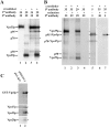
Vps35p and Vps29p are part of a multimeric membrane associated complex. (A) A cleared lysate from [35S]methionine-labeled wild-type cells (SEY 6210) was either left untreated (lanes 1 and 2) or treated with cross-linker (lanes 3 and 4) as described in Materials and Methods. After cross-linking, the proteins were precipitated, and either Vps35p (lanes 1 and 3) or Vps29p (lanes 2 and 4) was immunoprecipitated. The immunoprecipitates were subjected to SDS-PAGE on an 8% gel, and the proteins were visualized by fluorography. Strikingly, the pattern of bands seen in the cross-linked samples (lanes 3 and 4) was very similar, indicating that Vps35p and Vps29p interact with a common set of proteins. (B) P100 membranes were isolated from wild-type (SEY 6210) [35S]methionine-labeled cells and resuspended into 100 μl of lysis buffer. The cross-linker DTSSP was added, and the samples were allowed to cross-link for 30 min at room temperature. After cross-linking, precipitation by adding TCA, and resuspending the proteins, Vps35p (lanes 1, 2, 4, 6, and 7) or Vps29p (lane 3) was immunoprecipitated from the lysate. These primary immunoprecipitates were then resuspended into 100 μl urea buffer, and in the case of lanes 2, 4, 6, and 7, reducing agent was added. 900 μl of immunoprecipitation buffer was added, and the samples were further immunoprecipitated with antibodies against either Vps35p (lane 1), Vps29p (lanes 2, 3, and 4), Vps30p (lane 6), or Vps5 (lane 7). The supernatant from the sample in lane 1 (after the primary immunoprecipitation) was retained, and antibodies against Vps5p were added (lane 5). Proteins were resolved on an 8% SDS polyacrylamide gel. Vps35p could be cross-linked to Vps29p, and the proteins designated as p95 and p70 were found to have an identical migration to Vps5p and Vps17p. Immunoprecipitation of Vps5p from cross-linked lysates demonstrated the presence of this protein in a complex that also contained Vps35p and Vps29p. (C) GST-Vps5p expressed in yeast interacts with Vps17p, Vps35p, and Vps29p. vps5Δ cells (BHY152) expressing either pMSGST-5 or pEG (KT; empty vector) (Mitchell et al., 1993) were lysed in Hepes lysis buffer containing 0.5% Triton. The lysate was cleared by centrifugation and then incubated with glutathione-sepharose for 15 min. After several washes, the bound proteins were eluted and precipitated. The proteins were resolubilized in SDS-PAGE sample buffer, separated on an 8% polyacrylamide gel, and then transferred to nitrocellulose for Western blotting. GST-Vps5p was able to associate with Vps17p, Vps35p, and Vps29p (lane 2). GST alone did not associate with Vps17p, Vps35p, or Vps29p (lane 1).
Figure 3
Figure 3
Identification of p50 and assembly of the multimeric complex in mutant strains. (A) Lysates from either wild-type or vps26Δ cells that had been pulse-labeled with [35S]methionine were either treated with cross-linker (lanes 2 and 3) or left untreated (lane 1). After cross-linking, proteins were precipitated, washed with acetone, and resuspended into immunoprecipitation buffer. Vps35p was immunoprecipitated, and the proteins were resolved on an 8% SDS polyacrylamide gel. The band referred to as p50 (Vps26p) is missing in lysates prepared from vps26Δ cells, and yet Vps35p is still able to associate with Vps29p, Vps5p, and Vps17p. (B) Wild-type cells (SEY 6210) or vps35 (EMY18), vps30 (JCY3000), vps29 (PSY1-29), vps17 (KKY11), or vps5 (BHY152) mutants were converted to spheroplasts, labeled with [35S]methionine, chased, and then lysed with the Hepes lysis buffer. The lysate was cleared by centrifugation at 2,000 rpm in a microfuge, and then cross-linker was added to a final concentration of 2 mM. Cross-linking proceeded at room temperature for 30 min, and was then stopped by adding 100 μl of TCA and transferring the samples to ice. The proteins were precipitated and washed with acetone, and a lysate in immunoprecipitation buffer was prepared as before. Vps35p or Vps29p was immunoprecipitated from the lysates using double immunoprecipitations under nonreducing conditions. Interaction of Vps35p with Vps5p/Vps17p required Vps29p, and likewise Vps29p could not interact with Vps5p/Vps17p in a vps35 mutant.
Figure 4
Figure 4
Vps26p is required for the correct localization of Vps10p. Wild-type (SEY 6210) or vps26 (MSY2600) mutant cells were converted to spheroplasts and labeled with [35S]methionine for 15 min. Excess cold methionine/cysteine was then added, and the cells were chased for 45 min, after which the cells were lysed in Hepes buffer and the lysate was spun at 2,000 rpm to remove unbroken cells. Differential centrifugation (13,000 g, then 100,000 g) was then used to separate larger membranes such as vacuoles (P13) from small membrane compartments such as Golgi, endosomes, vesicles (P100), and cytosol (S100). The fractions were treated with TCA to precipitate the proteins that were then washed with acetone and resuspended into immunoprecipitation buffer. Vps10p was recovered from the different fractions by immunoprecipitation, and was resolved by SDS-PAGE. The proteins were visualized by fluorography. Deletion of VPS26 causes a dramatic shift in the distribution of Vps10p to a vacuolar membrane fraction.
Figure 5
Figure 5
Colocalization of Vps35p and Vps5p in a dense membrane fraction. P100 membranes from [35S]methionine-labeled wild-type cells (SEY 6210) were isolated by differential centrifugation. The membranes were then loaded onto a 10–60% sucrose gradient and spun to equilibrium. Fractions collected from the gradient were precipitated with TCA and washed with acetone, and various proteins were recovered by immunoprecipitation. Vps35p and Vps29p are associated with two discrete pools of membranes. The lighter pool contains the endosomal t-SNARE Pep12p, while the denser population of membranes appears denser than the late-Golgi marker Kex2p. Vps35p (and Vps29p) colocalizes with Vps5p and Vps17p in the denser fractions.
Figure 6
Figure 6
Analysis of the size of the membranes on which Vps35p and Vps5p associate. (A) 100,000 g membranes were prepared from wild-type cells carrying a myc-tagged version of Vps10p on the chromosome (MSY10-21). The membranes were resuspended into Hepes lysis buffer and loaded onto a sephacryl S1000 column preequilibrated with Hepes lysis buffer. The membranes were eluted using the Hepes lysis buffer, and ∼2.5-ml fractions were collected. Fractions were analyzed for the presence of Vps35p and Vps5p (and other proteins) by Western blotting. The membranes with which Vps35p and Vps5p are associated elute after membranes that contain Vps10p (and Kex2p), but before COPI/coatomer-coated vesicles. (B) A collage of images of anti-HA labeled membranes. A P100 membrane fraction from wild-type cells (SEY 6210) expressing pAH31 (HA-tagged VPS5) (Nothwehr and Hindes, 1997) was fixed with 2% paraformaldehyde and then prepared for cryosectioning. Frozen thin sections were labeled with anti-HA monoclonal antibodies, which were followed by anti-mouse antibodies coupled to 5-nm colloidal-gold. Labeling was predominately restricted to vesicular and tubulovesicular structures. Bar, 100 nm.
Figure 6
Figure 6
Analysis of the size of the membranes on which Vps35p and Vps5p associate. (A) 100,000 g membranes were prepared from wild-type cells carrying a myc-tagged version of Vps10p on the chromosome (MSY10-21). The membranes were resuspended into Hepes lysis buffer and loaded onto a sephacryl S1000 column preequilibrated with Hepes lysis buffer. The membranes were eluted using the Hepes lysis buffer, and ∼2.5-ml fractions were collected. Fractions were analyzed for the presence of Vps35p and Vps5p (and other proteins) by Western blotting. The membranes with which Vps35p and Vps5p are associated elute after membranes that contain Vps10p (and Kex2p), but before COPI/coatomer-coated vesicles. (B) A collage of images of anti-HA labeled membranes. A P100 membrane fraction from wild-type cells (SEY 6210) expressing pAH31 (HA-tagged VPS5) (Nothwehr and Hindes, 1997) was fixed with 2% paraformaldehyde and then prepared for cryosectioning. Frozen thin sections were labeled with anti-HA monoclonal antibodies, which were followed by anti-mouse antibodies coupled to 5-nm colloidal-gold. Labeling was predominately restricted to vesicular and tubulovesicular structures. Bar, 100 nm.
Figure 7
Figure 7
Immunolocaliztion of Vps5p by EM. vps4 cells (SEY 4-1) harboring the plasmid pAH31 (HA-tagged VPS5) (Nothwehr and Hindes, 1997) were fixed and prepared for cryoelectron microscopy. (A) Frozen thin sections were labeled with the anti-HA antibody to localize Vps5p, and were followed by 5 nm anti-mouse colloidal-gold. Labeling was found to be restricted to discrete regions of the prevacuolar endosomal membrane (Ec), and appeared most concentrated at the swollen rim of the cisterna (B). Also shown are the nucleus (n) and plasma membrane (pm). Bar, 100 nm.
Figure 8
Figure 8
Sizing the components of the multimeric complex reveals the existence of two distinct subcomplexes. P100 membranes prepared from wild-type cells (SEY 6210) were stripped by resuspending the pellet in Hepes lysis buffer containing 250 mM NaCl. The membranes were pelleted by centrifugation at 100,000 g. The supernatant was loaded onto a sephacryl S300 column equilibrated with lysis buffer plus 250 mM NaCl. Fractions were precipitated with TCA and analyzed by Western blotting. The bulk of Vps5p and Vps17p remain associated with each other and form a complex with a predicted size of 400–500 kD. Vps35p remains associated with Vps29p in an ∼230-kD complex.
Figure 9
Figure 9
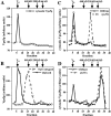
Cytosolic Vps5p assembles into a large (>10 6 D) complex. (A) Cytosol (100,000 g supernatant) prepared from wild-type cells (SEY 6210) lysed in Hepes lysis buffer was fractionated on a sephacryl S300 column. Fractions collected were precipitated and analyzed for the presence of Vps5p by Western blotting. Resulting autoradiograms were scanned and quantitated using National Institutes of Health Image (version 1.61) software. Vps5p is found almost entirely in the void volume fraction, suggesting that it is part of a complex of >106 D. (B) Vps5p that had been stripped from P100 membranes using 250 mM NaCl (as in Fig. 8) was dialyzed against Hepes lysis buffer to remove the salt. The supernatant was briefly spun at 13,000 g, and was then fractionated on a sephacryl S300 column. Fractions were collected, precipitated, and analyzed by Western blotting. The autoradiogram was quantitated as above. Vps5p is able to assemble into a large (>106-D) complex, and elutes in the void volume fraction (solid line). This contrasts with the size of Vps5p when fractionated in the continued presence of 250 mM NaCl (broken line; this data is the quantitation of the Vps5p signal in Fig. 8). In C, the fractionation of cytosolic Vps35p is analyzed. Vps35p is found in the void volume fraction in cytosol prepared from wild-type cells (SEY 6210) (solid line). However, when cytosol is prepared from vps29Δ cells (PSY 1-29), the bulk of Vps35p now elutes in a later fraction (broken line), indicating that Vps29p function is required for Vps35p to assemble into a large complex. Similarly in D, Vps29p in wild-type (SEY 6210) cytosol will partially fractionate in the void volume fraction (solid line), but when cytosol is prepared from vps35Δ (EMY 18) cells, all the detectable Vps29p is found in later fractions. Thus, Vps35p and Vps29p appear to assemble together into a large complex that elutes in the void volume from a sephacryl S300 column.
Figure 10
Figure 10
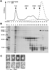
Recombinant Vps5p shows self-assembly activity. (A) Affinity-purified recombinant GST-Vps5p and GST alone was dialyzed against the Hepes lysis buffer before being loaded onto a sephacryl S300 gel filtration column. The elution profile of both GST-Vps5p (solid line) or GST (broken line) is shown. (B) The GST-Vps5p–containing fractions from the sephacryl S300 column were analyzed by SDS-PAGE. Lane M denotes the molecular mass markers, and lane L is the GST-Vps5p before fractionation on the sephacryl S300 column. The void volume fraction (6) contains predominately full-length GST-Vps5p, while the abundant breakdown products of the recombinant GST-Vps5p elute later. (C) The void volume fraction was adsorbed to a formvar/carbon-coated grid and negatively stained with uranyl acetate for examination by electron microscopy (upper four micrographs). In the lower four micrographs, the void volume fraction was labeled with anti-Vps5p antisera and 5 nm anti-rabbit colloidal gold before negative staining. The GST-Vps5p was found to have assembled into a homogeneous population of 15–20-nm spherical particles.
Figure 11
Figure 11
A schematic model for the mechanism of retromer-mediated vesicle formation at the endosome. (A) The proposed sequence of events involves Vps35p interacting with cytoplasmic tails of cargo such as Vps10p and Kex2p. Vps29p then activates Vps35p, causing clustering of the Vps35p and cargo. Vps5p/ Vps17p then assemble onto the membrane to drive vesicle formation, possibly with Vps17p providing the membrane attachment activity. (B) Schematic of the two subcomplexes of the retromer that assemble together onto the endosome membrane. The inner shell of the retromer coat would be the Vps35p/Vps29p/Vps26p subcomplex, while Vps5p/Vps17p form the outer shell.

References

    1. Babst M, Sato TK, Banta LM, Emr SD. Endosomal transport function in yeast requires a novel AAA-type ATPase, Vps4p. EMBO (Eur Mol Biol Organ) J. 1997;16:1820–1831. - PMC - PubMed
    1. Bachhawat AK, Suhan J, Jones EW. The yeast homologue of Hβ58, a mouse gene essential for embryogenesis performs a role in the delivery of proteins to the vacuole. Genes Dev. 1994;8:1379–1387. - PubMed
    1. Bankaitis VA, Johnson LM, Emr SD. Isolation of yeast mutants defective in protein targeting to the vacuole. Proc Natl Acad Sci USA. 1986;83:9075–9079. - PMC - PubMed
    1. Banta LM, Robinson JS, Klionsky DJ, Emr SD. Organelle assembly in yeast: characterization of yeast mutants defective in vacuolar biogenesis and protein sorting. J Cell Biol. 1988;107:1369–1383. - PMC - PubMed
    1. Becherer KA, Rieder SE, Emr SD, Jones EW. Novel syntaxin homologue, Pep12p, required for the sorting of lumenal hydrolases to the lysosome-like vacuole in yeast. Mol Biol Cell. 1996;7:579–594. - PMC - PubMed

Publication types

MeSH terms