. 1998 Aug 10;142(3):735-50.
doi: 10.1083/jcb.142.3.735.

Interaptin, an actin-binding protein of the alpha-actinin superfamily in Dictyostelium discoideum, is developmentally and cAMP-regulated and associates with intracellular membrane compartments

Affiliations

Interaptin, an actin-binding protein of the alpha-actinin superfamily in Dictyostelium discoideum, is developmentally and cAMP-regulated and associates with intracellular membrane compartments

F Rivero et al. J Cell Biol. .

Abstract

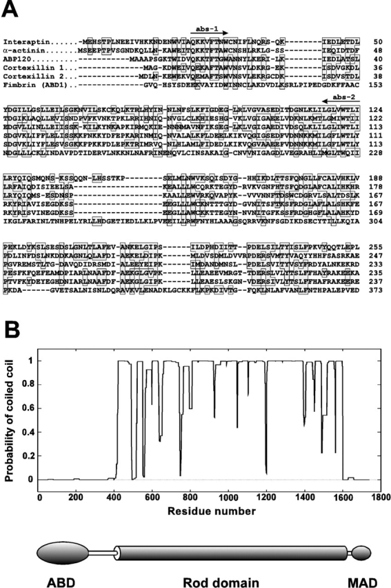
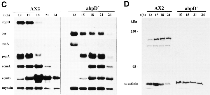
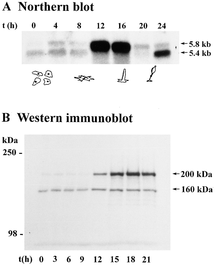
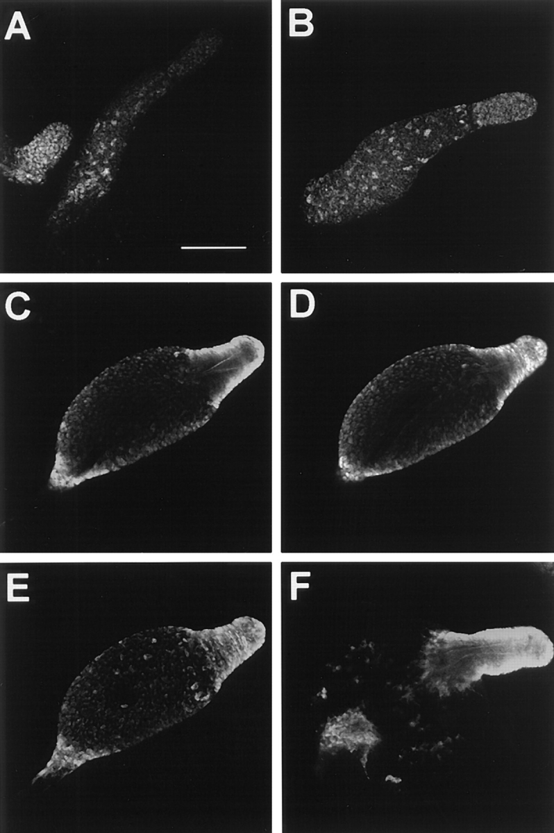
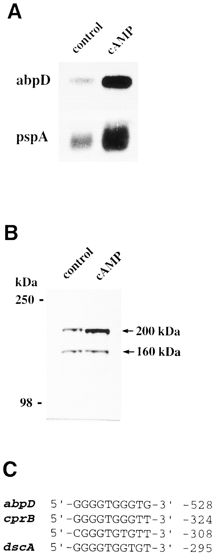
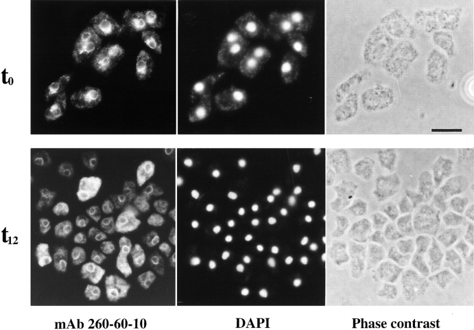
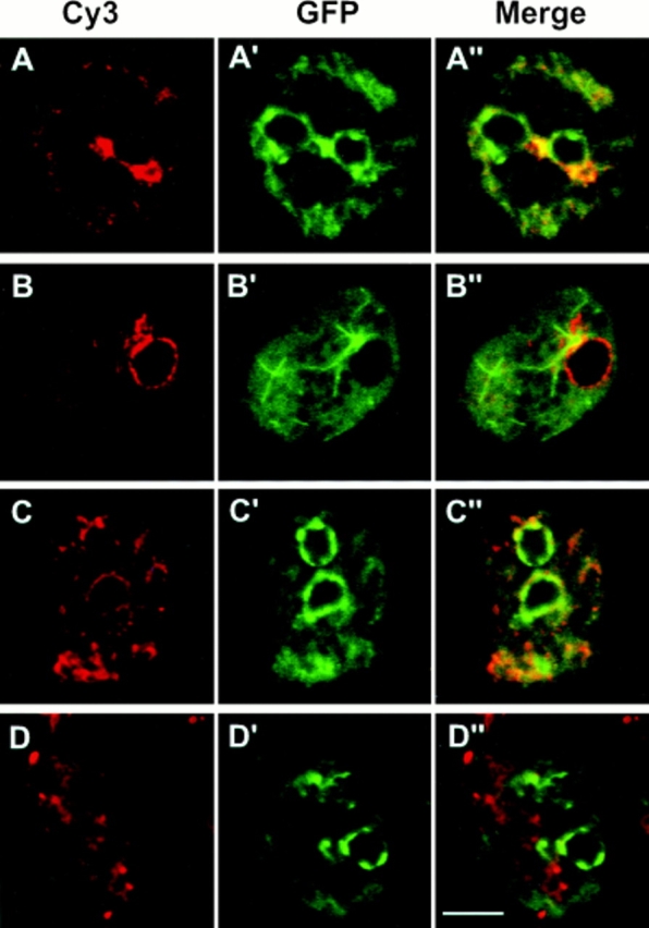
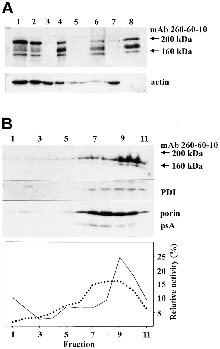
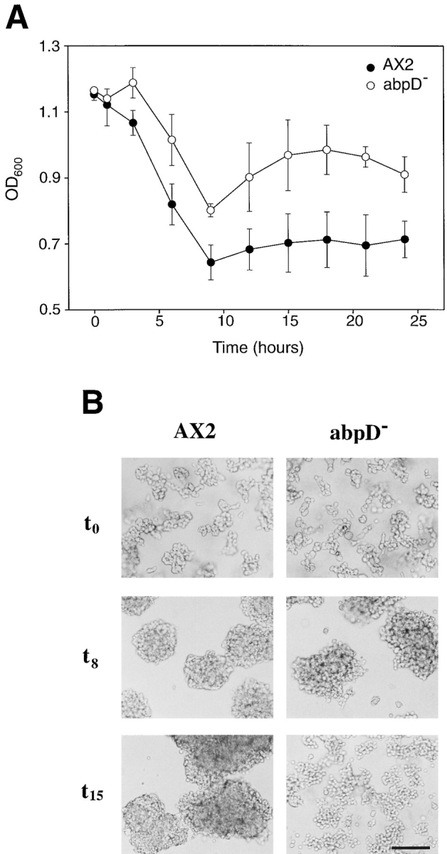
In a search for novel members of the alpha-actinin superfamily, a Dictyostelium discoideum genomic library in yeast artificial chromosomes (YAC) was screened under low stringency conditions using the acting-binding domain of the gelation factor as probe. A new locus was identified and 8.6 kb of genomic DNA were sequenced that encompassed the whole abpD gene. The DNA sequence predicts a protein, interaptin, with a calculated molecular mass of 204,300 D that is constituted by an actin-binding domain, a central coiled-coil rod domain and a membrane-associated domain. In Northern blot analyses a cAMP-stimulated transcript of 5.8 kb is expressed at the stage when cell differentiation occurs. Monoclonal antibodies raised against bacterially expressed interaptin polypeptides recognized a 200-kD developmentally and cAMP-regulated protein and a 160-kD constitutively expressed protein in Western blots. In multicellular structures, interaptin appears to be enriched in anterior-like cells which sort to the upper and lower cups during culmination. The protein is located at the nuclear envelope and ER. In mutants deficient in interaptin development is delayed, but the morphology of the mature fruiting bodies appears normal. When starved in suspension abpD- cells form EDTA-stable aggregates, which, in contrast to wild type, dissociate. Based on its domains and location, interaptin constitutes a potential link between intracellular membrane compartments and the actin cytoskeleton.


Figures

Figure 3
Figure 3
Structural features of interaptin. (A) Alignment of the ABD of interaptin with the ABD of other members of the α-actinin superfamily described in Dictyostelium. For fimbrin only the first ABD has been considered. The alignment was generated using the Clustal W program. Dashes indicate gaps introduced in the sequence for optimal alignment. Residues that are identical between interaptin and any of the other members of the α-actinin superfamily are boxed. Arrows indicate the sequences used to design the degenerate primers for cloning the abpD gene. GeneBank accession numbers: interaptin, AF057019; α-actinin, Y00689; ABP-120, X15430; cortexillin 1, L49527; cortexillin 2, L46371; fimbrin, L36202. (B). Probability of forming α-helical coiled-coil structures. Matrix MTIDK of the COILS version 2.1 algorithm (Lupas et al., 1991) was used with a window size of 28 and weighting. High values are reached through the central portion of the protein, defining a rod domain. The proposed structure of interaptin is shown below. The protein consists of three functional domains connected by serine rich stretches: an NH2-terminal ABD (Met-1 to Leu-255), a central rod domain (Lys-417 to Gln-1597) and a COOH-terminal MAD (Asn-1624 to Thr-1737).
Figure 1
Figure 1
Strategy of cloning and genomic organization of the abpD gene. Five overlapping genomic clones (G1 to G5) encompassing 8.6 kb were isolated and sequenced in both directions. Additionally, a cDNA clone (C5) was obtained after screening a λZAP cDNA library. The coding sequence is interrupted by two introns (shaded) of 417 and 82 base pairs. The abpD locus has been mapped to chromosome 4 of the Dictyostelium genome. DNA fragments used as probes for Northern and Southern blot analyses are depicted as P1 to P5. DNA fragments used for construction of lacZ fusions are depicted as L1 and L2. R, EcoRI restriction sites.
Figure 2
Figure 2
Deduced amino acid sequence of interaptin. The sequence predicts a protein of 204.3 kD. Internal repeats in the rod domain are underlined. A putative tyrosine phosphorylation site is boxed. A stretch of nonpolar amino acids at the COOH terminus is double underlined. Interaptin has received the GeneBank accession number AF057019.
Figure 10
Figure 10
Generation of an abpD mutant by homologous recombination. (A) A construct was made in which the blasticidin resistance cassette (Bsr) was inserted in a genomic clone between the ABD and a portion of the rod domain. The arrows at the bottom indicate the position of the oligonucleotide primers used for the PCR screening of mutants. (B) Southern blot analyses demonstrate that a gene disruption event has occurred. Genomic DNA was digested with EcoRV and blots probed with the 32P-labeled DNA fragments indicated below. With probe 1 insertion of the Bsr cassette causes the shift of a 0.75-kb band to a 2.2-kb band in the mutant. With probe 2 an additional band of ∼6 kb is apparent in the mutant which contains sequences of the pUC118 transformation vector, as shown after hybridization with a pUC probe. (C and D) Development of abpD mutant. AX2 and mutant cells were allowed to develop on nitrocellulose filters. At the indicated time points samples were taken for RNA extraction and Western blot analysis. Northern blots containing 30 μg RNA per lane (C) were probed with probe P1 to demonstrate that no RNA message is present in the mutant, whereas the blasticidin resistance gene (bsr) is expressed. Similar result was obtained with probe P4. Blots were also hybridized with probes for developmentally regulated genes. csaA codes for the cell adhesion molecule contact site A, responsible for cell–cell contacts in early aggregates (Noegel et al., 1986). pspA encodes the prespore-specific cell surface protein psA (Early et al., 1988). ecmA and ecmB encode the prestalk-specific extracellular matrix proteins ST340 and ST310, respectively (Jermyn et al., 1987). Myosin is a control for comparable loading. The abpD mutant shows a delay of ∼3 h in the developmental pattern as compared with wild type. In Western blot analysis (D) the 200-kD band is absent, and the 160-kD band appears as an extremely faint band in the abpD mutant. The blot was subsequently probed with α-actinin–specific mAb 47-62-17 (Schleicher et al., 1988) to show comparable loading.
Figure 10
Figure 10
Generation of an abpD mutant by homologous recombination. (A) A construct was made in which the blasticidin resistance cassette (Bsr) was inserted in a genomic clone between the ABD and a portion of the rod domain. The arrows at the bottom indicate the position of the oligonucleotide primers used for the PCR screening of mutants. (B) Southern blot analyses demonstrate that a gene disruption event has occurred. Genomic DNA was digested with EcoRV and blots probed with the 32P-labeled DNA fragments indicated below. With probe 1 insertion of the Bsr cassette causes the shift of a 0.75-kb band to a 2.2-kb band in the mutant. With probe 2 an additional band of ∼6 kb is apparent in the mutant which contains sequences of the pUC118 transformation vector, as shown after hybridization with a pUC probe. (C and D) Development of abpD mutant. AX2 and mutant cells were allowed to develop on nitrocellulose filters. At the indicated time points samples were taken for RNA extraction and Western blot analysis. Northern blots containing 30 μg RNA per lane (C) were probed with probe P1 to demonstrate that no RNA message is present in the mutant, whereas the blasticidin resistance gene (bsr) is expressed. Similar result was obtained with probe P4. Blots were also hybridized with probes for developmentally regulated genes. csaA codes for the cell adhesion molecule contact site A, responsible for cell–cell contacts in early aggregates (Noegel et al., 1986). pspA encodes the prespore-specific cell surface protein psA (Early et al., 1988). ecmA and ecmB encode the prestalk-specific extracellular matrix proteins ST340 and ST310, respectively (Jermyn et al., 1987). Myosin is a control for comparable loading. The abpD mutant shows a delay of ∼3 h in the developmental pattern as compared with wild type. In Western blot analysis (D) the 200-kD band is absent, and the 160-kD band appears as an extremely faint band in the abpD mutant. The blot was subsequently probed with α-actinin–specific mAb 47-62-17 (Schleicher et al., 1988) to show comparable loading.
Figure 4
Figure 4
Developmental regulation of the abpD gene. Dictyostelium amebae were starved on nitrocellulose filters. At the indicated time points samples were taken for RNA extraction or preparation of total cell homogenates. (A) RNA blots containing 30 μg of RNA per lane were probed with probe P3 (see Fig. 1). Expression is highest at the mound and preculminant stage; very low levels are present at other stages of the developmental period. (B) Total cell homogenates of 4 × 10⁵ cells were resolved in 6% polyacrylamide gels and blotted onto PVDF filters. Blots were incubated with mAb 260-60-10. This mAb recognizes two bands, a 160-kD constitutive band and a 200-kD developmentally regulated band.
Figure 6
Figure 6
Investigation of cell type–specific expression of abpD. Cells were allowed to develop on agar, and multicellular structures were transferred to coverslips, fixed with methanol at room temperature, and then incubated with mAb 260-60-10. Optical sections were taken with a confocal laser scanning microscope. (A) Interaptin-rich cells appear scattered in the rear of a slug, resembling anterior-like cells. On the left, intensely stained tip of an early culminant. (B) Early culminant. Staining is enriched at the tip and in cells scattered along the culminant. (C and D) Two confocal sections, 16 μm apart, through a late culminant. Interaptin-rich cells accumulate at the upper and lower cups. The conus is apparent in C, and the stalk tube appears weakly lined by surrounding fluorescence in D. (E) Maximum projection image of a late culminant. 16 confocal sections 8 μm apart were taken. Images were superimposed and for each pixel the one with the highest intensity of all sections is represented. (F) Sorus of a mature fruiting body. Staining is most intense at the upper and lower cup and in few scattered cells in the spore region. Interaptin positive cells appear to line the stalk tube at the upper cup. Bar: (A–E) 100 μm; (F) 60 μm.
Figure 5
Figure 5
Expression of the abpD gene is regulated by cAMP. Cells were allowed to develop on agar to the stage of tight mounds, disaggregated and incubated for 2 h in suspension in the presence of 5 mM cAMP. Cells were then collected for RNA extraction and Western blot analysis. (A) A Northern blot containing 30 μg RNA per lane was hybridized with probe P3. For control pspA, a known cAMP-stimulated gene, was used. (B) Total cell homogenates of 4 × 10⁵ cells were resolved in 6% polyacrylamide gels and blotted onto PVDF. Blots were incubated with mAb 260-60-10. Only the 200-kD band is stimulated by cAMP. (C) Comparison of G-rich sequence elements upstream of cAMP-regulated genes. Numbers indicate the position relative to the ATG start codon. cprB, the gene coding for cysteine proteinase 2, is cAMP inducible and contains two of these elements (Pears and Williams, 1987); dscA, the gene coding for discoidin 1A, is repressed by cAMP (Poole and Firtel, 1984).
Figure 7
Figure 7
Immunofluorescence labeling with interaptin-specific mAb 260-60-10 of vegetative (t 0) and developed (t 12) cells. Developed cells were disaggregated before fixation with methanol. Perinuclear and Golgi-like staining are apparent in vegetative cells, along with a weak punctate staining throughout the cytoplasm. DAPI, a fluorescent dye that binds to DNA, was used to confirm the perinuclear localization. The same pattern is observed in developed cells, but the cytoplasmic staining is more intense in a subpopulation of cells. Bar, 10 μm.
Figure 8
Figure 8
Subcellular localization of interaptin. Confocal sections through double labeled vegetative cells. (A) Immunostaining with mAb 260-60-10 of cells expressing a MAD-GFP fusion (A′). The merge (A′′) indicates that the COOH-terminal domain is responsible for the subcellular localization of interaptin. (B) Immunostaining with mAb 260-60-10 of cells expressing a GFP–α-tubulin fusion (B′). The merge (B′′) shows an enrichment of immunostaining in the centrosomal region. (C) Immunostaining with mAb 221-135-1 (an ER marker; Monnat et al., 1997) of cells expressing a MAD-GFP fusion (C′). (D) Immunostaining with mAb 221-35-2 (a marker of membranes of the contractile vacuole and the endo/lysosomal system; Jenne et al., 1998) of cells expressing a MAD-GFP fusion (D′). Merges C′′ and D′′ indicate that interaptin localizes to vesicles of the ER compartment rather than to vesicles of the contractile vacuole or the endo/lysosomal system. Bar, 5 μm.
Figure 9
Figure 9
Extraction and subcellular fractionation experiments on developed cells. Multicellular structures equivalent to 3 × 10⁸ cells were washed, disaggregated, lysed and processed as described in Materials and Methods. Proteins were resolved in 6 or 12% polyacrylamide gels, blotted onto PVDF and incubated with the mAbs against the proteins indicated. (A) Salt and alkali extraction of a membrane fraction. Lane 1, total cell homogenate; lane 2, total cell lysate. Lanes 3 and 4, cytosolic and membrane (including nuclei) fraction, respectively, after 120,000 g centrifugation of a total cell lysate. Lanes 5 and 6, supernatant and pellet, respectively, after extraction of the membrane fraction of lane 4 with 1 M KCl. Lanes 7 and 8, supernatant and pellet, respectively, after extraction of the membrane fraction of lane 4 with 0.1 M NaOH. Blots were incubated with mAb 260-60-10 for interaptin and act-1 (Simpson et al., 1984) for actin. Both the 200- and the 160-kD proteins are recovered almost quantitatively in the membrane pellet after lysis and extraction with salt or alkali. The 200-kD band was partially degraded. (B) Sucrose gradient fractionation of a membrane (including nuclei) pellet. Samples were centrifuged to equilibrium on 30–50% (wt/vol) sucrose gradients atop 84% (wt/vol) cushions (fraction 11). After centrifugation 1 ml fractions were collected from the top and analyzed. 5–10% of each fraction was analyzed in Western blots using mAbs 260-60-10 for interaptin, 221-135-1 for PDI (an ER marker; Monnat et al., 1997), mAb100 for porin (a mitochondrial protein; Troll et al., 1992) and MUD-1 for psA (a protein of prespore vesicles; Gregg et al., 1982). The enzymes assayed were alkaline phosphatase (a marker for plasma membrane and the contractile vacuole; dotted line) and acid phosphatase (a marker for lysosomes; solid line). A fraction of the acid phosphatase has been released during the lysis step and appears at the top of the gradient. The 200-kD (almost completely degraded) and the 160-kD protein were enriched in the highest density fractions, in a pattern similar to PDI and distinct from all other markers tested.
Figure 11
Figure 11
Alterations in the pattern of agglutination of the abpD mutant. Axenically grown AX2 and abpD cells were washed and starved in Soerensen's phosphate buffer at a density of 1 × 10⁷ cells/ml with shaking at 160 rpm. At the time points indicated optical density (600 nm wave length) was measured in a spectrophotometer (A). Each point represents the average ± SD of three independent experiments. The pattern observed in the mutant after 9 h of starvation indicates a dissociation of the aggregates previously formed as shown in the photographs (B), and occurs at the time when the abpD gene begins to be expressed. Differences between AX2 and abpD cells were significant (P < 0.001, Student's t test) from 3 h on. Bar, 100 μm.

Similar articles

Cited by

References

    1. Adachi H, Hasebe T, Yoshinaga K, Ohta T, Sutoh K. Isolation of Dictyostelium discoideumcytokinesis mutants by restriction enzyme-mediated integration of the blasticidin S resistance marker. Biochem Biophys Res Commun. 1994;205:1808–1814. - PubMed
    1. Beck KA, Buchanan JA, Malhotra V, Nelson WJ. Golgi spectrin: identification of an erythroid β-spectrin homolog associated with the Golgi complex. J Cell Biol. 1994;127:707–723. - PMC - PubMed
    1. Beck KA, Buchanan JA, Nelson WJ. Golgi membrane skeleton: identification, localization and oligomerization of a 195 kD ankyrin isoform associated with the Golgi complex. J Cell Sci. 1997;110:1239–1249. - PubMed
    1. Berthold J, Stadler J, Bozzaro S, Fichtner B, Gerisch G. Carbohydrate and other epitopes of the contact site A glycoprotein of Dictyostelium discoideumas characterized by monoclonal antibodies. Cell Differ. 1985;16:187–202. - PubMed
    1. Bennett H, Condeelis J. Isolation of an immunoreactive analogue of brain fodrin that is associated with the cell cortex of Dictyosteliumamoebae. Cell Motil Cytoskel. 1988;11:303–317. - PubMed

Publication types

MeSH terms

Associated data