. 1998 Aug 10;142(3):751-61.
doi: 10.1083/jcb.142.3.751.

Spindle self-organization and cytokinesis during male meiosis in asterless mutants of Drosophila melanogaster

Affiliations

Spindle self-organization and cytokinesis during male meiosis in asterless mutants of Drosophila melanogaster

S Bonaccorsi et al. J Cell Biol. .

Abstract

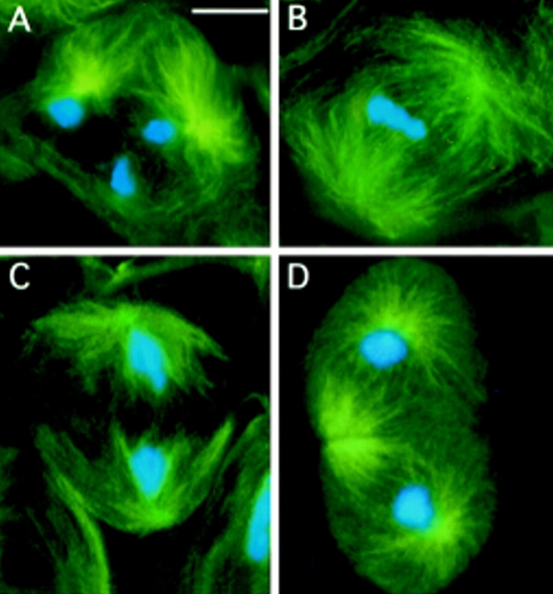
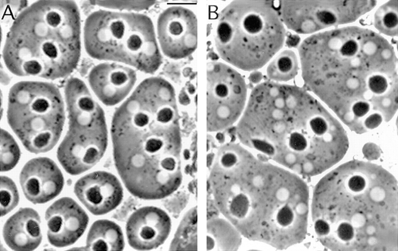
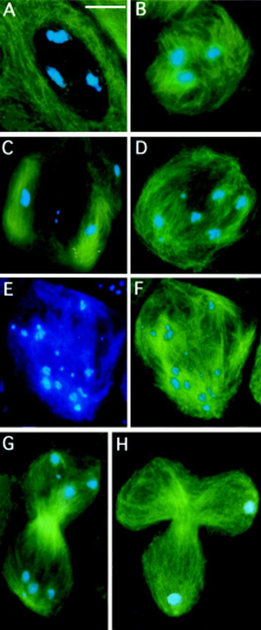
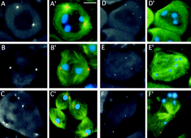
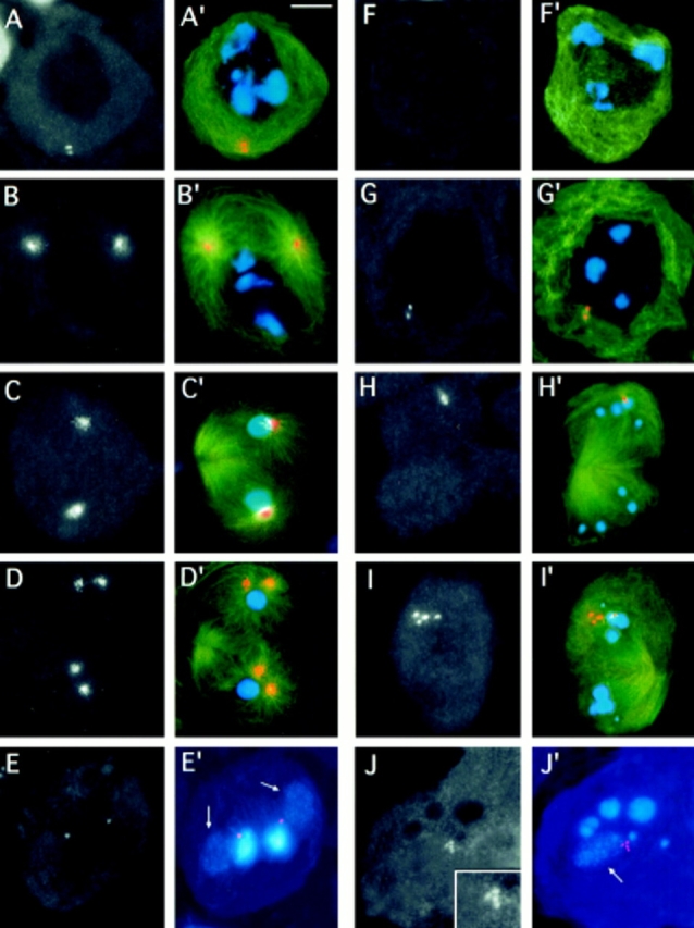
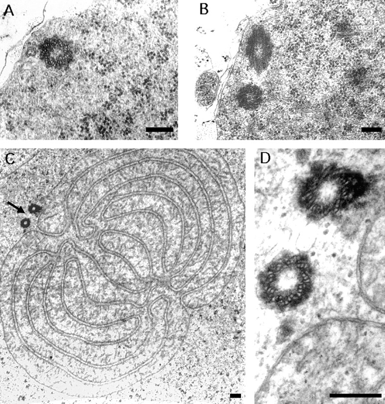
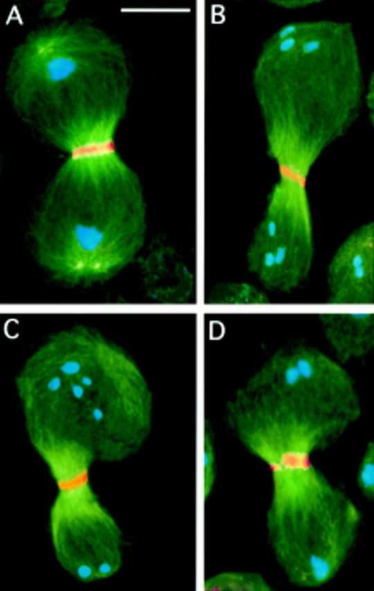
While Drosophila female meiosis is anastral, both meiotic divisions in Drosophila males exhibit prominent asters. We have identified a gene we call asterless (asl) that is required for aster formation during male meiosis. Ultrastructural analysis showed that asl mutants have morphologically normal centrioles. However, immunostaining with antibodies directed either to gamma tubulin or centrosomin revealed that these proteins do not accumulate in the centrosomes, as occurs in wild-type. Thus, asl appears to specify a function required for the assembly of centrosomal material around the centrioles. Despite the absence of asters, meiotic cells of asl mutants manage to develop an anastral spindle. Microtubules grow from multiple sites around the chromosomes, and then focus into a peculiar bipolar spindle that mediates chromosome segregation, although in a highly irregular way. Surprisingly, asl spermatocytes eventually form a morphologically normal ana-telophase central spindle that has full ability to stimulate cytokinesis. These findings challenge the classical view on central spindle assembly, arguing for a self-organization of this structure from either preexisting or newly formed microtubules. In addition, these findings strongly suggest that the asters are not required for signaling cytokinesis.


Figures

Figure 2
Figure 2
First meiotic division in wild-type (Oregon R) males. Cells were stained for tubulin (green) and DNA (by Hoechst 33258; blue). (A) Prometaphase I (stage M1; see Cenci et al.[1994] for stage designation). (B) metaphase I (stage M3). (C) Early anaphase I (stage M4a). (D) Telophase I (stage M5). Note the prominent asters in all phases of meiotic cell division. Bar, 10 μm.
Figure 1
Figure 1
Abnormal spermatids in asl mutants. Live testis squashes were viewed by phase contrast microscopy to examine onion stage spermatids. (A) Regular spermatids from Oregon-R controls with nuclei (white structures) and nebenkern (dark structures) of similar sizes. (B) Spermatids from asl mutants showing nuclei and nebenkern of various sizes. See text for details on the origin of these aberrant spermatids. Bar, 10 μm.
Figure 3
Figure 3
First meiotic division in asl mutant males. Cells were stained for tubulin (green) and DNA (blue). (A) A prometaphase I–like figure (at an M1-like stage as judged by the degree of chromatin condensation) with no asters. (B) Microtubule nucleation around the bivalents; this type of meiotic stage is never seen in the wild-type. (C) A metaphase I–like figure in which two large bivalents are associated with minispindles, and another large bivalent (upper right) is surrounded by microtubules that are not clearly polarized. Note that the tiny fourth chromosomes that have just begun to segregate are associated with very few microtubules. (D) An anaphase I–like stage in which three pairs of homologs (including the fourth chromosomes) have segregated, while a large bivalent (bottom left) is still unseparated. (E and F) An anaphase I–like figure showing segregation of sister chromatids; E shows only the chromosomes, while F shows both the chromosomes and the microtubules. See text for further explanation. (G) A telophase I figure showing a morphologically normal central spindle and scattered chromosomes at the poles. (H) A telophase I with a tripartite central spindle where the chromosomes have segregated only to two poles; in this cell the chromosomes are atypically congregated into discrete telophase nuclei. Bar, 10 μm.
Figure 4
Figure 4
Failure of centrosome assembly in asl mutants. Cells were stained for α tubulin (green), γ tubulin (orange), and DNA (blue). Panels in black and white show only γ tubulin immunofluorescence; color panels show merged images. (A–C′) wild-type; (A, A′) prometaphase I; (B, B) early anaphase I; and (C, C′) telophase I showing well-organized centrosomes that accumulate γ tubulin. In one of the telophases shown in C and C, centrosomes have already started to separate in preparation for the second meiotic division. (D– F′) asl mutants; (D, D) prometaphase I–like figure; (E, E′) anaphase I; and (F, F) telophase I, showing no γ tubulin accumulations at the cell poles. Note that γ tubulin is dispersed in small aggregates that do not appear to have the ability to nucleate microtubules. Bar, 10 μm.
Figure 5
Figure 5
Centrosomin immunostaining of asl testes. Cells were stained for α tubulin (green), centrosomin (orange), and DNA (blue). Panels in black and white show only centrosomin immunofluorescence. Color panels show merged images; tubulin immunofluorescence was not merged in the color images shown in E′ and J′. (A– E) wild-type; (A, A′) mature primary spermatocyte at the S5 stage showing a pair of centrosomes just under the plasma membrane; (B, B′) prometaphase I; (C, C′) telophase I; and (D, D) late telophase I showing prominent centrosomin-decorated centrosomes. Note that in the telophase shown in D and D, the centrosomes have already started to separate. (E and E) Two spermatids, each consisting of a nucleus and a Nebenkern. The weak fluorescence of the Nebenkern (arrows) is due to the mitochondrial DNA they con-tain. Note that the anticentrosomin antibodies detect the basal body located between the nucleus and the Nebenkern. (F–J) asl mutants; (F and F) mature primary spermatocyte at the S5 stage showing no centro– somin accumulations; (G and G) prometaphase I showing a doublet of centrosomin- enriched bodies near the nuclear envelope; (H and H) telophase I with a pair of centrosomin-enriched bodies at one of the poles; (I and I) telophase I showing a quartet of centrosomin- enriched bodies at one of its poles. (J and J) Highly irregular spermatid associated with four centrosomin-containing entities (enlarged in the insert of J; the arrow points at the Nebenkern). Bar, 10 μm.
Figure 6
Figure 6
Presence of morphologically normal centrioles in asl1mutants. (A) cross-section through the proximal part of a centriole in a mature primary spermatocyte. (B) A pair of centrioles lying at approximately right angles, located at the periphery of a primary spermatocyte. (C) Cross-section through a Nebenkern of an onion-stage spermatid, irregularly associated with two basal bodies (arrow). (D) Higher magnification of the basal bodies in C, consisting of nine peripheral triplets of tubules. (A) 28,000×; (B) 22,000×; (C) 13,000×; (D) 60,000×. Bars, 0.2 μm.
Figure 7
Figure 7
Central spindle formation and cytokinesis in asl mutants. Cells were immunostained for tubulin (green); DNA (blue); and either actin (orange; A and B), anillin (orange; C), or KLP3A (orange; D). (A) A wild-type telophase I and (B) an asl telophase I showing similar actin bands in the middle of their central spindles. (C and D) asl telophase I figures showing normal anillin (C) and KLP3A (D) accumulations in the spindle midzone. Note that in the cells in B and C, the central spindle is asymmetrically located with respect to the cell poles. Bar, 10 μm.

References

    1. Albertson DG, Thomson JN. Segregation of holocentric chromosomes at meiosis in the nematode Caenorhabditis elegans. . Chromosome Res. 1993;1:15–26. - PubMed
    1. Ault JG, Rieder CL. Centrosome and kinetochore movement during mitosis. Curr Opin Cell Biol. 1994;6:41–49. - PubMed
    1. Callaini G, Whitfield WG, Riparbelli MG. Centriole and centrosome dynamics during the embryonic cell cycles that follow the formation of the cellular blastoderm in Drosophila. . Exp Cell Res. 1997;234:183–190. - PubMed
    1. Cao L-G, Wang Y-L. Signals from the spindle midzone are required for the stimulation of cytokinesis in cultured epithelial cells. Mol Biol Cell. 1996;7:225–232. - PMC - PubMed
    1. Cenci G, Bonaccorsi S, Pisano C, Verni F, Gatti M. Chromatin and microtubule organization during premeiotic, meiotic, and early postmeiotic stages of Drosophila melanogasterspermatogenesis. J Cell Sci. 1994;107:3521–3534. - PubMed

Publication types