. 1998 Aug 10;142(3):787-801.
doi: 10.1083/jcb.142.3.787.

Mitotic centromere-associated kinesin is important for anaphase chromosome segregation

Affiliations

Mitotic centromere-associated kinesin is important for anaphase chromosome segregation

T Maney et al. J Cell Biol. .

Abstract

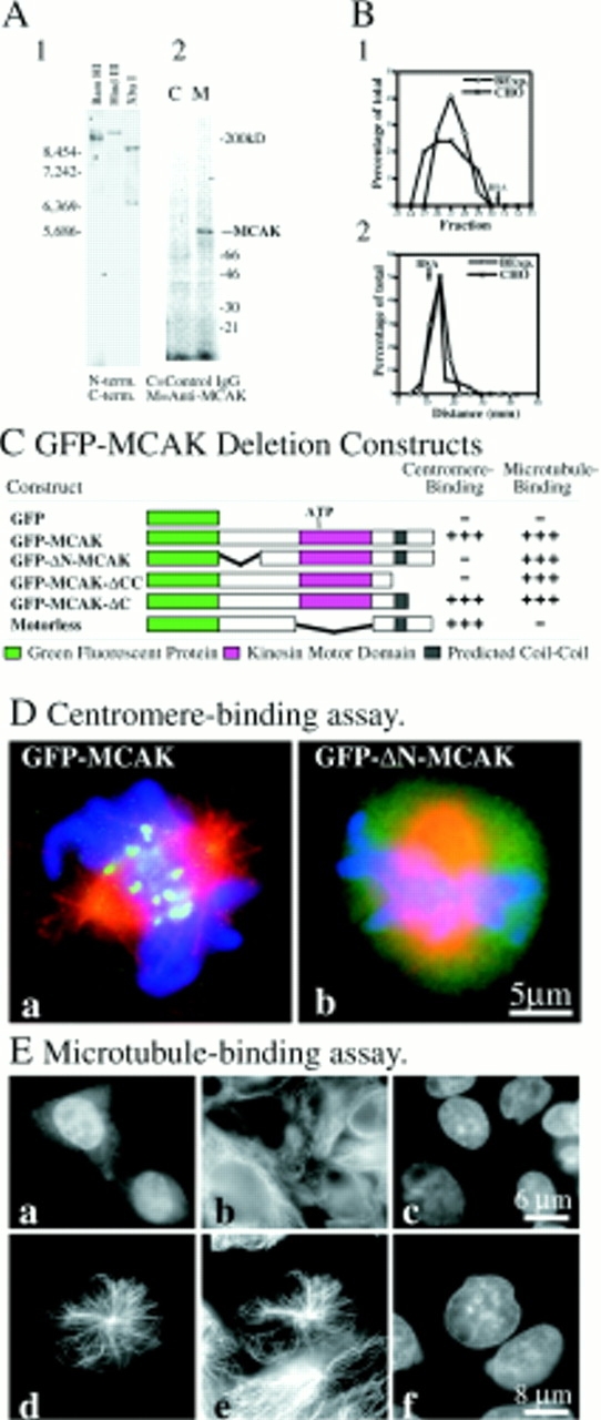
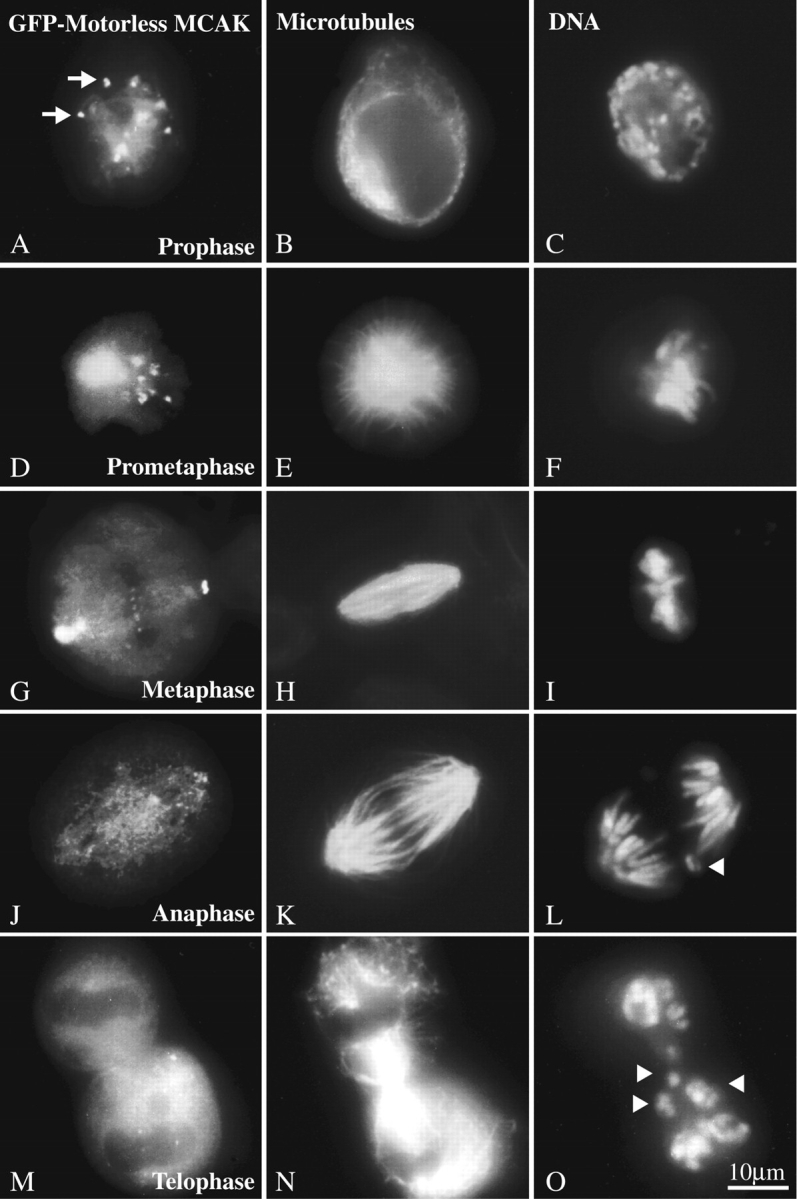
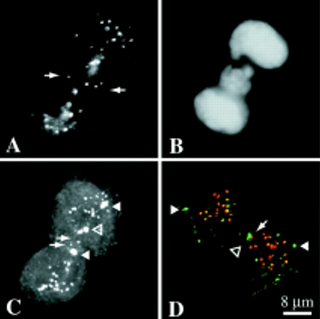
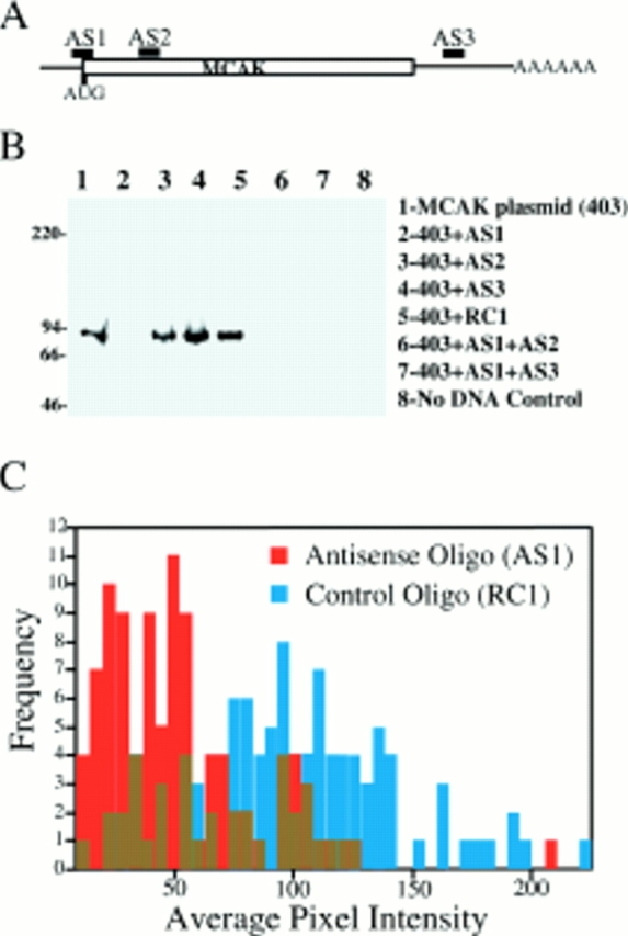
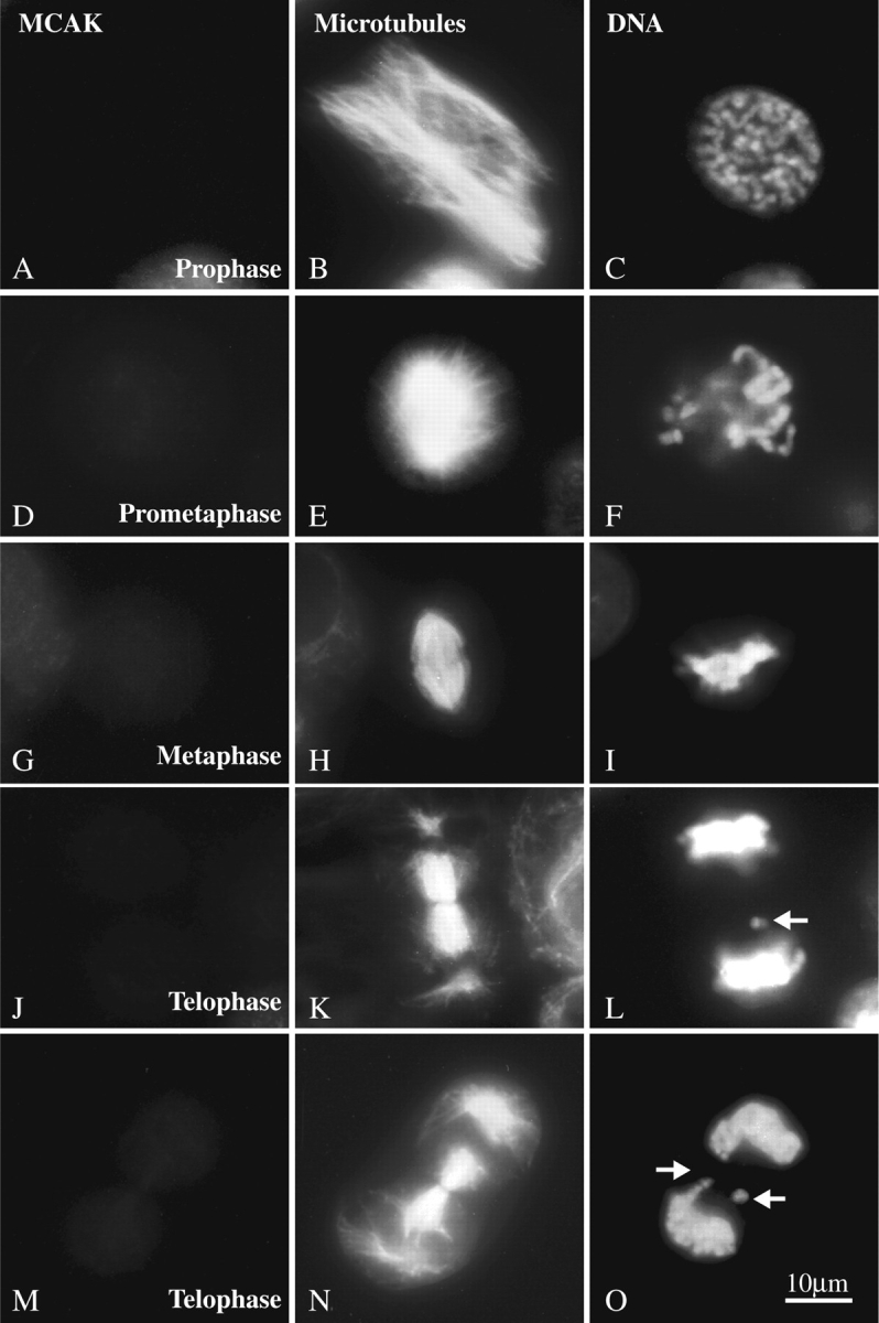
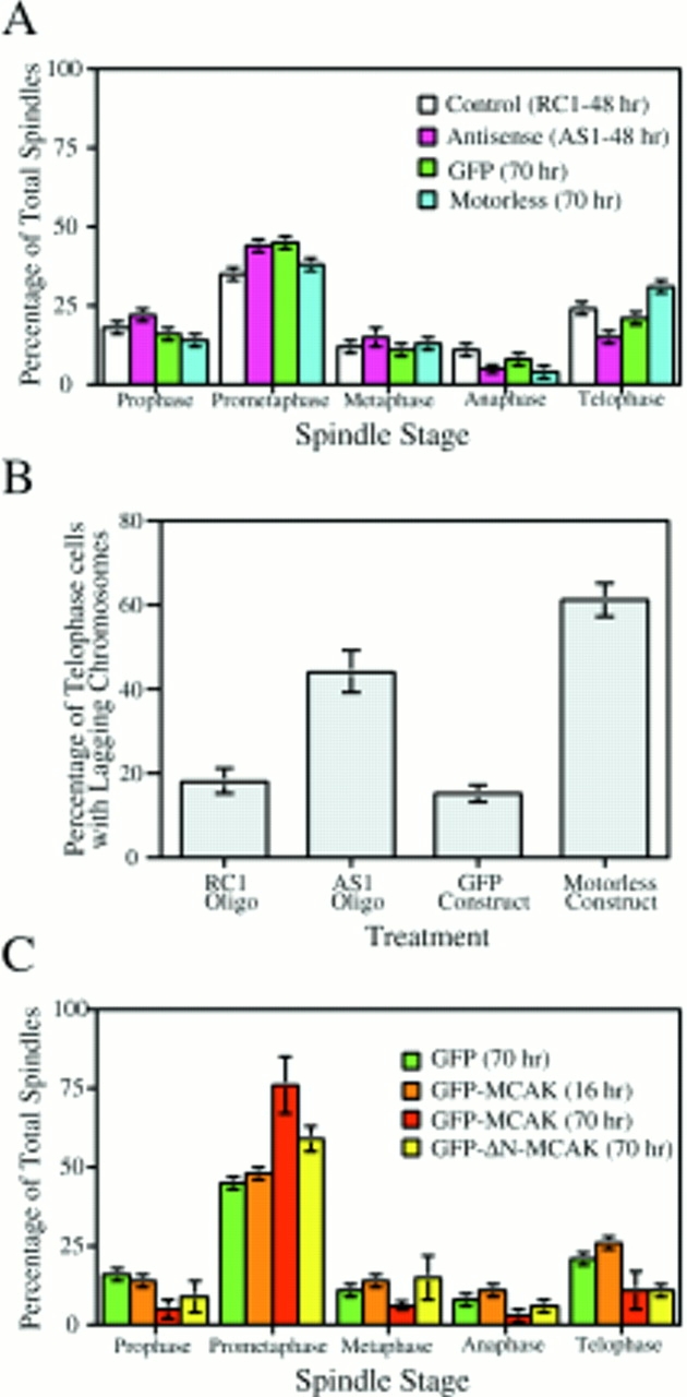
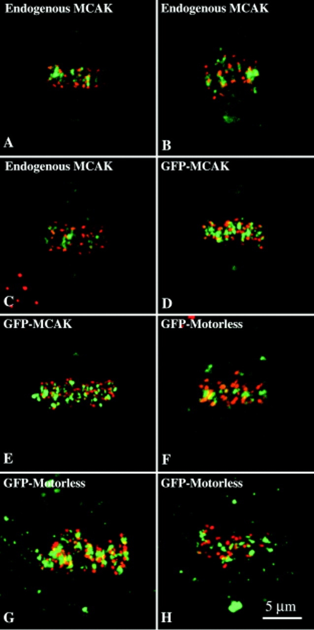
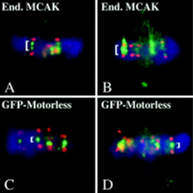
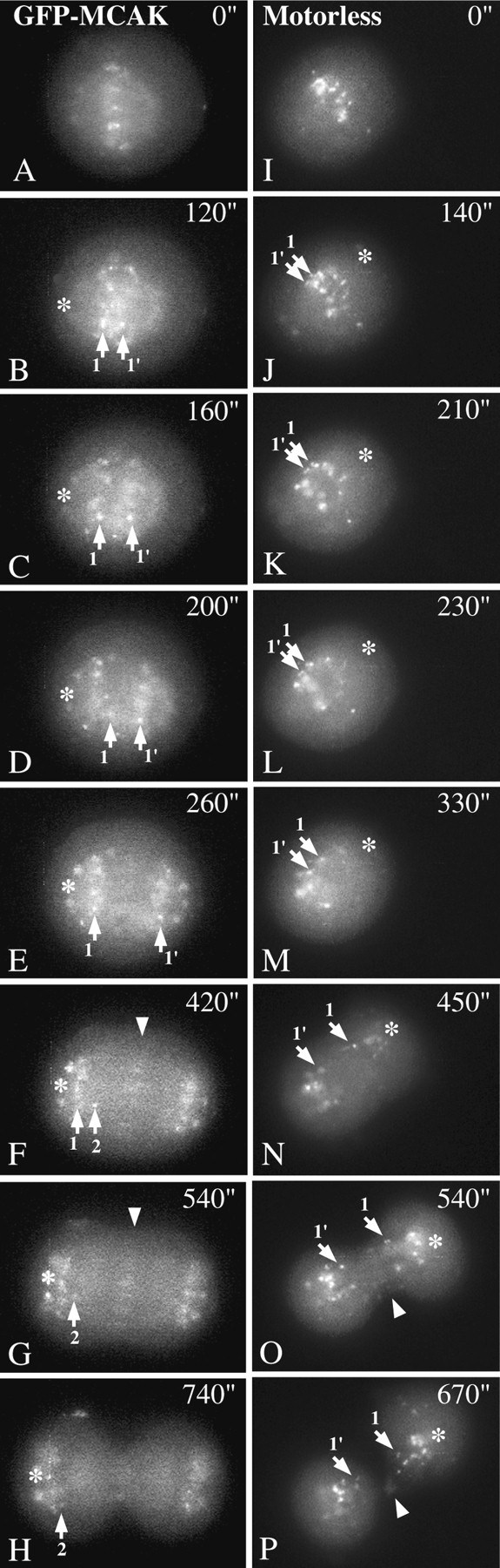
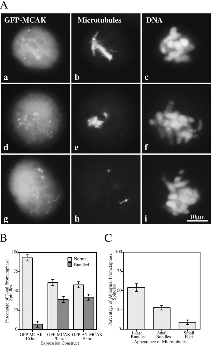
Mitotic centromere-associated kinesin (MCAK) is recruited to the centromere at prophase and remains centromere associated until after telophase. MCAK is a homodimer that is encoded by a single gene and has no associated subunits. A motorless version of MCAK that binds centromeres but not microtubules disrupts chromosome segregation during anaphase. Antisense-induced depletion of MCAK results in the same defect. MCAK overexpression induces centromere-independent bundling and eventual loss of spindle microtubule polymer suggesting that centromere-associated bundling and/or depolymerization activity is required for anaphase. Live cell imaging indicates that MCAK may be required to coordinate the onset of sister centromere separation.


Figures

Figure 1
Figure 1
Molecular and functional analysis of MCAK. (A) MCAK is a homodimer, encoded by a single gene with no associated subunits. (1) Genomic CHO-K1 DNA digested with BamHI, HindIII, or XbaI and hybridized to P32-labeled probe synthesized from DNA sequences for the NH2-terminal or COOH-terminal regions of MCAK, exclusive of the motor domain. Hybridizations of the combined NH2-terminal and COOH-terminal regions indicate that MCAK is encoded by a single gene. (2) Cell lysates from [S35]methionine-labeled CHO-K1 cells immunoprecipitated with affinity-purified anti-MCAK polyclonal antibodies (M) or nonimmune purified IgG. (B) Hydrodynamic analysis of MCAK (1) Gel exclusion chromatography of Baculovirus-expressed MCAK (BExp-MCAK) and CHO lysate superimposed. Gel filtration standards have been omitted for clarity. CHO MCAK was detected by Western immunoblot. (2) Sucrose density gradient of BExp-MCAK and CHO lysate run in parallel and superimposed. Standards have been omitted for clarity. Intensities of scanned gels and blots have been plotted against the fraction number (in the case of 1) and the distance migrated (in the case of 2). MCAK in CHO cell lysates behaves similarly to purified His-tagged MCAK expressed in the baculovirus expression system. (C) GFP-MCAK deletion mutant analysis. NH2-terminal fusion of GFP to MCAK does not affect binding of expression fusion protein to mitotic centromeres during cell division. However, deletion of the NH2 terminus of MCAK or the COOH terminus, inclusive of the predicted coiled-coil domain, abolishes centromere binding. Cells lysed before fixation in the absence of ATP exhibit rigor binding of MCAK to microtubules. Constructs were assayed for microtubule-binding capability using this assay. (D) Examples of centromere-binding assay. (a) Full-length GFP-MCAK expressed for 16 h in CHO cells exhibits a spindle localization pattern indistinguishable from endogenous protein. (b) GFP-ΔN-MCAK fails to localize to mitotic centromeres. GFP-MCAK and GFP-ΔN-MCAK, green; microtubules, red; DNA, blue. (E) Microtubule-binding assay. (a–c) Normal interphase CHO cells transfected with GFP-MCAK and fixed 16 h later. Diffuse cytoplasmic and nuclear label is evident. (a) GFP-MCAK. (b) Microtubules. (c) DNA. (d–f) Interphase cell transfected with GFP-MCAK and lysed before fixation. Constructs were assayed for the ability to bind microtubules in this manner. (d) GFP-MCAK. (e) Microtubules. (f) DNA. Bars: (D) 5 μm; (E, a–c) 6 μm; (E, d–f) 8 μm.
Figure 2
Figure 2
The GFP-motorless deletion exhibits a lagging chromosome phenotype at the onset of anaphase. Like endogenous MCAK, motorless GFP-MCAK localizes to nuclei, mitotic centromeres, and centrosomes. (A, D, G, J, M) Motorless GFP-MCAK; (B, E, H, K, N) microtubules; (C, F, I, L, O) DNA. GFP-motorless appears on centromeres (A, arrows) at prophase (A–C). Spindle assembly and chromosome distribution appear to be unaffected through prometaphase (D–F) and metaphase alignment (G–I). Lagging chromosomes (arrowheads) appear during anaphase (J–L) and telophase (M–O). Bar, 10 μm.
Figure 3
Figure 3
Lagging chromosomes have undergone sister chromosome separation. (a and b) Fluorescence microscopy of a telophase cell transfected with GFP-motorless. Single dots of centromere-bound GFP-motorless (a) are visible (arrows). (b) DNA. (c) Confocal projected Z-series of a motorless GFP-motorless transfectant in late telophase. GFP-motorless (shown) exhibits lagging centromeres (arrows), spindle poles (arrowheads), and midbody (open arrowhead). In the confocal micrograph, one daughter cell has rotated, bringing the centrosome between the cleavage furrow and the chromosomes. Single centromeres can be seen in the vicinity of the cleavage furrow (arrows). (d) Confocal projected Z-series of a late telophase cell transfected with GFP-motorless. The merged image consists of CREST sera label (red) and GFP-motorless (green). Spindle poles (filled arrowheads) and midbody (open arrowhead) are also indicated. Several lagging centromeres are visible with one (arrow) residing close to the cleavage furrow. All centromeres appear to have separated. Bar, 8 μm.
Figure 4
Figure 4
Antisense-induced depletion of MCAK protein. (A and B) Potency of phosphorothioate oligos for the inhibition of MCAK translation in vitro. (A) Diagram of the location of three antisense oligonucleotides within MCAK mRNA transcript. AS1, start codon; AS2, within the amino acid coding region; AS3, within the 3′ untranslated region. (B) Transcription/translation of MCAK in pBluescript II SK (construct 403) using T3 polymerase and [S35]methionine. Oligos were added to the reaction in a 10-fold excess over DNA template. Oligonucleotide AS1 was the only oligo effective in inhibiting translation in vitro. (C) AS1-induced depletion of endogenous MCAK. 100 pmol of antisense (AS1) or control (RC1) oligos were scrape-loaded into CHO cells in combination with digoxigenin-UTP. Digoxigenin-positive cells were scored immunofluorescently for MCAK intensity.
Figure 5
Figure 5
Effect of endogenous MCAK depletion on chromosome segregation. Mitotic cells triple-labeled with anti-MCAK (A, D, G, J, M); anti-tubulin (B, E, H, K, N); and Hoechst (C, F, I, L, O). Endogenous MCAK was below the level of detection by immunofluorescence microscopy. Spindle assembly and chromosome alignment appeared normal during prophase (A–C), prometaphase (D–F), and metaphase (G–I). However, many telophase cells (J–O) with lagging chromosomes (arrows) were identified. Bar, 10 μm.
Figure 6
Figure 6
Spindle profiles of transfectants and antisense-loaded mitotic cells. (A) Spindle profiles of control (RC1) and antisense (AS1) loaded cells (48 h after loading) were not significantly different from each other or from control GFP-transfected or GFP-motorless–MCAK transfected cells (70 h after transfection). Inset, Spindle stages based on live video analysis. P, prophase; PM, prometaphase; M, metaphase; A, anaphase. (B) Telophase cells of AS1-loaded and GFP-motorless–transfected cells have significantly increased numbers of lagging chromosomes relative to control telophase cells. (C) Transfected GFP-MCAK constructs that contain the MCAK motor domain induce mitotic arrest in a prometaphase-like configuration after 70 h of expression. For scrape-loaded cells the number of mitotic cells scored is >1,000; in the case of transfected cells the number of mitotic cells scored is >300.
Figure 7
Figure 7
Metaphase alignment in GFP-motorless-transfected cells. Projected confocal Z-series of merged images of metaphase cells double- labeled with CREST sera (red) and either endogenous MCAK (A–C, green); GFP-MCAK (D and E, green); or GFP-motorless (F–H, green). Metaphase alignment of GFP-motorless (F–H) does not differ substantively from control cells (A–E). Bar, 5 μm.
Figure 8
Figure 8
Metaphase distribution of MCAK differs slightly from GFP-motorless. Control metaphase spindles (A and B) showing endogenous MCAK label (green) and CREST label (red). GFP-motorless-transfected cells (B and D) showing GFP-motorless (green) versus CREST label (red). In control metaphase cells, MCAK appears to extend throughout the inner centromere regions (A and B; large brackets) whereas GFP-motorless consistently appears compressed (C and D; small brackets).
Figure 9
Figure 9
Live cell imaging of GFP-MCAK– and GFP-motorless–transfected cells. GFP-MCAK–transfected cells (A–H) were tracked from an arbitrary time at metaphase (A, t = 0′′) until telophase (H). All chromosomes in this cell segregated completely despite the fact that one centromere suffered a transient reversal (D; 1) or anti-poleward oscillation during chromosome-to-pole movement. GFP-motorless–transfected cells (I-P) were tracked from an arbitrary point during metaphase (I, t = 0′′) to telophase (P). In this cell, one pair of sister centromeres (1, 1′) fail to separate at the onset of anaphase (K) and continue to oscillate. Eventually the sister separate late (M), however, they never make it to the spindle pole. Both late-separating sister chromosomes end up lagging close to the cleavage furrow at cytokinesis (O and P). Other lagging centromeres that were not traced because they did not remain in the plane of focus throughout anaphase are also visible (O and P). Both GFP-MCAK and GFP-motorless appear on the spindle midzone during anaphase B (F and G, arrowhead; O, arrowhead).
Figure 10
Figure 10
Pseudoprometaphase-arrest by MCAK overexpression results from spindle microtubule abnormalities. (A) Examples of typical prometaphase-arrested spindles. GFP-MCAK (a, d, g); microtubules (b, e, h); DNA (c, f, i). Spindles microtubules consist of large bundles (a–c); small bundles (d–f) or small to nonexistent foci of tubulin staining (g–i). (B) Characterization of prometaphase spindles in 16-h vs. 70-h GFP-MCAK and GFP-ΔN-MCAK transfectants. Significant increases in abnormal prometaphase spindles occur between 16 and 70 h after transfection. (n = 200 prometaphase cells.) (C) Quantitation of microtubule disappearance in pseudoprometaphase-arrested spindles (GFP-MCAK overexpression). (n = 150 abnormal prometaphase spindles.) Bar, 10 μm.

Similar articles

Cited by

References

    1. Ault JG, DeMarco AJ, Salmon ED, Rieder CL. Studies on the ejection properties of asters: astral microtubule turnover influences the oscillatory behavior and positioning of mono-oriented chromosomes. J Cell Sci. 1991;99:701–710. - PubMed
    1. Berger B, Wilson DB, Wolf E, Tonchev T, Milla M, Kim PS. Predicting coiled coils by use of pairwise residue correlations. Proc Natl Acad Sci USA. 1995;92:8259–8263. - PMC - PubMed
    1. Bloom K. The centromere frontier: Kinetochore components, microtubule-based motility, and the CEN-value paradox. Cell. 1993;73:621–624. - PubMed
    1. Brinkely BR, Cartwright J. Ultrastructural analysis of mitotic spindle elongation in mammalian cells in vitro: Direct microtubule counts. J Cell Biol. 1971;50:416–431. - PMC - PubMed
    1. Brown KD, Wood KW, Cleveland DW. The kinesin-like protein CENP-E is kinetochore-associated throughout poleward chromosome segregation during anaphase-A. J Cell Sci. 1996;109:961–969. - PubMed

Publication types

Substances