. 1998 Aug 10;142(3):827-35.
doi: 10.1083/jcb.142.3.827.

Microphthalmia gene product as a signal transducer in cAMP-induced differentiation of melanocytes

Affiliations

Microphthalmia gene product as a signal transducer in cAMP-induced differentiation of melanocytes

C Bertolotto et al. J Cell Biol. .

Abstract

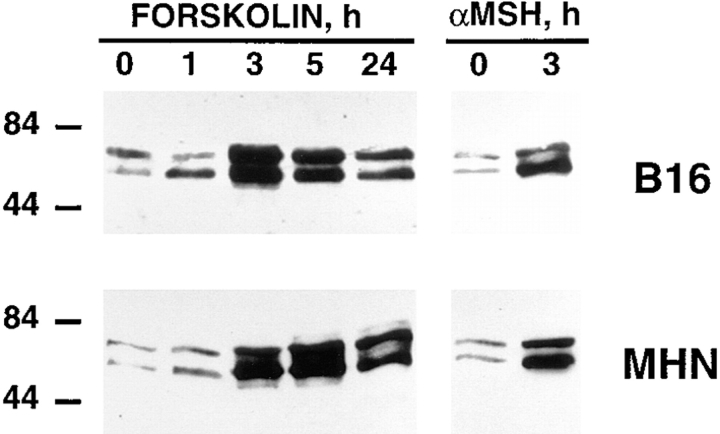
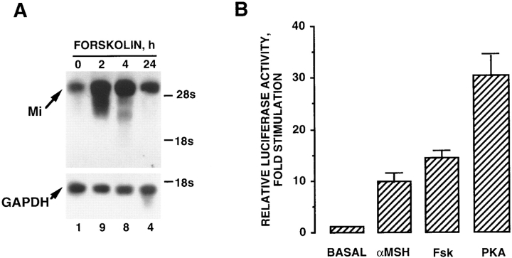
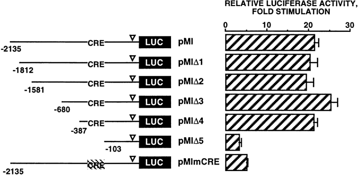
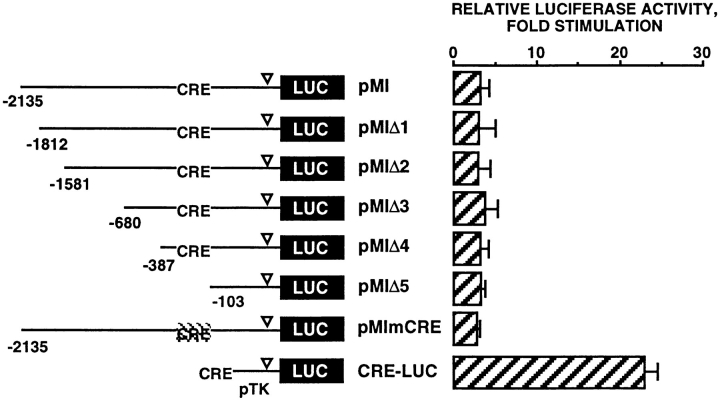
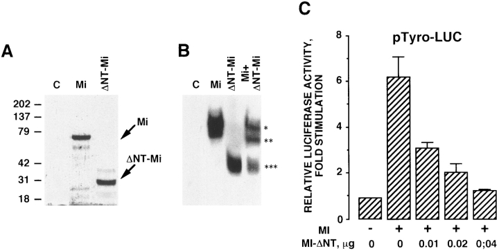
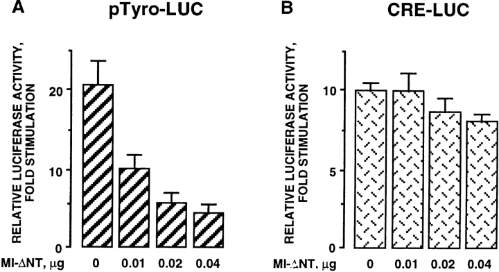
Melanocyte differentiation characterized by an increased melanogenesis, is stimulated by alpha-melanocyte-stimulating hormone through activation of the cAMP pathway. During this process, the expression of tyrosinase, the enzyme that controls melanin synthesis is upregulated. We previously showed that cAMP regulates transcription of the tyrosinase gene through a CATGTG motif that binds microphthalmia a transcription factor involved in melanocyte survival. Further, microphthalmia stimulates the transcriptional activity of the tyrosinase promoter and cAMP increases the binding of microphthalmia to the CATGTG motif. These observations led us to hypothesize that microphthalmia mediates the effect of cAMP on the expression of tyrosinase. The present study was designed to elucidate the mechanism by which cAMP regulates microphthalmia function and to prove our former hypothesis, suggesting that microphthalmia is a key component in cAMP-induced melanogenesis. First, we showed that cAMP upregulates the transcription of microphthalmia gene through a classical cAMP response element that is functional only in melanocytes. Then, using a dominant-negative mutant of microphthalmia, we demonstrated that microphthalmia is required for the cAMP effect on tyrosinase promoter. These findings disclose the mechanism by which cAMP stimulates tyrosinase expression and melanogenesis and emphasize the critical role of microphthalmia as signal transducer in cAMP-induced melanogenesis and pigment cell differentiation.


Figures

Figure 1
Figure 1
Forskolin treatment stimulates microphthalmia gene expression in B16 mouse melanoma cells. Immunofluorescence labeling was performed with the anti-microphthalmia mAb. Unstimulated B16 mouse melanoma cells (A), B16 mouse melanoma cells stimulated with 20 μM forskolin for 3 h (B), 5 h (C), and 24 h (D). Bar, 10 μm.
Figure 2
Figure 2
cAMP-elevating agents increases microphthalmia expression in B16 mouse melanoma cells and in normal human melanocytes. 20 μg of proteins from B16 mouse melanoma cells (top panel) or from normal human melanocytes (bottom panel), non-stimulated or treated for the indicated times with 20 μM forskolin or 1 μM α-MSH, were subjected to Western blot analysis using the anti-microphthalmia mAb. Molecular masses, indicated on the left, are expressed in kD.
Figure 3
Figure 3
cAMP-elevating agents regulate microphthalmia expression through a transcriptional mechanism. (A) Northern blot analysis for microphthalmia and GAPDH mRNA transcripts from B16 cells exposed for the indicated times to 20 μM forskolin. (B) B16 cells were transfected with 0.35 μg of pMI, 0.1 μg of empty pCDNA3, and 0.05 μg of pCMVβ′-GAL. Then, cells were treated for 12 h with 20 μM forskolin or 1 μM α-MSH. To study the effects of PKA expression, B16 cells were transfected with 0.35 μg of pMI, 0.1 μg of pCDNA3 encoding PKA, and 0.05 μg of pCMVβGAL. Luciferase activity was normalized by the β-galactosidase activity and the results were expressed as fold stimulation of the basal luciferase activity from unstimulated cells. Data are means ± SE of five experiments performed in triplicate.
Figure 4
Figure 4
The effect of forskolin on microphthalmia promoter activity is mediated by a consensus CRE motif (−147/−140). B16 cells were transfected with 0.45 μg of pMI, pMIΔ1, pMIΔ2, pMIΔ3, pMIΔ4, pMIΔ5, or pMImCRE and 0.05 μg of pCMVβGAL. After 12 h with forskolin, luciferase activity was normalized by the β-galactosidase activity and the results were expressed as fold stimulation of the basal luciferase activity from unstimulated cells. Data are means ± SE of five experiments performed in triplicate. ▿ indicates the TATA box position.
Figure 5
Figure 5
The CRE motif (−147/−140) binds CREB and confers cAMP sensitivity to the minimal thymidine kinase promoter. (A) Gel shift experiments were performed with CREB-1 bZIP (254–327) using a labeled oligonucleotide containing the CRE site of the microphthalmia promoter in control conditions or in presence of an excess (50-fold) of unlabeled homologous oligonucleotide. (B) B16 nuclear extracts from control (−) or forskolin-treated cells (+) were incubated with the same labeled CRE probe or with a labeled oligonucleotide in which the CRE site was mutated (mCRE). When indicated, reactions were carried out in the presence of an excess (50-fold) of unlabeled homologous CRE probe or with 0.6 μl of an anti-CREB antibody. (C) B16 cells were transfected with 0.35 μg of pTK, pTKCRE, or pTKmCRE and 0.05 μg of pCMVβGAL. After 12 h with forskolin, luciferase activity was normalized by the β-galactosidase activity and the results were expressed as fold stimulation of the basal luciferase activity from unstimulated cells. Data are means ± SE of three experiments performed in triplicate.
Figure 6
Figure 6
The CRE motif do not confer cAMP sensitivity to microphthalmia promoter in NIH3T3 fibroblast cells. NIH3T3 fibroblast cells were transfected with 0.45 μg of pMI, pMIΔ1, pMIΔ2, pMIΔ3, pMIΔ4, pMIΔ5, pMImCRE, or CRE-LUC and 0.05 μg of pCMVβGAL. After 12 h with forskolin, luciferase activity was normalized by the β-galactosidase activity and the results were expressed as fold stimulation of the basal luciferase activity from unstimulated cells. Data are means ± SE of five experiments performed in triplicate. ▿ indicates the TATA box position.
Figure 7
Figure 7
Mi-ΔNT, a form of microphthalmia lacking the NH2-terminal transactivation domain, exerts dominant-negative effects on the wild-type microphthalmia. (A) Microphthalmia and Mi-ΔNT were in vitro translated in the presence of [35S]methionine, and then analyzed by gel electrophoresis. Molecular masses, indicated on the left, are expressed in kD. (B) For gel shift assay, 2 μl of in vitro–translated microphthalmia, Mi-ΔNT, or microphthalmia plus Mi-ΔNT were incubated with labeled tyrosinase M-box. *, Wild-type microphthalmia homodimer; ***, Mi-ΔNT homodimer; and **, microphthalmia plus Mi-ΔNT heterodimer. Autoradiogram was exposed overnight at −80°C. (C) B16 cells were transfected with 0.4 μg of pTyro, 0.01 μg of pCDNA3 encoding microphthalmia, 0.05 μg of pCMVβGAL, and different amounts of pCDNA3 encoding Mi-ΔNT. After 24 h, luciferase activity was normalized by the β-galactosidase activity and the results were expressed as fold stimulation of the basal luciferase activity from unstimulated cells. Data are means ± SE of five experiments performed in triplicate.
Figure 8
Figure 8
Regulation of tyrosinase promoter activity by forskolin is mediated by microphthalmia. B16 cells were transfected with (A) 0.4 μg of pTyro or (B) 0.4 μg of a CRE-LUC reporter plasmid, 0.05 μg of pCMVβGAL, and different amounts of pCDNA3 encoding Mi-ΔNT. After 24 h with forskolin, luciferase activity was normalized by the β-galactosidase activity and the results were expressed as fold stimulation of the basal luciferase activity from unstimulated cells. Data are means ± SE of five experiments performed in triplicate.

References

    1. Abdel-Malek A, Swope BV, Amornsiripanitch N, Nordlund JJ. In vitro modulation of proliferation and melanization of S91 melanoma cells by prostaglandins. Cancer Res. 1987;47:3141–3146. - PubMed
    1. Arany Z, Newsome D, Olread D, Livingston DM, Eckner R. A family of transcriptional adaptator proteins targeted by E1A oncoprotein. Nature. 1995;374:81–84. - PubMed
    1. Arias J, Alberts AS, Brindle P, Claret FX, Smeal T, Karim M, Ferasmisco J, Montminy M. Activation of cAMP and mitogen responsive genes relies on a commun nuclear factor. Nature. 1994;370:226–229. - PubMed
    1. Bannister AJ, Kouzarides T. CBP-induces stimulation of c-fos activity is abrogates by E1A. EMBO (Eur Mol Biol Organ) J. 1995;14:4758–4762. - PMC - PubMed
    1. Barber JI, Townsend D, Olds DP, King RA. Dopachrome oxydoreductase: a new enzyme in the pigment pathway. J Invest Dermatol. 1984;83:145–149. - PubMed

Publication types

MeSH terms