. 1998 Aug 10;142(3):837-46.
doi: 10.1083/jcb.142.3.837.

Adhesive but not lateral E-cadherin complexes require calcium and catenins for their formation

Affiliations

Adhesive but not lateral E-cadherin complexes require calcium and catenins for their formation

N A Chitaev et al. J Cell Biol. .

Abstract

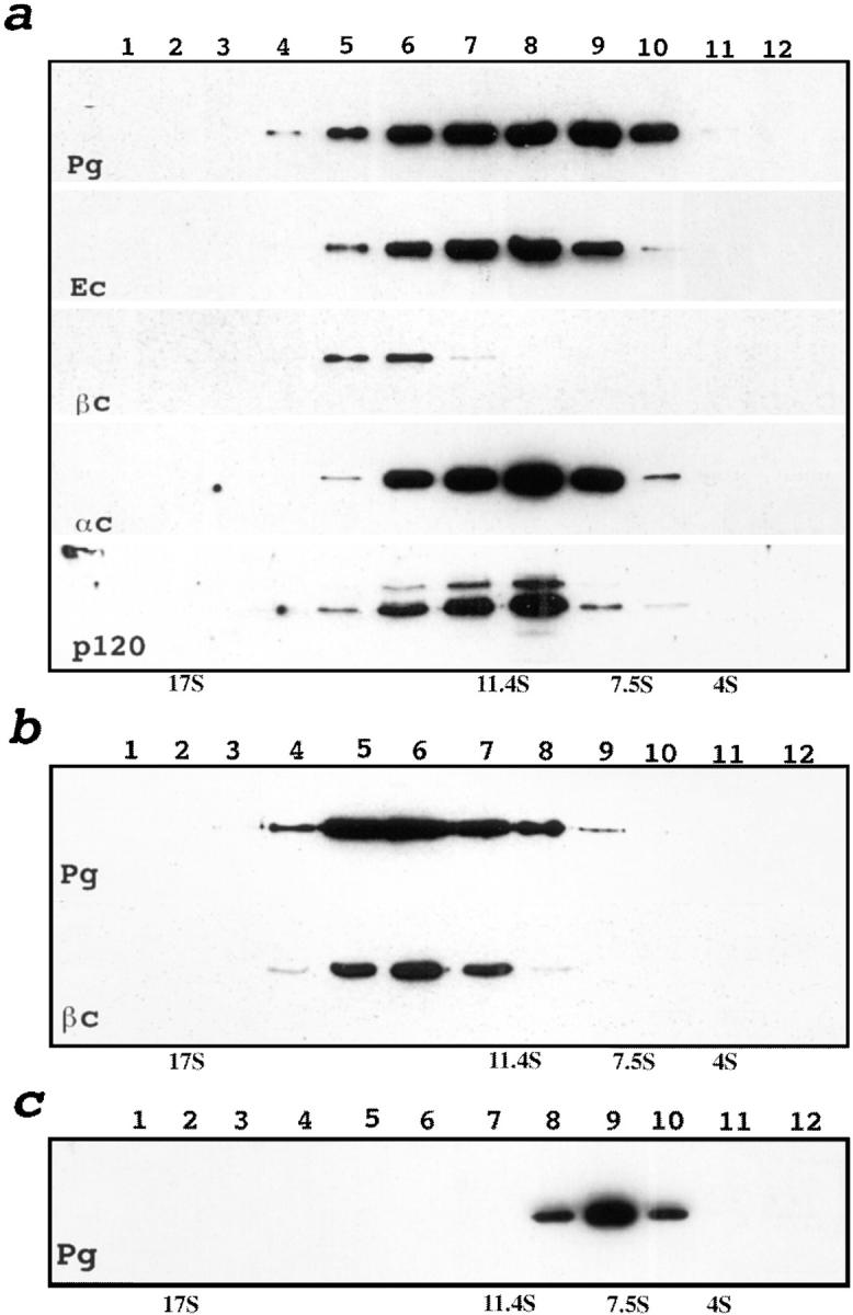
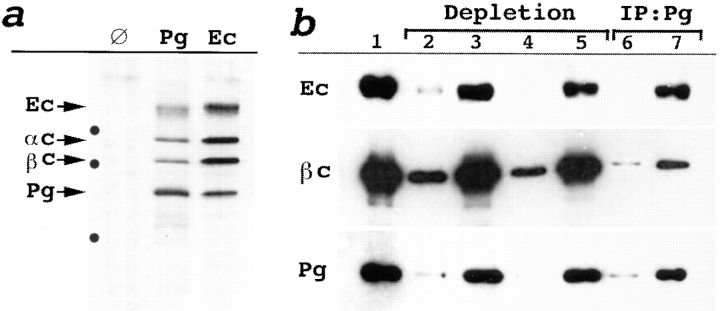
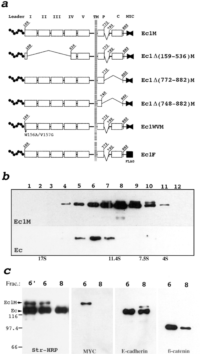
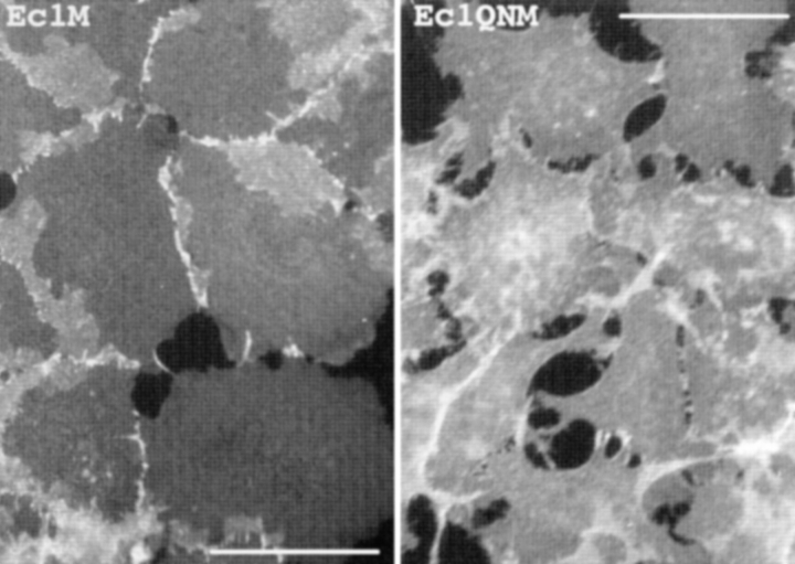
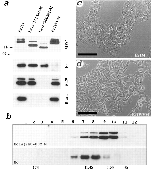
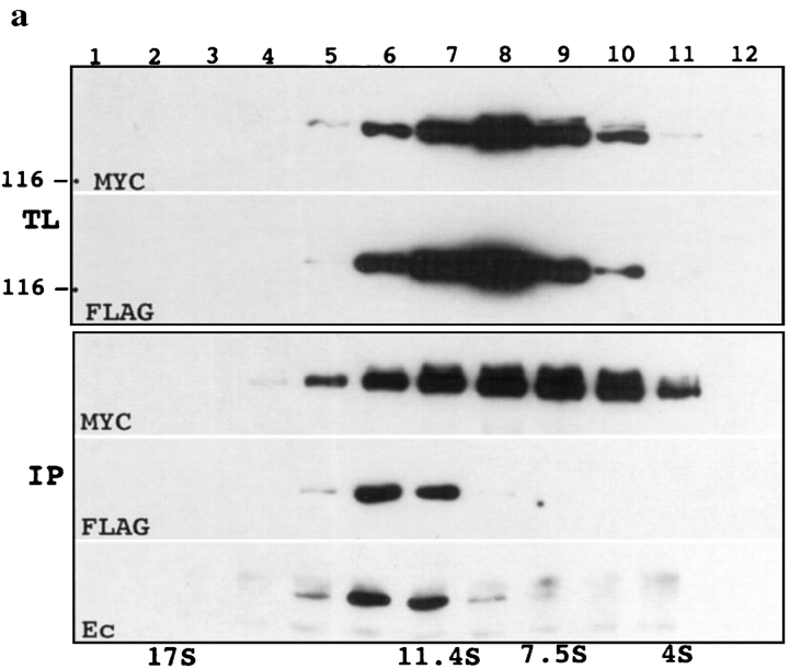
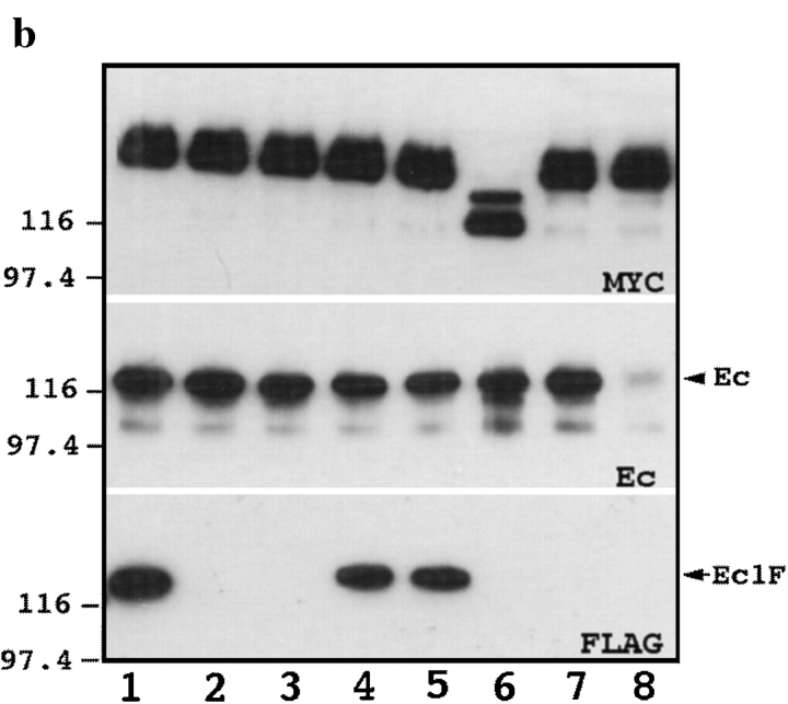
We examined intercadherin interactions in epithelial A-431 cells producing endogenous E-cadherin and recombinant forms of E-cadherin tagged either by myc or by flag epitopes. Three distinct E-cadherin complexes were found. The first is a conventional E-cadherin-catenin complex consisting of one E-cadherin molecule linked either to beta-catenin/alpha-catenin or to plakoglobin/alpha-catenin dimers. The second is a lateral E-cadherin complex incorporating two E-cadherin- catenin conventional complexes combined in parallel fashion via dimerization of the NH2-terminal extracellular domain of E-cadherin. The third complex is likely to contain two E-cadherin-catenin conventional complexes derived from two opposing cells and arranged in an antiparallel fashion. Formation of the antiparallel but not lateral complex strictly depends on extracellular calcium and E-cadherin binding to catenins. Double amino acid substitution Trp156Ala/Val157Gly within the extracellular NH2-terminal E-cadherin domain completely abolished both lateral and antiparallel inter-E-cadherin association. These data support an idea that the antiparallel complex has the adhesion function. Furthermore, they allow us to suggest that antiparallel complexes derive from lateral dimers and this complex process requires catenins and calcium ions.


Figures

Figure 1
Figure 1
Sedimentation analysis of plakoglobin-associated complexes. (a) Total lysates of A-431 cells were subjected to sucrose gradient centrifugation (refer to Materials and Methods). Each fraction was coimmunoprecipitated with anti-plakoglobin mAb 11E4, separated by SDS-PAGE, and then analyzed by immunoblotting with plakoglobin (Pg), E-cadherin (Ec), β-catenin (βc), α-catenin (αc), and p120 (p120) antibodies. Note the plakoglobin–β-catenin complex appears in fractions 5–7. Two bands in the blot developed by anti-p120 mAb represent distinct splice forms of this protein. (b and c) Fraction 6 (b) or 9 (c) of the gradient was dialyzed for 6 h against lysis buffer and loaded onto a new gradient. After refractionation each fraction was analyzed as in a. Anti-β-catenin staining shows the stability of the plakoglobin/ β-catenin complex upon recentrifugation. The peak distribution of protein standards in a parallel gradient of known S values (bovine serum albumin, 4 S; rabbit IgG, 7.5 S; catalase, 11.4 S; apoferritin, 17 S) is shown at the bottom.
Figure 2
Figure 2
Coimmunoprecipitation and immunoblot analysis of proteins collected from fraction 6 (13 S). (a) Autoradiograms of [35S]methionine proteins coimmunoprecipitated with mock (∅), anti-Pg (Pg), and anti–E-cadherin (Ec) antibodies and separated by SDS-PAGE. Identified proteins (Ec, E-cadherin; αc, α-catenin; βc, β-catenin; Pg, plakoglobin) are shown by arrows. Molecular weight markers are indicated by dots from top to bottom: 116, 97.4, and 67 kD. P-cadherin apparently comigrates with E-cadherin in the anti-plakoglobin immunoprecipitate. (b) Western blot analysis of the total proteins from fraction 6 developed with anti–E-cadherin (Ec), β-catenin (βc), and plakoglobin (Pg) antibodies before (lane 1), and after depletion with anti–E-cadherin C20820 (first round, lane 2; second round, lane 4) or anti-desmoglein (first round, lane 3; second round, lane 5) mAbs. The samples depleted with anti–E-cadherin (lane 6) or anti-desmoglein (lane 7) mAbs were coimmunoprecipitated with anti-plakoglobin mAbs (IP:Pg) and analyzed for the presence of E-cadherin, β-catenin, and plakoglobin. Depletion with E-cadherin antibody (lane 6), but not desmoglein antibody (lane 7), reduces recovery of the plakoglobin–β-catenin complex.
Figure 3
Figure 3
Schematic representation of human E-cadherin gene constructs (a) and sedimentation/coimmunoprecipitation analysis showing E-cadherin dimerization (b and c). (a) Five extracellular cadherin-like repeats (numbered I–V), the intracellular p120-binding site (P), the catenin-binding domain (C), and myc (fused solid triangles) or flag (solid square) epitopes are indicated. Deletions are depicted by brackets. Leader propeptide (Leader) is shown by the dotted line. Numbers show positions of the corresponding amino acids. (b) Fractions of the total lysate of the A-431 cells producing Ec1M were separated in a sucrose gradient. The fractions were then coimmunoprecipitated with anti-myc mAb and analyzed by immunoblotting with anti-myc and anti–E-cadherin mAbs for the presence of Ec1M (Ec1M) and endogenous E-cadherin (Ec), respectively. Sedimentation of the Ec1M–E-cadherin complex is the same as the plakoglobin–β-catenin complex (refer to Fig. 1 a). (c) Surface proteins of Ec1M-expressing cells were biotinylated and then the lysate was separated by sucrose gradient and fractions were immunoprecipitated by anti–E-cadherin mAb. Only the 13 S (fraction 6) and 9 S (fraction 8) fractions are shown. Immunoprecipitates were analyzed by immunoblotting using streptavidin-HRP conjugate (Str., HRP), anti-myc (myc), anti– E-cadherin, and anti–β-catenin antibodies. Note that anti–E-cadherin antibody coimmunoprecipitates biotinylated Ec1M only in fraction 6. This coimmunoprecipitation is stable after dissociation of the labeled cells into single-cell suspension (lane 6′). The absence of catenin biotinylation in E-cadherin–catenin complexes indicates specific incorporation of biotin only into surface proteins. Positions of endogenous E-cadherin (Ec) and Ec1M are indicated by arrows. Molecular weight markers are shown in kD.
Figure 6
Figure 6
Immunofluorescent microscopy of cells expressing Ec1M (Ec1M) and Ec1QNM (Ec1QNM) stained with anti-myc antibody shows cell contact localization of Ec1M versus Ec1QNM protein. Note the strong morphological abnormalities of the Ec1QNM-expressing cells. Bar, 40 μm.
Figure 4
Figure 4
Analysis of E-cadherin mutants stably expressed in A-431 cells. (a) Western blot of coimmunoprecipitates obtained using anti-myc 9E10 antibody from A-431 cells transfected to produce Ec1M, Ec1Δ(772–882)M, Ec1Δ(748–882)M, and Ec1WVM. Blots were developed with anti-myc (myc), anti– E-cadherin (Ec), anti-p120 (p120), and anti–β-catenin (β-cat) antibodies. (b) Sucrose gradient centrifugation of the lysate obtained from Ec1Δ(748–882)M-expressing cells. Each fraction of the gradient was coimmunoprecipitated with anti-myc antibody and analyzed by immunoblotting. Staining with anti-myc, Ec1Δ(748–882)M, and anti–E-cadherin (Ec) shows the distribution profile of the Ec1Δ(748–882)M protein, and its association with endogenous E-cadherin. Note that sedimentation of the hybrid complex shifted from 13 to 9 S. The upper band on Ec1Δ(748–882)M apparently represents precursor protein. (c and d) Phase-contrast photomicrographs of A-431 cells expressing Ec1M (c, Ec1M), and Ec1WVM (d, Ec1WVM) proteins showing a strong dominant-negative effect of the Ec1WVM mutant on cell morphology. Bars, 100 μm.
Figure 5
Figure 5
Analysis of the adhesive E-cadherin–catenin complex. (a) Two independent A-431 cell subclones producing either Ec1F or Ec1M were cocultivated overnight and subjected to sucrose gradient centrifugation. Immunoblot analysis of the total lysates (TL) of each gradient fraction (numbered at the top) with anti-myc (MYC), or anti-flag (FLAG) antibodies, shows that both proteins were similarly distributed along the gradient and were present approximately in the same amounts. Anti-myc immunoprecipitates (IP) obtained from each fraction were separated by SDS-PAGE, transferred to nitrocellulose, and then stained with anti-myc (MYC), anti-flag (FLAG) and anti-E-cadherin (Ec) antibody. Note that the same sucrose gradient fractions contain E-cadherin–Ec1M and Ec1M–Ec1F complexes. (b) Cells expressing Ec1F were cocultivated as in a with cells producing Ec1M (lanes 1–5) or with its mutants (lanes 6–8). In lane 2, cells before lysis were treated with 10 mM EGTA at 37°C; in lanes 3 and 4, cells after treatment as in lane 2 were further incubated for 30 min in low calcium or in normal medium, respectively; in lane 5, 10 mM EGTA was added into the lysis buffer; in lanes 6–8 Ec1F-expressing cells were cocultured with cells expressing Ec1Δ(768– 879)M (lane 6); Ec1QNM (lane 7); and Ec1WVM (lane 8). Total lysates of these cocultures were coimmunoprecipitated with anti-myc antibody and assayed for presence of the myc-tagged forms of E-cadherin (MYC), endogenous E-cadherin (E-cadherin), or Ec1F by epitope-specific antibody.
Figure 5
Figure 5
Analysis of the adhesive E-cadherin–catenin complex. (a) Two independent A-431 cell subclones producing either Ec1F or Ec1M were cocultivated overnight and subjected to sucrose gradient centrifugation. Immunoblot analysis of the total lysates (TL) of each gradient fraction (numbered at the top) with anti-myc (MYC), or anti-flag (FLAG) antibodies, shows that both proteins were similarly distributed along the gradient and were present approximately in the same amounts. Anti-myc immunoprecipitates (IP) obtained from each fraction were separated by SDS-PAGE, transferred to nitrocellulose, and then stained with anti-myc (MYC), anti-flag (FLAG) and anti-E-cadherin (Ec) antibody. Note that the same sucrose gradient fractions contain E-cadherin–Ec1M and Ec1M–Ec1F complexes. (b) Cells expressing Ec1F were cocultivated as in a with cells producing Ec1M (lanes 1–5) or with its mutants (lanes 6–8). In lane 2, cells before lysis were treated with 10 mM EGTA at 37°C; in lanes 3 and 4, cells after treatment as in lane 2 were further incubated for 30 min in low calcium or in normal medium, respectively; in lane 5, 10 mM EGTA was added into the lysis buffer; in lanes 6–8 Ec1F-expressing cells were cocultured with cells expressing Ec1Δ(768– 879)M (lane 6); Ec1QNM (lane 7); and Ec1WVM (lane 8). Total lysates of these cocultures were coimmunoprecipitated with anti-myc antibody and assayed for presence of the myc-tagged forms of E-cadherin (MYC), endogenous E-cadherin (E-cadherin), or Ec1F by epitope-specific antibody.

References

    1. Birchmeier W, Behrens J. Cadherin expression in carcinomas: role in formation of cell junctions and the prevention of invasiveness. Biochim Biophys Acta. 1994;1198:11–26. - PubMed
    1. Blaschuk OW, Sullivan R, David S, Pouliot Y. Identification of a cadherin cell adhesion recognition sequence. Dev Biol. 1990;139:227–229. - PubMed
    1. Boukamp P, Petrussevska RT, Breitkreutz D, Hornung J, Markham A, Fusenig NE. Normal keratinization in a spontaneously immortalized aneuploid human keratinocyte cell line. J Cell Biol. 1988;106:761–771. - PMC - PubMed
    1. Brieher WM, Yap AS, Gumbiner BM. Lateral dimerization is required for the homophilic binding activity of C-cadherin. J Cell Biol. 1996;135:487–496. - PMC - PubMed
    1. Bussemakers MJ, van Bokhoven A, Mees SG, Kemler R, Schalken JA. Molecular cloning and characterization of the human E-cadherin cDNA. Mol Biol Rep. 1993;17:123–128. - PubMed

Publication types