. 1998 Aug 10;142(3):873-81.
doi: 10.1083/jcb.142.3.873.

The fibronectin domain ED-A is crucial for myofibroblastic phenotype induction by transforming growth factor-beta1

Affiliations

The fibronectin domain ED-A is crucial for myofibroblastic phenotype induction by transforming growth factor-beta1

G Serini et al. J Cell Biol. .

Abstract

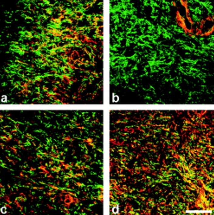
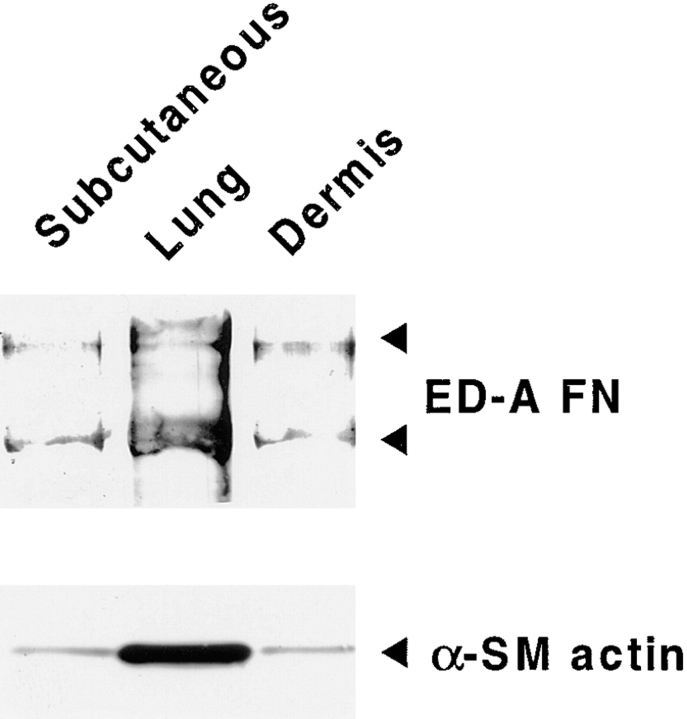
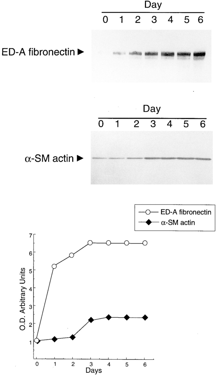
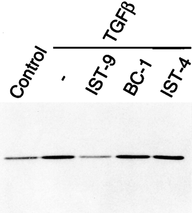
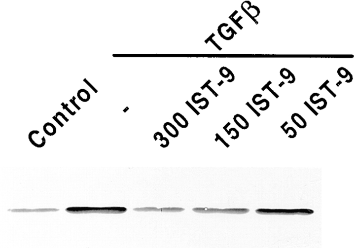
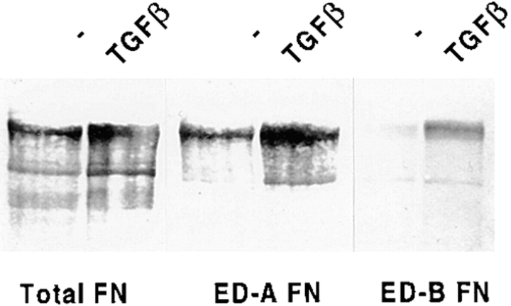
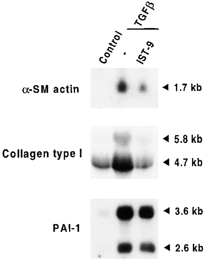
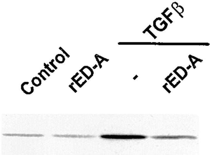
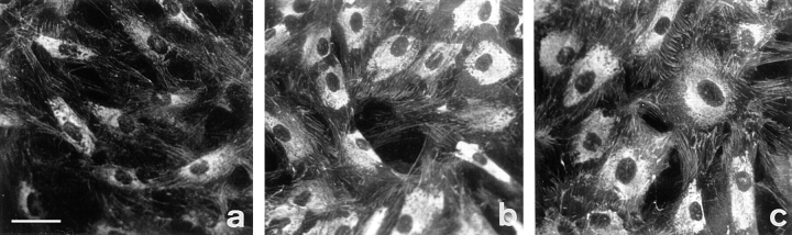
Transforming growth factor-beta1 (TGFbeta1), a major promoter of myofibroblast differentiation, induces alpha-smooth muscle (sn) actin, modulates the expression of adhesive receptors, and enhances the synthesis of extracellular matrix (ECM) molecules including ED-A fibronectin (FN), an isoform de novo expressed during wound healing and fibrotic changes. We report here that ED-A FN deposition precedes alpha-SM actin expression by fibroblasts during granulation tissue evolution in vivo and after TGFbeta1 stimulation in vitro. Moreover, there is a correlation between in vitro expression of alpha-SM actin and ED-A FN in different fibroblastic populations. Seeding fibroblasts on ED-A FN does not elicit per se alpha-SM actin expression; however, incubation of fibroblasts with the anti-ED-A monoclonal antibody IST-9 specifically blocks the TGFbeta1-triggered enhancement of alpha-SM actin and collagen type I, but not that of plasminogen activator inhibitor-1 mRNA. Interestingly, the same inhibiting action is exerted by the soluble recombinant domain ED-A, but neither of these inhibitory agents alter FN matrix assembly. Our findings indicate that ED-A-containing polymerized FN is necessary for the induction of the myofibroblastic phenotype by TGFbeta1 and identify a hitherto unknown mechanism of cytokine-determined gene stimulation based on the generation of an ECM-derived permissive outside in signaling, under the control of the cytokine itself.


Figures

Figure 1
Figure 1
α-SM actin and ED-A FN expression in granulation tissues at different times after wounding examined by confocal laser scanning microscopy. Sections of 4- (a and b), 7- (c), and 12-d-old (d) granulation tissue were double stained for total actin (a, red) or α-SM actin (b–d, red) and ED-A FN (a–d, green). (a) Fibroblastic cells showing an important cytoplasmic staining for total actin are already present and interact with ED-A FN (yellow staining, corresponding to the overlay of red and green staining) which appears de novo in huge amounts as early as 4 d after wounding within granulation tissue stroma. (b) At 4 d α-SM actin is localized only in connection with SM-cells of small blood vessels, but not in connection with fibroblasts. (c) A 7-d-old wound tissue shows focal α-SM actin staining of fibroblasts within an ED-A FN-rich extracellular network. (d) 12-d-old granulation tissue fibroblastic cells show wide positivity for α-SM actin. α-SM actin and ED-A FN are colocalized (c and d; yellow); areas of colocalization are more abundant in 12- (d) than in 7-d-old (c) granulation tissue. Bar, 50 μM.
Figure 2
Figure 2
ED-A FN and α-SM actin expression in cultured rat fibroblasts isolated from different tissues. Passage 5 rat fibroblast obtained from subcutaneous tissue, lung, and dermis were cultured for 4 d in absence of serum, then ED-A FN or α-SM actin expression were evaluated by Western blot analysis on equal amounts of total protein extracts. The levels of ED-A FN expression parallel those of α-SM actin.
Figure 3
Figure 3
Time course analysis of α-SM actin and ED-A FN expression modulation by TGFβ1 in cultured human subcutaneous fibroblasts. Fibroblasts were incubated with 10 ng/ml of TGFβ1 and total proteins were extracted after different time of continuous stimulation. Immunoblotting after SDS-PAGE shows that TGFβ1 induces a clear-cut α-SM actin increase only after 72 h of treatment, whereas ED-A FN response to TGFβ1 precedes and then parallels α-SM actin increase. Tracks were loaded with equal amounts of total proteins. These data are the means of five independent experiments; the SEMs, which are not represented in the figure, were always lower than 5% of the values.
Figure 4
Figure 4
ED-A FN antibodies block TGFβ1 induction of α-SM actin in cultured human subcutaneous fibroblasts. Cells were seeded on simple gelatin or on gelatin containing blocking monoclonal antibodies against different FN type III domains and then stimulated with TGFβ1 (10 ng/ml) for 3 d; α-SM actin was then analyzed by Western blotting on equal amounts of total proteins. Note that inhibition of α-SM actin induction by TGFβ1 was obtained only when fibroblasts were plated on gelatin containing 300 μg/ml of IST-9.
Figure 5
Figure 5
The inhibition of TGFβ1 induction of α-SM actin by IST-9 is dose dependent. Cells were seeded on simple gelatin or on gelatin containing different amounts (50, 150, and 300 μg/ml) of IST-9 and then stimulated with TGFβ1 (10 ng/ml) for 3 d; α-SM actin was then analyzed by Western blotting on equal amounts of total proteins.
Figure 6
Figure 6
TGFβ1 induces the insertion of both ED-A and ED-B domain within cellular FN. Passage 5 human subcutaneous fibroblasts were stimulated with TGFβ1 (10 ng/ml) for 3 d; total FN, ED-A FN, and ED-B FN were then evaluated by Western blot analysis on equal amounts of total proteins. Treatment with TGFβ1 determined an increase of total FN and of both ED-A and ED-B FN isoforms when compared with control cells.
Figure 7
Figure 7
ED-A FN antibodies block TGFβ1 induction of α-SM actin and collagen type I, but not of PAI-1 mRNA in cultured human subcutaneous fibroblasts. Under the same experimental conditions described in Fig. 4, cells treated with TGFβ1 for 24 h contain higher levels of α-SM actin, collagen type I, and PAI-1 mRNA when compared with untreated cells; treatment with IST-9 mAb dramatically inhibits α-SM actin and collagen type I induction, but not that of PAI-1.
Figure 8
Figure 8
Addition of rED-A domain blocks TGFβ1 induction of α-SM actin in cultured human subcutaneous fibroblasts. Cells were seeded on simple gelatin or on gelatin containing 300 μg/ml of rED-A domain and then stimulated or not with TGFβ1 (10 ng/ml) for 3 d; α-SM actin was then analyzed by Western blotting on equal amount of total protein extracts. Treatment of fibroblasts with rED-A slightly lowered basal α-SM actin expression levels and inhibited α-SM actin induction by TGFβ1.
Figure 9
Figure 9
FN matrix assembly evaluation on TGFβ1 stimulated fibroblasts after IST-9 mAb treatment. Subcutaneous fibroblasts were plated on simple gelatin (a and b) or on gelatin containing IST-9 mAb (c) and then stimulated with TGFβ1 (10 ng/ml) for 3 d (b and c). Cells were then stained with an anti-total FN rabbit polyclonal antibody. When compared with control (a), TGFβ1 stimulation determines an increase in FN expression and assembly by fibroblasts (b). IST-9 mAb treatment (c) does not interfere with TGFβ1 action. Bar, 25 μM.

References

    1. Allen-Hoffmann BL, Crankshaw CL, Mosher DF. Transforming growth factor β increases cell surface binding and assembly of exogenous (plasma) fibronectin by normal human fibroblasts. Mol Cell Biol. 1988;8:4234–4242. - PMC - PubMed
    1. Balza E, Borsi L, Allemmanni G, Zardi L. Transforming Growth Factor β regulates the levels of different fibronectin isoforms in normal cultured fibroblasts. FEBS (Fed Eur Biochem Soc) Lett. 1988;228:42–44. - PubMed
    1. Berndt A, Kosmehl H, Mandel U, Gabler U, Luo X, Celada D, Zardi L, Katenkamp D. TGFβ and bFGF synthesis and localization in Dupuytren's disease (nodular palmar fibrosis) relative to cellular activity, myofibroblast phenotype and oncofetal variants of fibronectin. Histochemical J. 1995;27:1014–1020. - PubMed
    1. Bochaton-Piallat ML, Gabbiani G, Pepper MS. Plasminogen activator expression in rat arterial smooth muscle cells depends on their phenotype and is modulated by cytokines. Circ Res. 1998;82:1086–1093. - PubMed
    1. Border WA, Ruoslahti E. Transforming growth factor-β in disease: the dark side of tissue repair. J Clin Invest. 1992;90:1–7. - PMC - PubMed

Publication types