. 1998 Aug;117(4):1143-52.
doi: 10.1104/pp.117.4.1143.

Characterization of a maize tonoplast aquaporin expressed in zones of cell division and elongation

Affiliations

Characterization of a maize tonoplast aquaporin expressed in zones of cell division and elongation

F Chaumont et al. Plant Physiol. 1998 Aug.

Abstract

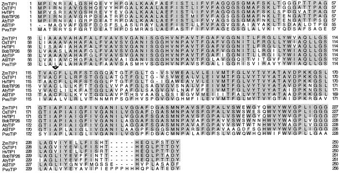
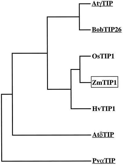
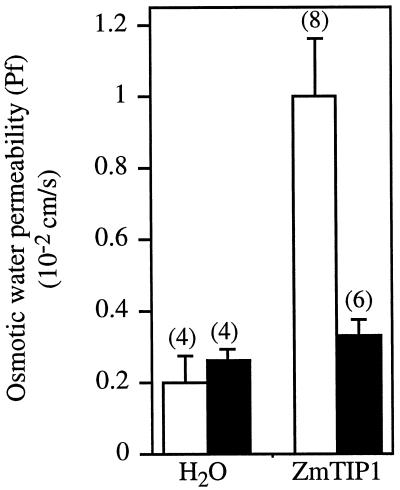
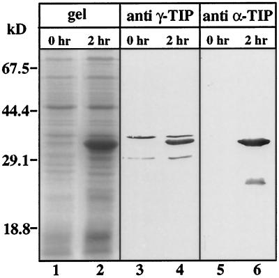
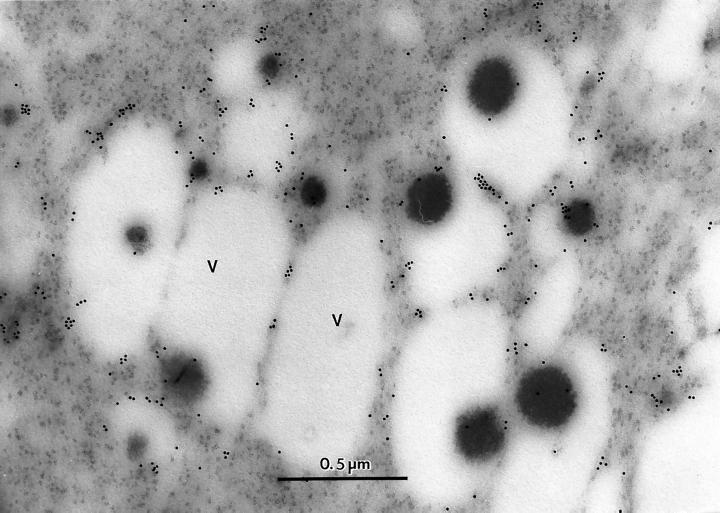
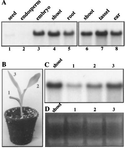
We studied aquaporins in maize (Zea mays), an important crop in which numerous studies on plant water relations have been carried out. A maize cDNA, ZmTIP1, was isolated by reverse transcription-coupled PCR using conserved motifs from plant aquaporins. The derived amino acid sequence of ZmTIP1 shows 76% sequence identity with the tonoplast aquaporin gamma-TIP (tonoplast intrinsic protein) from Arabidopsis. Expression of ZmTIP1 in Xenopus laevis oocytes showed that it increased the osmotic water permeability of oocytes 5-fold; this water transport was inhibited by mercuric chloride. A cross-reacting antiserum made against bean alpha-TIP was used for immunocytochemical localization of ZmTIP1. These results indicate that this and/or other aquaporins is abundantly present in the small vacuoles of meristematic cells. Northern analysis demonstrated that ZmTIP1 is expressed in all plant organs. In situ hybridization showed a high ZmTIP1 expression in meristems and zones of cell enlargement: tips of primary and lateral roots, leaf primordia, and male and female inflorescence meristems. The high ZmTIP1 expression in meristems and expanding cells suggests that ZmTIP1 is needed (a) for vacuole biogenesis and (b) to support the rapid influx of water into vacuoles during cell expansion.


Figures

Figure 1
Figure 1
Comparison of ZmTIP1 sequence with other plant TIPs. Amino acid sequences were compared with the Clustal W multiple alignment program (Thompson et al., 1994). The amino acid sequences were obtained from the following sources: ZmTIP1 (this work); OsTIP1 (Liu et al., 1994); HvTIP1 (Schünmann and Ougham, 1996); BobTIP26 (Barrieu et al., 1998); Atγ-TIP (Hofte et al., 1992); Atδ-TIP (Daniels et al., 1996); and Pvα-TIP (Johnson et al., 1990). Identical amino acid residues common to at least three sequences are shaded. Numbering refers to the respective amino acid sequence. The position of the Cys residue responsible for the mercury sensitivity of Atγ-TIP and Atδ-TIP is noted by an arrowhead in the consensus line.
Figure 2
Figure 2
Dendogram of the comparison between ZmTIP1 and other plant TIPs. Amino acid sequences from Figure 1 were compared using the program PILEUP (Genetics Computer Group, Madison, WI). Underlined sequences have been identified as aquaporins.
Figure 3
Figure 3
Osmotic water permeability (Pf) values of individual ZmTIP1 cRNA-injected oocytes derived from volume change measurements made over two independent preparations of oocytes. White bars, Control (no mercuric chloride); black bars, assay performed in the presence of 3 mm mercuric chloride with a 10-min preincubation. Data are expressed as the mean ± se, with the number of replicates indicated next to each bar in parentheses.
Figure 4
Figure 4
Coomassie blue-stained gel and immunoblot of extracts of E. coli expressing GST-C-ZmTIP1. Total E. coli protein extract before (lanes 1, 3, and 5) and after (lanes 2, 4, and 6) 2 h of induction of GST-C-ZmTIP1 expression by 2 mm isopropyl β-d-thiogalactopyranoside was fractionated by SDS-PAGE. Polypeptides were visualized with Coomassie blue (lanes 1 and 2) or transferred to nitrocellulose and immunostained using Arabidopsis γ-TIP antiserum (lanes 3 and 4) or bean α-TIP (lanes 5 and 6). The positions of molecular mass standards are indicated.
Figure 5
Figure 5
Immunocytochemical localization of ZmTIP1 in maize embryos. The gold particles are primarily at the interface of the cytoplasm and the vacuole (V). The vacuoles apparently contain aggregated protein. Magnification is ×60,000.
Figure 6
Figure 6
Genomic Southern analysis. Total maize genomic DNA (15 μg per lane) was digested with EcoRI, HindIII, and BamHI and hybridized with labeled 3′-untranslated region of ZmTIP1 cDNA. The positions of the Mr markers are indicated.
Figure 7
Figure 7
Gel-blot analysis of ZmTIP1 mRNA in different vegetative and reproductive organs. Total RNA (20 μg) was extracted from the indicated organs (A, C, and D) and from 10-d-old maize plantlet leaves (1–3 in B–D), and separated by gel electrophoresis in the presence of ethidium bromide (D). After transfer the blots were hybridized with ZmTIP1 probe (A and C). Total shoot RNA was used as a control to compare the signal intensity in the different blots.
Figure 8
Figure 8
Localization of ZmTIP1 mRNA by in situ hybridization. The controls, hybridized with sense RNA, are shown in C, E, and I; all other panels were hybridized with antisense RNA. A, Longitudinal section of 3-d-old root tip; B, longitudinal section in the zone of lateral root initiation; C, control, same section as shown in A; D and E, median sections of 3-d-old plumule; F, median section of an immature tassel; G, close-up of F; H and I, median section of an immature ear. Ca, Root cap; DZ, division zone; EZ, elongation zone; Gl, glume; Le, lemma; LF, lower floret; Lo, lodicule; LP, leaf primordium; S, stamen; VB, vascular bundle; TZ, transition zone; UF, upper floret. White and black arrows in B indicate RNA transcript signal in the lateral root meristems and the vascular bundle, respectively.

Similar articles

Cited by

References

    1. Baluska F, Volkmann D, Barlow PW. Specialized zones of development in roots. View from the cellular level. Plant Physiol. 1996;112:3–4. - PMC - PubMed
    1. Barrieu F, Chaumont F, Chrispeels M. High expression of the tonoplast aquaporin ZmTIP1 in epidermal and conducting tissues of maize. Plant Physiol. 1998;117:1153–1163. - PMC - PubMed
    1. Barrieu F, Thomas D, Marty-Mazars D, Charbonnier M, Marty F. Tonoplast intrinsic proteins from cauliflower (Brassica oleracea L. var. botrytis): immunological analysis, cDNA cloning and evidence for expression in meristematic tissues. Planta. 1998;204:335–344. - PubMed
    1. Cone KC, Burr FA, Burr B. Molecular analysis of the maize anthocyanin regulatory locus C1. Proc Natl Acad Sci USA. 1986;83:9631–9635. - PMC - PubMed
    1. Daniels MJ, Chaumont F, Mirkov TE, Chrispeels MJ. Characterization of a new vacuolar membrane aquaporin sensitive to mercury at a unique site. Plant Cell. 1996;8:587–599. - PMC - PubMed

Publication types

MeSH terms

Associated data