. 1998 Aug;117(4):1153-63.
doi: 10.1104/pp.117.4.1153.

High expression of the tonoplast aquaporin ZmTIP1 in epidermal and conducting tissues of maize

Affiliations

High expression of the tonoplast aquaporin ZmTIP1 in epidermal and conducting tissues of maize

F Barrieu et al. Plant Physiol. 1998 Aug.

Abstract

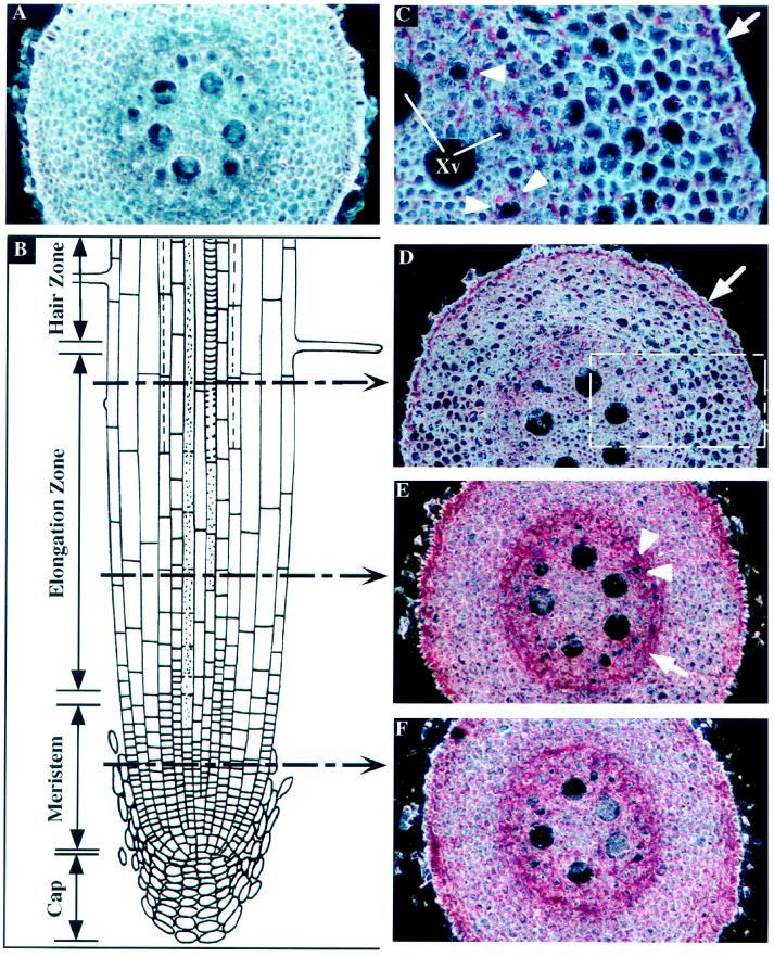
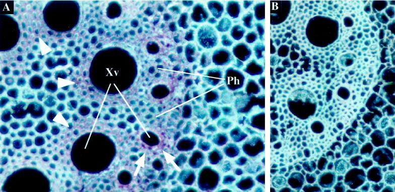
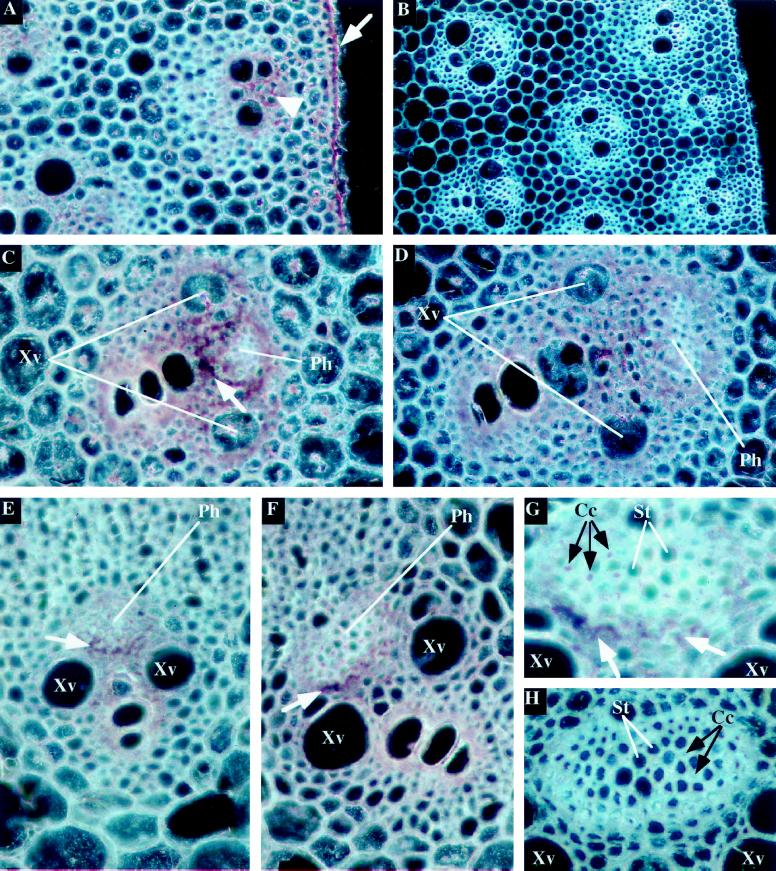
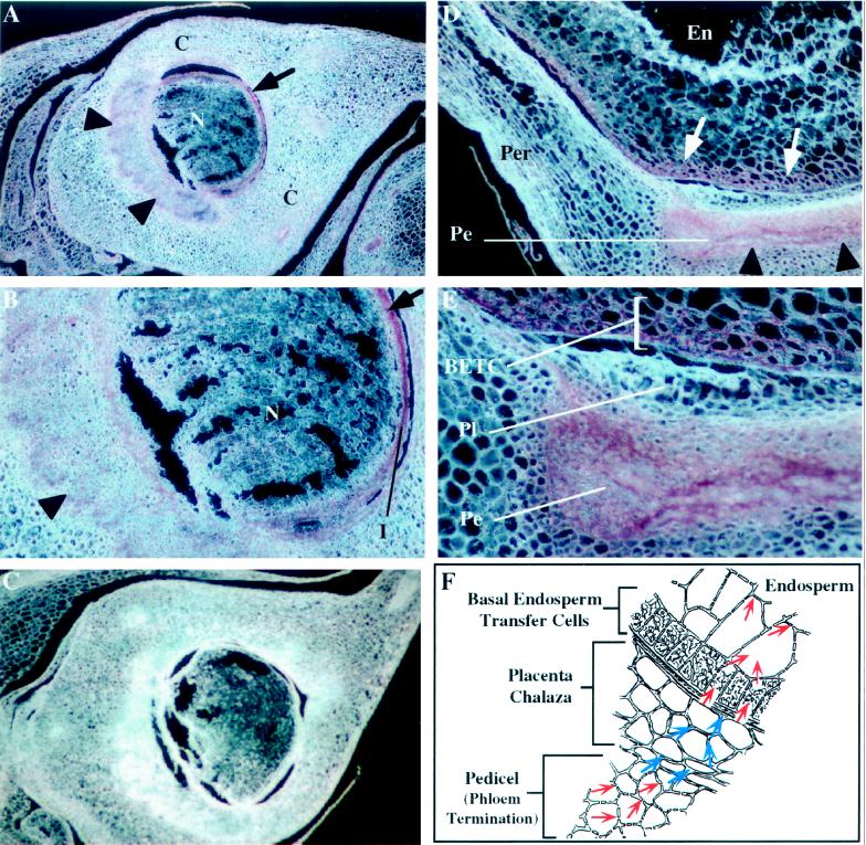
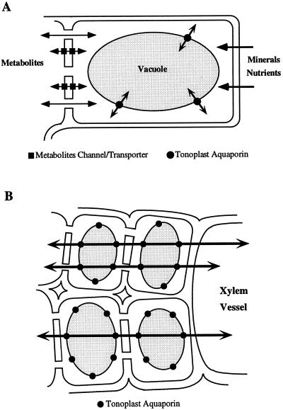
Aquaporins are integral membrane proteins of the tonoplast and the plasma membrane that facilitate the passage of water through these membranes. Because of their potentially important role in regulating water flow in plants, studies documenting aquaporin gene expression in specialized tissues involved in water and solute transport are important. We used in situ hybridization to examine the expression pattern of the tonoplast aquaporin ZmTIP1 in different organs of maize (Zea mays L.). This tonoplast water channel is highly expressed in the root epidermis, the root endodermis, the small parenchyma cells surrounding mature xylem vessels in the root and the stem, phloem companion cells and a ring of cells around the phloem strand in the stem and the leaf sheath, and the basal endosperm transfer cells in developing kernels. We postulate that the high level of expression of ZmTIP1 in these tissues facilitates rapid flow of water through the tonoplast to permit osmotic equilibration between the cytosol and the vacuolar content, and to permit rapid transcellular water flow through living cells when required.


Figures

Figure 1
Figure 1
In situ localization of ZmTIP1 mRNA in maize root tip. Transverse sections of the root tip were hybridized with ZmTIP1 sense (A) or antisense (C–F) digoxigenin-labeled RNA probes and photographed under dark-field conditions. The transcript signal is red. A, Control transverse section in the middle of the elongation zone hybridized with a ZmTIP1 sense probe. B, Schematic representation of a longitudinal section of a root tip. Discontinuous arrows indicate the approximate sites of transverse sections presented in D, E, and F. C, High magnification of the area boxed in D. Arrow indicates the expression of ZmTIP1 in epidermal cells. Arrowheads indicate the probe accumulation in the parenchyma cells that surround the small early metaxylem vessels. Xv, Xylem vessels. D, Transverse section at the end of the elongation zone. Arrow indicates the expression of ZmTIP1 in epidermal cells. E, Transverse section in the middle of the elongation zone. Arrow indicates ZmTIP1 expression in the endodermis/pericycle. Arrowheads indicate the probe accumulation around the early metaxylem vessels. F, Transverse sections in the meristematic zone.
Figure 2
Figure 2
In situ localization of ZmTIP1 mRNA in mature maize root. Transverse sections of the root (10–12 cm from the tip) were hybridized with ZmTIP1 antisense (A) or sense (B) digoxigenin-labeled RNA probes and photographed under dark-field conditions. The transcript signal is red. A, Expression of ZmTIP1 in the parenchyma cells of early (arrows) and late (arrowheads) xylem vessels. Xv, Xylem vessels; Ph, phloem strand. B, Control section hybridized with a ZmTIP1 sense probe.
Figure 3
Figure 3
In situ localization of ZmTIP1 mRNA in the vascular bundles and epidermis of stems and in the vascular bundles of leaves. Transverse sections of stems (A–D) and leaf sheaths (E–H) of 5-week-old maize plants were hybridized with ZmTIP1 antisense (A, C, D, E, F, and G) or sense (B and H) digoxigenin-labeled RNA probes and photographed under dark-field conditions. The transcript signal is red. Cc, Companion cells; Ph, phloem strand; St, sieve tubes; Xv, xylem vessels. A, Expression of ZmTIP1 in the epidermis (arrow) and in cells close to the vascular bundles (arrowhead) of maize stem. B, Control section of maize stem hybridized with a ZmTIP1 sense probe. C, Expression of ZmTIP1 in cells close to a peripheral vascular bundle of the stem. Arrow indicates the high accumulation of ZmTIP1 transcripts in parenchyma cells around the phloem bundle. D, Expression of ZmTIP1 in cells close to a central vascular bundle of the stem. E and F, Expression of ZmTIP1 in cells close to small (E) and large (F) vascular bundles of maize leaf. Arrows indicate the high concentration of the probe in parenchyma cells located between the phloem strand and the xylem vessels. G, High magnification of the phloem strand presented in F showing expression of ZmTIP1 in parenchyma cells (white arrows) and companion cells (black arrows). H, Control section of a leaf phloem strand hybridized with a ZmTIP1 sense probe.
Figure 4
Figure 4
In situ localization of ZmTIP1 mRNA in developing pistils and caryopses of maize. Longitudinal sections of nonfertilized maize ears 7-cm long (A–C) and developing maize caryopses 14 d after pollination (D and E) were hybridized with ZmTIP1 antisense (A, B, D, and E) or sense (C) digoxigenin-labeled RNA probes and photographed under dark-field conditions. The transcript signal is red. C, Carpel wall; En, endosperm; I, integuments; N, Nucellus; Pe, Pedicel; Per, Pericarp; Pl, Placenta-chalaza. A, Section of a developing pistil showing expression of ZmTIP1 in the termination zone of the vascular bundle under the ovule (arrowheads) and around the nucellus (arrow). B, High magnification of the developing ovule presented in A showing expression of ZmTIP1 in the vascular tissue (arrowhead) and in the outer layer(s) of the nucellus (arrow). C, Control section of a developing pistil hybridized with a ZmTIP1 sense probe. D, Expression of ZmTIP1 in the basal region of the developing caryopse. The ZmTIP1 transcripts are detected in the pedicel area (arrowheads) and in a zone of the endosperm that is adjacent to the pedicel (arrows). E, High magnification of the pedicel area presented in D showing expression of ZmTIP1 in the basal endosperm transfer cells (BETC). F, Schematic representation of the three main tissues shown in E (adapted from Thorne, 1985). Red arrows within the cells indicate probable symplastic intercellular transport of assimilates and water. Blue arrows over the cell walls indicate apoplastic movement of assimilates and water.
Figure 5
Figure 5
Schematic representation of two roles for tonoplast aquaporins. A, Tonoplast aquaporins are needed for cytoplasmic osmotic equilibration in cells that can experience rapid fluxes of metabolites or mineral nutrients. B, Tonoplast aquaporins permit rapid transcellular flow and increase the effective cross-section of the cytoplasm for symplastic flow.

Similar articles

Cited by

References

    1. Barrieu F, Thomas D, Marty-Mazars D, Charbonnier M, Marty F. Tonoplast intrinsic proteins from cauliflower (Brassica oleracea L. var. botrytis): immunological analysis, cDNA cloning and evidence for expression in meristematic tissues. Planta. 1998;204:335–344. - PubMed
    1. Canny MJ. Vessel contents during transpiration: embolisms and refilling. Am J Bot. 1997;84:1223–1230. - PubMed
    1. Chaumont F, Barrieu F, Herman EM, Chrispeels MJ. Characterization of a maize tonoplast aquaporin expressed in zones of cell division and elongation. Plant Physiol. 1998;117:1143–1152. - PMC - PubMed
    1. Chrispeels MJ, Maurel C. Aquaporins: the molecular basis of facilitated water movement through living plant cells. Plant Physiol. 1994;105:9–13. - PMC - PubMed
    1. Daniels MJ, Chaumont F, Mirkov TE, Chrispeels MJ. Characterization of a new vacuolar membrane aquaporin sensitive to mercury at a unique site. Plant Cell. 1996;8:587–599. - PMC - PubMed

LinkOut - more resources