. 1998 Aug;117(4):1171-8.
doi: 10.1104/pp.117.4.1171.

Chemotropic and contact responses of phytophthora sojae hyphae to soybean isoflavonoids and artificial substrates

Affiliations

Chemotropic and contact responses of phytophthora sojae hyphae to soybean isoflavonoids and artificial substrates

PF Morris et al. Plant Physiol. 1998 Aug.

Abstract

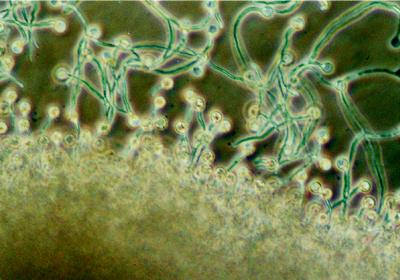
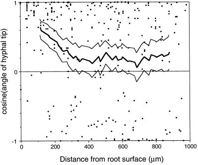
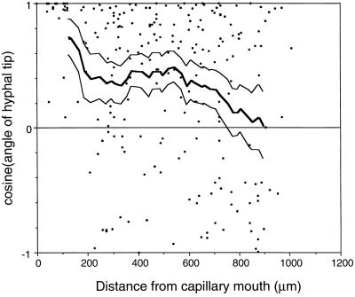
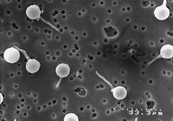
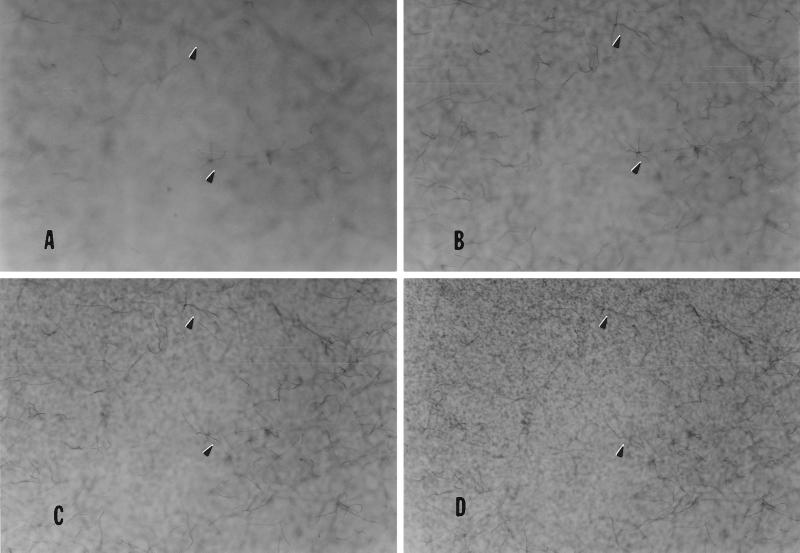
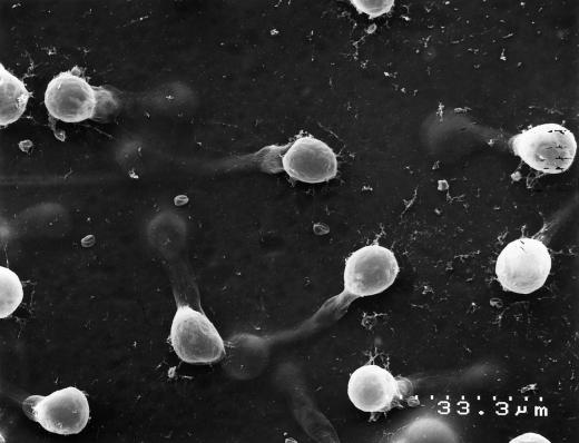
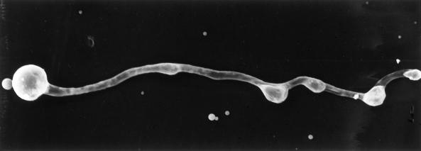
We have investigated the role of the isoflavones daidzein and genistein on the chemotropic behavior of germinating cysts of Phytophthora sojae. Hyphal germlings were shown to respond chemotropically to daidzein and genistein, suggesting that hyphal tips from zoospores that have encysted adjacent to the root may use specific host isoflavones to locate their host. Observations of the contact response of hyphal germlings were made on several different substrates in the presence and absence of isoflavones. Hyphal tips of germlings detected and penetrated pores in membranes and produced multiple appressoria on smooth, impenetrable surfaces. Hyphae that successfully penetrated the synthetic membrane were observed to grow away from the membrane surface. The presence of isoflavones in the medium surrounding the hyphal germlings did not appear to alter any of those habits. Daidzein and genistein did not inhibit germination or initial hyphal growth at concentrations up to 20 &mgr;M.


Figures

Figure 1
Figure 1
Chemotropism of hyphae toward a capillary tube containing genistein (magnification ×165). Zoospores encysted around the capillary tube and the majority of cysts produced hyphae that grew in the direction of the source. Hyphae farthest from the source changed direction to grow toward the capillary tube.
Figure 2
Figure 2
Chemotropic growth of hyphae toward soybean root tips. Zoospores were induced to encyst in a random orientation. Approximately 3 h after the introduction of the root tip into the chemotaxis chamber, the angles of hyphal tips were measured relative to a line drawn perpendicular to the root surface. ▪, Cosine of growth angle; thick line, cosines averaged over a 200-μm window at 20-μm increments; thin lines, upper and lower 95% significance limits for cosine means calculated using a t test. The number of data points in each window ranged from 38 to 74. Chemotropism was considered significant when the lower confidence limit was greater than 0.
Figure 3
Figure 3
Chemotropic growth of hyphae toward capillary tubes containing 20 μm genistein. Zoospores were induced to encyst in a random orientation. Approximately 3 h after the introduction of the capillary into the chemotaxis chamber, the angles of hyphal tips were measured relative to a line drawn from the counterpoint of the mouth of the capillary. •, Cosine of growth angle; thick line, cosines averaged over a 200-μm window at 20-μm increments; thin line, upper and lower 95% significance limits for cosine means calculated using a t test. The number of data points in each window ranged from 30 to 66. Chemotropism was considered significant when the lower confidence limit was greater than 0.
Figure 4
Figure 4
Hyphal germlings growing along and penetrating pores of a PET membrane. Zoospores were induced to encyst on the membrane by application of an agar plug containing isoflavones to the opposite side of the membrane. Germinating hyphae penetrated pores detected by the hyphal tip. The distance indicated is the distance between two hatch marks.
Figure 5
Figure 5
Hyphae growing away from a membrane after having penetrated a pore (magnification ×27). Zoospores were induced to encyst on a PET membrane. Hyphae that had successfully penetrated through pores were visualized by staining with 3-[4,5-dimethylthiazol-2-yl]-2,5-diphenyltetrazollium bromide. The plane of focus in each picture (A–D) is progressively closer to the membrane surface and is slightly above the membrane in D. Arrows point to hyphae that are more visible in frames A, B, or C, indicating that they must be growing away from the membrane surface (D).
Figure 6
Figure 6
Germination of cysts on a surface of cross-linked collagen overlying a PET membrane. Zoospores were induced to encyst on the collagen by application of an agar plug containing isoflavones to the opposite side of the membrane. Germinating hyphae penetrated the collagen layer and subsequently formed appressorial structures that are believed to be located over membrane pores. The distance indicated is the distance between two hatch marks.
Figure 7
Figure 7
Multiple appressoria were produced by a germinating hyphae on clear plastic wrap (magnification ×1000). Zoospores were induced to encyst on a smooth surface. The hyphal germlings produced multiple appressoria as they grew along the surface.

References

    1. Akiyama T, Ishida J, Nakagawa S, Ogawara H, Watanabee S, Itoh N, Shibuya M, Fukami Y. Genistein, a specific inhibitor of tyrosine-specific protein kinases. J Biol Chem. 1987;262:5592–5595. - PubMed
    1. Allen EA, Hazen BE, Hoch HC, Kwon Y, Leinhos ME, Staples RC, Stumpf MA, Terhune BT. Appressorium formation in response to topographical signals by 27 rust species. Phytopathology. 1991;81:323–331.
    1. Carlile MJ (1983) Motility, taxis, and tropism in Phytophthora. In DC Erwin, S Bartnicki-Garcia, PH Tsao, eds, Phytophthora: Its Biology, Taxonomy, Ecology, and Pathology. American Phytopathological Society Press, St. Paul, MN, pp 95–107
    1. Coley-Smith JR. White rot disease of Allium: problems of soil-borne diseases in microcosm. Plant Pathol. 1990;39:214–222.
    1. Dharmatilake AJ, Bauer WD. Chemotaxis of Rhizobium meliloti towards nodulation gene-inducing compounds from alfalfa roots. Appl Environ Microbiol. 1992;58:1153–1158. - PMC - PubMed

LinkOut - more resources