. 1998 Aug;117(4):1445-61.
doi: 10.1104/pp.117.4.1445.

Direct evidence for rapid degradation of Bacillus thuringiensis toxin mRNA as a cause of poor expression in plants

Affiliations

Direct evidence for rapid degradation of Bacillus thuringiensis toxin mRNA as a cause of poor expression in plants

E J De Rocher et al. Plant Physiol. 1998 Aug.

Abstract

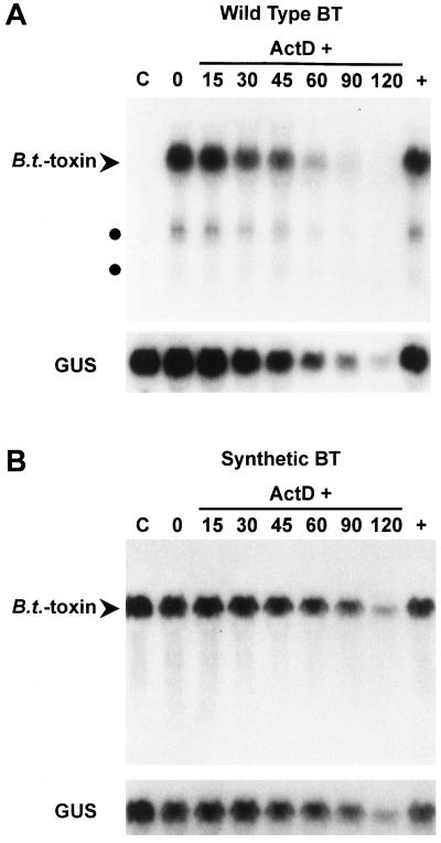
It is well established that the expression of Bacillus thuringiensis (B.t.) toxin genes in higher plants is severely limited at the mRNA level, but the cause remains controversial. Elucidating whether mRNA accumulation is limited transcriptionally or posttranscriptionally could contribute to effective gene design as well as provide insights about endogenous plant gene-expression mechanisms. To resolve this controversy, we compared the expression of an A/U-rich wild-type cryIA(c) gene and a G/C-rich synthetic cryIA(c) B.t.-toxin gene under the control of identical 5' and 3' flanking sequences. Transcriptional activities of the genes were equal as determined by nuclear run-on transcription assays. In contrast, mRNA half-life measurements demonstrated directly that the wild-type transcript was markedly less stable than that encoded by the synthetic gene. Sequences that limit mRNA accumulation were located at more than one site within the coding region, and some appeared to be recognized in Arabidopsis but not in tobacco (Nicotiana tabacum). These results support previous observations that some A/U-rich sequences can contribute to mRNA instability in plants. Our studies further indicate that some of these sequences may be differentially recognized in tobacco cells and Arabidopsis.


Figures

Figure 1
Figure 1
Schematic representation of the method used to construct the synthetic B.t.-toxin gene. A two-step PCR approach was used to generate synthetic versions of each of four segments of a cryIA(c) gene using sets of overlapping oligonucleotides that incorporated sequence changes according to the criteria described in the text. As shown for one of the four segments, the alternating sense and antisense polarity of the overlapping modified-sequence oligonucleotides indicated by the directions of the arrows allowed the oligonucleotides to anneal to each other and serve as primers for DNA synthesis in PCR. After the first set of 10 PCR cycles, the addition of 30-mer terminal primers followed by a further 25 PCR cycles preferentially amplified those molecules spanning the full segment. PCR products for each segment were cloned and then assembled by standard cloning methods to generate the complete synthetic B.t.-toxin-coding region.
Figure 2
Figure 2
Schematic diagram of B.t.-toxin and control gene constructions. Expression of wild-type B.t.-toxin, synthetic B.t.-toxin, and globin genes in transient expression assays and stably transformed cell lines and plants was under the control of 2X35S. For transient expression in maize, constructs included an ADH1 intron. Polyadenylation signals were provided by the 3′ UTR from the pea Rubisco small subunit rbcS-E9 gene in all constructs. Plasmid numbers for each construct are indicated at the left.
Figure 3
Figure 3
RNA gel-blot analysis of relative expression levels of the synthetic and wild-type B.t.-toxin genes in plant cells. Accumulation of synthetic (SYN) and wild-type (WT) B.t.-toxin mRNAs was compared in transiently transformed maize and tobacco cells and in stably transformed tobacco cells and Arabidopsis plants. A, Plasmids containing wild-type or synthetic B.t.-toxin genes were electroporated into maize BMS and tobacco BY-2 protoplasts. Plasmids containing a human β-globin gene were coelectroporated in both types of experiments to serve as an internal standard. The positions of the B.t.-toxin and globin transcripts are indicated. B, Tobacco BY-2 cells and Arabidopsis plants were stably transformed with constructs containing wild-type or synthetic B.t.-toxin genes and a GUS gene serving as an internal standard. The positions of the B.t.-toxin and GUS transcripts are indicated.
Figure 4
Figure 4
Estimation of the minimum difference in mRNA accumulation between wild-type (WT) and synthetic (SYN) B.t.-toxin genes. Tobacco BY-2 protoplasts were electroporated with plasmids containing either wild-type or synthetic B.t.-toxin genes and plasmids containing a human β-globin gene as an internal standard. RNA from protoplasts expressing the synthetic B.t.-toxin gene was diluted in 2-fold steps up to 64-fold with total RNA from untransformed tobacco cells. Each lane was loaded with 20 μg of RNA. The positions of the B.t.-toxin and globin transcripts are indicated by arrowheads. The blot was hybridized with a probe specific for the rbcS-E9 3′ UTR common to all three transcripts. Dilution factors are indicated by numbers above the lanes.
Figure 5
Figure 5
Accumulation of B.t.-toxin protein in plants expressing the wild-type and synthetic B.t.-toxin genes as determined by SDS-PAGE analysis of protein extracts from pools of independent Arabidopsis lines stably transformed with the wild-type (WT) or synthetic (SYN) B.t.-toxin genes. Purified cryIA(c) protein (predicted molecular mass approximately 133 kD) expressed in E. coli was included as a positive control (C) for anti-cryIA(c) antibody binding. Positions of molecular mass standards (in kD) are indicated between the panels. A, Immunoblot incubated with anti-cryIA(c) polyclonal antibodies. B, Immunoblot of duplicate lanes incubated with anti-cryIA(c) polyclonal antibodies and overdeveloped to demonstrate the lack of detectable B.t. toxin in plants transgenic for the wild-type B.t.-toxin gene.
Figure 6
Figure 6
Measurement of relative half-lives of wild-type and synthetic B.t.-toxin transcripts (BT). Wild-type and synthetic B.t.-toxin mRNA decay was measured over a time course in stably transformed tobacco BY-2 suspension cell cultures. Cells were harvested before transient CHX treatment (C), after the 2-h treatment followed by removal of CHX (0), at intervals after the addition of ActD (15, 30, 45, 60, 90, and 120 min), and after continuous treatment with CHX and no ActD (+). A, RNA gel-blot analysis of a time-course experiment using a cell line expressing the wild-type B.t.-toxin gene. Full-length wild-type B.t.-toxin transcripts are indicated by the arrowhead, and short polyadenylated B.t.-toxin transcripts are indicated by dark circles. The blot was stripped and rehybridized with a probe specific for GUS mRNA. B, RNA gel-blot analysis of a time-course experiment using a cell line expressing the synthetic B.t.-toxin gene. Full-length synthetic B.t.-toxin transcripts are indicated by the arrowhead. The blot was stripped and rehybridized with a probe specific for GUS mRNA.
Figure 7
Figure 7
Effects of wild-type B.t.-toxin sequences on synthetic B.t.-toxin mRNA accumulation. A, Schematic diagram of chimeric gene constructs used to test the effects of wild-type B.t.-toxin sequences on mRNA accumulation. The synthetic B.t.-toxin-coding region was divided into four segments using restriction sites conserved between the wild-type and synthetic coding regions. Each segment of the wild-type gene was substituted individually for the corresponding segment of the synthetic gene. B, Histogram summarizing the results of RNA gel-blot analysis of chimeric B.t.-toxin gene expression relative to the synthetic B.t.-toxin gene in pools of stably transformed tobacco BY-2 cell lines (white bars) and Arabidopsis plants (black bars). Hybridization signals for full-length B.t.-toxin mRNAs were quantitated and normalized using GUS-hybridization signals as an internal standard. Hybridization signals of the synthetic B.t.-toxin transcripts in tobacco and Arabidopsis were set equal to one to allow comparison of relative expression levels between tobacco and Arabidopsis. Data are presented as means and ses for three sets of tobacco and four sets of Arabidopsis pools.

References

    1. Adang MJ, Brody MS, Cardineau G, Eagan N, Roush RT, Shewmaker CK, Jones A, Oakes JV, McBride KE. The reconstruction and expression of a Bacillus thuringiensis cryIIIA gene in protoplasts and potato plants. Plant Mol Biol. 1993;21:1131–1145. - PubMed
    1. Adang MJ, Firoozabady E, Klein J, DeBoer D, Sekar V, Kemp JD, Murray EE, Rocheleau TA, Rashka K, Staffeld G, and others (1987) Expression of a Bacillus thuringiensis insecticidal crystal protein gene in tobacco plants. In CJ Arntzen, C Ryan, eds, Molecular Strategies for Crop Protection: UCLA Symposia on Molecular and Cellular Biology, New Series, Vol 46. Alan R. Liss, New York, pp 345–353
    1. Adang MJ, Staver MJ, Rocheleau TA, Leighton J, Barker RF, Thompson DV. Characterization of full-length and truncated plasmid clones and the crystal protein of Bacillus thuringiensis subsp. kurstaki HD-73 and their toxicity to Manduca sexta. Gene. 1985;36:289–300. - PubMed
    1. Barton KA, Whiteley HR, Yang N-S. Bacillus thuringiensis δ-endotoxin expressed in transgenic Nicotiana tabacum provides resistance to lepidopteran insects. Plant Physiol. 1987;85:1103–1109. - PMC - PubMed
    1. Bigelow CC, Channon M (1982) In GD Fasman, ed, CRC Handbook of Biochemistry and Molecular Biology, Ed 3: Proteins, Vol 1. CRC Press, Boca Raton, FL, pp 209–243

Publication types

Associated data

LinkOut - more resources