

Rapid identification of Candida dubliniensis by indirect immunofluorescence based on differential localization of antigens on C. dubliniensis blastospores and Candida albicans germ tubes
- PMID: 9705368
- PMCID: PMC105138
- DOI: 10.1128/JCM.36.9.2428-2433.1998
Rapid identification of Candida dubliniensis by indirect immunofluorescence based on differential localization of antigens on C. dubliniensis blastospores and Candida albicans germ tubes
Abstract
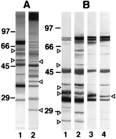
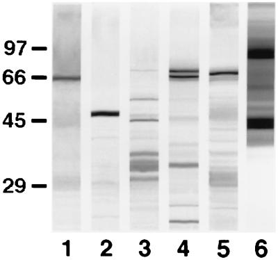
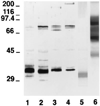
There is a clear need for the development of a rapid and reliable test for the identification of Candida dubliniensis and for the discrimination of this species from Candida albicans. In the present study we have investigated the potential use of C. dubliniensis-specific antigens as a basis for its identification. We produced an anti-C. dubliniensis serum which, after adsorption with C. albicans blastospores, was found to differentially label C. dubliniensis isolates in an indirect immunofluorescence test. In this test, the antiserum reacted with blastospores and germ tubes of C. dubliniensis and with blastospores of Candida krusei and Rhodotorula rubra but did not react with blastospores of several other Candida species including C. albicans. The antiserum also reacted with C. albicans germ tubes. The anti-C. dubliniensis adsorbed serum reacted with specific components of 25, 28, 37, 40, 52, and 62 kDa in the C. dubliniensis extract and with a variety of antigens from other yeast species. The antigens from non-C. dubliniensis yeasts showing reactivity with the anti-C. dubliniensis adsorbed serum are mostly expressed within the cell walls of these yeast species, and this reactivity does not interfere with the use of the anti-C. dubliniensis adsorbed serum in an indirect immunofluorescence test for the rapid identification of C. dubliniensis.
Figures

References
-
- Barturen B, Bikandi J, San Millán R, Moragues M D, Regúlez P, Quindós G, Pontón J. Variability in expression of antigens responsible for serotype specificity in Candida albicans. Microbiology. 1995;141:1535–1543. - PubMed
-
- Coleman, D., D. Sullivan, K. Haynes, M. Henman, D. Shanley, D. Bennett, G. Moran, C. McCreary, L. O’Neill, and B. Harrington. 1997. Molecular and phenotypic analysis of Candida dubliniensis: a recently identified species linked with oral candidosis in HIV-infected and AIDS patients. Oral Dis. 3(Suppl. 1):S96–S101. - PubMed
-
- Coleman D C, Sullivan D J, Bennett D E, Moran G P, Barry H J, Shanley D B. Candidiasis: the emergence of a novel species, Candida dubliniensis. AIDS. 1997;11:557–567. - PubMed
-
- Delgado W, Aguirre J M. Las micosis orales en la era del sida. Rev Iberoam Micol. 1997;14:14–22. - PubMed
Publication types
MeSH terms
Substances
LinkOut - more resources
Full Text Sources
Medical
