

Cooperative oligonucleotides mediating direct capture of hepatitis C virus RNA from serum
- PMID: 9705373
- PMCID: PMC105143
- DOI: 10.1128/JCM.36.9.2454-2459.1998
Cooperative oligonucleotides mediating direct capture of hepatitis C virus RNA from serum
Abstract
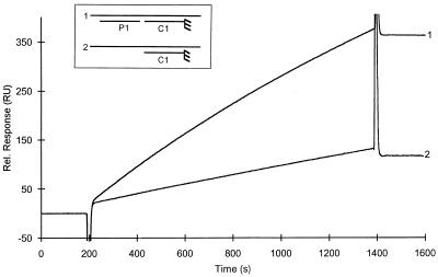
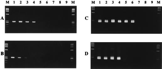
A novel method for direct capture of hepatitis C virus (HCV) RNA from clinical samples has been developed. This approach takes advantage of the cooperative interactions between adjacently hybridized oligonucleotides. Here, this cooperative effect was combined with solid-phase technology, whereby a capture probe was covalently coupled to magnetic beads and a second probe, which anneals adjacent to the capture probe site, was prehybridized in solution to the target. When these contiguously hybridized probes were used for the extraction of HCV RNA from clinical samples, the capture efficiency was increased up to 25-fold in comparison to capture with a single probe. The applicability of this sample preparation assay was further investigated by performing a comparative study with both a conventional guanidinium extraction method and a commercial quantitative assay.
Figures


Similar articles
-
Rapid detection of hepatitis C virus RNA by direct capture from blood.J Med Virol. 1994 Jan;42(1):22-8. doi: 10.1002/jmv.1890420105. J Med Virol. 1994. PMID: 8308516
-
Homogeneous quantitative assay of hepatitis C virus RNA by polymerase chain reaction in the presence of a fluorescent intercalater.Anal Biochem. 1995 Aug 10;229(2):207-13. doi: 10.1006/abio.1995.1404. Anal Biochem. 1995. PMID: 7485974
-
Capture of single-stranded DNA assisted by oligonucleotide modules.Anal Biochem. 1998 Jan 15;255(2):195-203. doi: 10.1006/abio.1997.2472. Anal Biochem. 1998. PMID: 9451504
-
[Serum levels of HCV-RNA determined by branched DNA (bDNA) probe assay in chronic hepatitis C: method and clinical significance on interferon (IFN) therapy].Nihon Rinsho. 1994 Jul;52(7):1747-53. Nihon Rinsho. 1994. PMID: 7521414 Review. Japanese.
-
"Real-time" polymerase chain reaction.Gastroenterology. 1999 Mar;116(3):763-4. doi: 10.1016/s0016-5085(99)70202-7. Gastroenterology. 1999. PMID: 10029634 Review. No abstract available.
Cited by
-
Laboratory evaluation of a fully automated chemiluminescence immunoassay for rapid detection of HBsAg, antibodies to HBsAg, and antibodies to hepatitis C virus.J Clin Microbiol. 2004 Feb;42(2):610-7. doi: 10.1128/JCM.42.2.610-617.2004. J Clin Microbiol. 2004. PMID: 14766824 Free PMC article.
-
LNA-enhanced detection of single nucleotide polymorphisms in the apolipoprotein E.Nucleic Acids Res. 2002 Oct 1;30(19):e100. doi: 10.1093/nar/gnf099. Nucleic Acids Res. 2002. PMID: 12364617 Free PMC article.
-
Lateral flow microarrays: a novel platform for rapid nucleic acid detection based on miniaturized lateral flow chromatography.Nucleic Acids Res. 2007;35(10):e74. doi: 10.1093/nar/gkm269. Epub 2007 May 3. Nucleic Acids Res. 2007. PMID: 17478499 Free PMC article.
-
Tandem oligonucleotide synthesis using linker phosphoramidites.Nucleic Acids Res. 2005 Apr 6;33(6):1940-8. doi: 10.1093/nar/gki333. Print 2005. Nucleic Acids Res. 2005. PMID: 15814811 Free PMC article.
-
Diagnostic tests for hepatitis C: recent trends in electrochemical immunosensor and genosensor analysis.World J Gastroenterol. 2014 Nov 14;20(42):15476-91. doi: 10.3748/wjg.v20.i42.15476. World J Gastroenterol. 2014. PMID: 25400433 Free PMC article. Review.
References
-
- Beaulieux F, See D M, Leparc-Goffart I, Aymard M, Lina B. Use of magnetic beads versus guanidinium thiocyanate-phenol-chloroform RNA extraction followed by polymerase chain reaction for the rapid, sensitive detection of enterovirus RNA. Res Virol. 1997;148:11–15. - PubMed
-
- Chomczynski P, Sacchi N. Single-step method of RNA isolation by acid guanidinium thiocyanate-phenol-chloroform extraction. Anal Biochem. 1987;162:156–159. - PubMed
Publication types
MeSH terms
Substances
LinkOut - more resources
Full Text Sources
Other Literature Sources
Medical
