. 1998 Sep;36(9):2535-41.
doi: 10.1128/JCM.36.9.2535-2541.1998.

Detection of a novel strain of porcine circovirus in pigs with postweaning multisystemic wasting syndrome

Affiliations

Detection of a novel strain of porcine circovirus in pigs with postweaning multisystemic wasting syndrome

I Morozov et al. J Clin Microbiol. 1998 Sep.

Abstract

Swine infectious agents, especially viruses, are potential public health risks associated with the use of pig organs for xenotransplantation in humans. Therefore, there is a need for better characterization of swine viruses and for the development of diagnostic tests for their detection. We report here isolation of a novel strain of porcine circovirus (PCV) from pigs with postweaning multisystemic wasting syndrome (PMWS). Affected pigs exhibited severe interstitial pneumonia and lymphoid depletion. The complete nucleotide sequence (1,768 nucleotides) of the genome of the PCV isolate was determined and compared with the sequence of the PCV strain isolated from PK-15 cells. Sequence comparison revealed significant differences between the two PCV strains, with an overall DNA homology of 76%. Two major open reading frames (ORFs) were identified. ORF1 was more conserved between the two strains, with 83% nucleotide homology and 86% amino acid homology. ORF2 was more variable, with nucleotide homology of 67% and amino acid homology of 65%. PCR and in situ hybridization demonstrated abundant viral DNA in various organs of pigs with PMWS. In situ hybridization demonstrated that this strain of PCV targets multiple organs and infects macrophages, lymphocytes, endothelial cells, and epithelial cells.


Figures
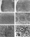
FIG. 1
FIG. 1
Photomicrographs of normal porcine tissues (A and C) and tissues from pigs with PMWS (B, D, E, and F). (A) Normal porcine lymph node with multiple distinct lymphoid follicles. (B) Lymph node from a pig with PMWS. The node is moderately depleted of lymphocytes and lacks follicles. (C) Peyer’s patch from a normal pig. The submucosa is entirely filled with lymphocytes. (D) Peyer’s patch from a pig with PMWS exhibits severe lymphoid depletion. (E) Lymph node from a pig with PMWS. Sinuses contain multinucleate giant cells. (F) Peyer’s patch from a pig with PMWS showing multiple macrophages in a moderately depleted Peyer’s patch filled with amphophilic intracytoplasmic inclusion bodies. Inset, higher magnification of a binucleate macrophage (outlined in center of panel) that is distended with globular intracytoplasmic inclusion bodies.
FIG. 2
FIG. 2
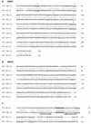
Comparative alignments of ORF1 and ORF2 predicted proteins (A and B) and origins of replication (C) of PCV ISU-31 and PCV PK-15. (A) Alignment of the ORF1-encoded proteins of PCV ISU-31 and PCV PK-15. Domains typical of Rep proteins (1 to 3) and the nucleotide binding site (4) are underlined. (B) Alignment of the ORF2-encoded proteins of PCV ISU-31 and PCV PK-15. (C) DNA alignment of replication origins of PCV ISU-31 and PCV PK-15. The inverted repeat of the putative stem-loop structure is underlined by arrows, the conserved nonanucleotide sequence is shown in boldface type, and the three 6-bp repeats are underlined.
FIG. 3
FIG. 3
Genome organizations of PCV ISU-31 (A) and PCV PK-15 (B). The two major ORFs are indicated by hatched boxes; black arrows show the positions and orientations of ORFs with the potential to encode proteins of more than 45 amino acids.
FIG. 4
FIG. 4
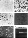
Photomicrograph of lymph node from a normal control pig (A) and a pig with PMWS (B and C) hybridized with an antisense RNA probe (A and B) and a sense RNA probe (C). Panels D and E show results of immunofluorescent staining of uninfected PK-15 cells (D) and cells infected with PCV ISU-31 (E) with a polyclonal serum, which was confirmed to be PCV positive by PCR, from a pig with PMWS. A serum dilution of 1:10 was used. (F) Detection of PCV in tissues of pigs with PMWS by PCR with primers PCV75 and PCV1073. Lane 1, molecular weight marker; lane 2, negative (no DNA) control; lane 3, PCR of DNA isolated from noninfected PK-15 cells; lanes 4 to 10, PCR of DNA samples isolated from lymph nodes of pigs with PMWS.

Similar articles

Cited by

References

    1. Albina E. Epidemiology of porcine reproductive and respiratory syndrome (PRRS): an overview. Vet Microbiol. 1997;55:309–316. - PubMed
    1. Allan G M, McNeilly F, Cassidy J P, Reilly G A C, Adair B, Ellis W A, McNulty M S. Pathogenesis of porcine circovirus, experimental infections of colostrum deprived piglets and examination of pig fetal material. Vet Microbiol. 1995;44:49–64. - PubMed
    1. Allan G M, Phenix K V, Todd D, McNulty M S. Some biological and physico-chemical properties of Porcine Circovirus. J Vet Med B. 1994;41:17–26. - PubMed
    1. Boevink P, Chu P W G, Keese P. Sequence of Subterranean Clover Stunt Virus DNA: affinities with the Geminiviruses. Virology. 1995;207:354–361. - PubMed
    1. Botner A. Diagnosis of PRRS. Vet Microbiol. 1997;55:295–301. - PubMed

MeSH terms

LinkOut - more resources