. 1998 Sep;36(9):2557-64.
doi: 10.1128/JCM.36.9.2557-2564.1998.

Development of rRNA-targeted PCR and in situ hybridization with fluorescently labelled oligonucleotides for detection of Yersinia species

Affiliations

Development of rRNA-targeted PCR and in situ hybridization with fluorescently labelled oligonucleotides for detection of Yersinia species

K Trebesius et al. J Clin Microbiol. 1998 Sep.

Abstract

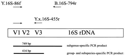
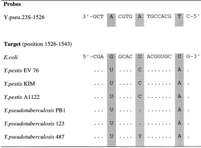
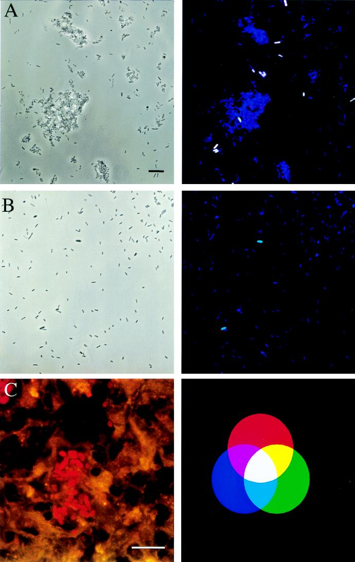
In this report, we present details of two rapid molecular detection techniques based on 16S and 23S rRNA sequence data to identify and differentiate Yersinia species from clinical and environmental sources. Near-full-length 16S rRNA gene (rDNA) sequences for three different Yersinia species and partial 23S rDNA sequences for three Y. pestis and three Y. pseudotuberculosis strains were determined. While 16S rDNA sequences of Y. pestis and Y. pseudotuberculosis were found to be identical, one base difference was identified within a highly variable region of 23S rDNA. The rDNA sequences were used to develop primers and fluorescently tagged oligonucleotide probes suitable for differential detection of Yersinia species by PCR and in situ hybridization, respectively. As few as 10(2) Yersinia cells per ml could be detected by PCR with a seminested approach. Amplification with a subgenus-specific primer pair followed by a second PCR allowed differentiation of Y. enterocolitica biogroup 1B from biogroups 2 to 5 or from other pathogenic Yersinia species. Moreover, a set of oligonucleotide probes suitable for rapid (3-h) in situ detection and differentiation of the three pathogenic Yersinia species (in particular Y. pestis and Y. pseudotuberculosis) was developed. The applicability of this technique was demonstrated by detection of Y. pestis and Y. pseudotuberculosis in spiked throat and stool samples, respectively. These probes were also capable of identifying Y. enterocolitica within cryosections of experimentally infected mouse tissue by the use of confocal laser scanning microscopy.


Figures

FIG. 1
FIG. 1
Positions and specificities of PCR primers and lengths of the amplification products generated with different PCR primers.
FIG. 2
FIG. 2
Alignment of the target region for probe Y.pseu.23S-1526. Numbering of the target positions corresponds to the E. coli numbering described in reference . Identical nucleotides are represented by dots.
FIG. 3
FIG. 3
Result of a seminested-PCR amplification of Yersinia rDNA by using primer pair Y.16S-86f–B.16S-794r for the first PCR (left panel) and primer pair Y.16S-86f–Y.e.eur.16S-455r for the second PCR (right panel) analyzed with a 1.5% agarose gel. One microliter of the first PCR mixture served as a template for the second PCR mixture. Lane 1, Y. pestis EV 76; 2, Y. pseudotuberculosis YPIII; 3, Y. enterocolitica Y-108-c; 4, Y. enterocolitica WA-314; 5, Y. intermedia; 6, Y. ruckeri; 7, H. alvei; 8, 1-kb ladder (Bethesda Research Laboratories, Eggenstein, Germany).
FIG. 4
FIG. 4
Detection of Yersinia species by in situ hybridization. Bars, 10 μm. Binding of at least two differently labelled probes results in distinct mixed colors, as shown in the additive-color illustration (C, right panel). Dual combinations of the red, green, and blue colors result in yellow (green and red), purple (red and blue), and turquoise (green and blue). White is a result of a combination of all three colors. (A and B) The same microscopic fields were viewed by phase-contrast microscopy (left panels) and by epifluorescence microscopy (right panels). Oligonucleotides Y.pseu.23S-1526-CT, Y.p.997-FLUOS, and B.16S-338-Cy5 were simultaneously applied to spiked stool (A) and throat (B) samples. As indicated by the white color, Y. pseudotuberculosis hybridized to all three labeled probes, whereas the turquoise color of the Y. pestis cells in the throat swab specimen is a result of the simultaneous binding of probes B.16S-338-Cy5 and Y.p.16S-997-FLUOS. (C) In situ detection of Y. enterocolitica in spleen sections of an infected mouse (left panel). Tissue sections were hybridized with probe Y.16S-69-CT and detected with the double-exposure option for the green and the red fluorescence of the Leica software package. Single bacterial cells are clearly visible within the spleen sections.

Similar articles

Cited by

References

    1. Aleksic S, Bockemühl J. Proposed revision of the Wauters et al. antigenic scheme for serotyping of Yersinia enterocolitica. J Clin Microbiol. 1984;20:99–102. - PMC - PubMed
    1. Altschul S F, Madden T L, Schaffer A A, Zhang J, Zhang Z, Miller W, Lipman D J. Gapped BLAST and PSI-BLAST: a new generation of protein database search programs. Nucleic Acids Res. 1997;25:3389–3402. - PMC - PubMed
    1. Amann R I, Krumholz L, Stahl D. Fluorescent oligonucleotide probing of whole cells for determinative, phylogenetic and environmental studies in microbiology. J Bacteriol. 1990;172:762–770. - PMC - PubMed
    1. Amann R I, Ludwig W, Schleifer K H. Phylogenetic identification and in situ detection of individual microbial cells without cultivation. Microbiol Rev. 1995;59:143–169. - PMC - PubMed
    1. Ben-Gurion R, Shafferman A. Essential virulence determinants of different Yersinia species are carried on a common plasmid. Plasmid. 1981;5:183–187. - PubMed

Publication types

MeSH terms

LinkOut - more resources